. 2021 Dec;39(12):1574-1580.
doi: 10.1038/s41587-021-00962-z. Epub 2021 Jul 5.

High-content single-cell combinatorial indexing

Affiliations

High-content single-cell combinatorial indexing

Ryan M Mulqueen et al. Nat Biotechnol. 2021 Dec.

Abstract

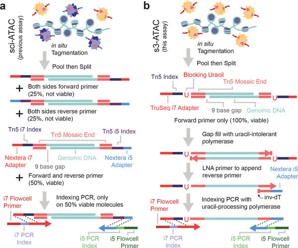
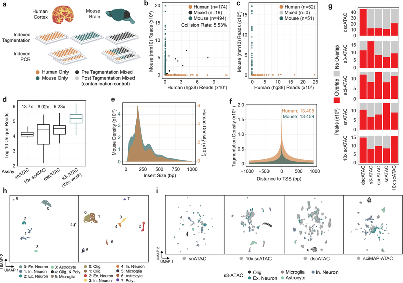
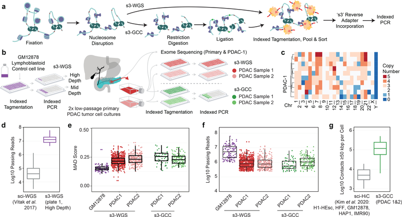
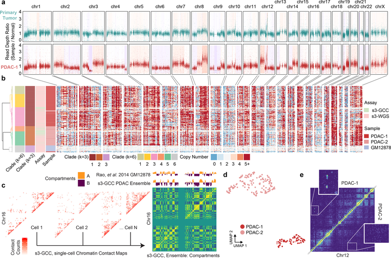
Single-cell combinatorial indexing (sci) with transposase-based library construction increases the throughput of single-cell genomics assays but produces sparse coverage in terms of usable reads per cell. We develop symmetrical strand sci ('s3'), a uracil-based adapter switching approach that improves the rate of conversion of source DNA into viable sequencing library fragments following tagmentation. We apply this chemistry to assay chromatin accessibility (s3-assay for transposase-accessible chromatin, s3-ATAC) in human cortical and mouse whole-brain tissues, with mouse datasets demonstrating a six- to 13-fold improvement in usable reads per cell compared with other available methods. Application of s3 to single-cell whole-genome sequencing (s3-WGS) and to whole-genome plus chromatin conformation (s3-GCC) yields 148- and 14.8-fold improvements, respectively, in usable reads per cell compared with sci-DNA-sequencing and sci-HiC. We show that s3-WGS and s3-GCC resolve subclonal genomic alterations in patient-derived pancreatic cancer cell lines. We expect that the s3 platform will be compatible with other transposase-based techniques, including sci-MET or CUT&Tag.


Figures

Figure 1 |
Figure 1 |. Symmetrical strand single-cell combinatorial indexing ATAC-seq (s3-ATAC) improves molecular capture rate.
(a) Schematic of standard sci-ATAC library construction. (b) Schematic of s3-ATAC library construction with intermediate steps of adapter switching leading to increased genomic molecule capture rate.
Figure 2 |
Figure 2 |. s3-ATAC on human cortex and mouse whole brain.
(a) Experimental flow through and plate layout for the mixed-species experiment, including tagmentation and PCR plate conditions per well. (b) Scatter plots of single-cell libraries with counts of unique reads aligned to mouse or human chromosomes in a chimeric reference genome. Points are colored to reflect species assignment (see Online Methods) in both pre-tagmentation mixing and (c) post-tagmentation mixing. (d) Comparison of unique read counts per cell, restricted to only properly-paired reads for s3-ATAC mouse whole-brain sampled cells to unique read counts produced for previously reported data sets (n=3,034, 4,117, 46,653, and 298 cells, for snATAC, 10x scATAC, dscATAC and ours, respectively). All comparisons to our data are significantly less (Welch’s two-sample t-test, p-values < 5.7×10−42, 3.1×10−40, 1.6×10−37, respectively). Fold improvement of our library complexity per method is listed above the method. Box plot represents median and center quartiles with whiskers at 10th and 90th percentile. (e) Insert size distribution of human and mouse libraries reflect nucleosome banding. (f) Enrichment of reads at transcription start sites (“TSS”) for human and mouse libraries with enrichment calculation following ENCODE standard practices. (g) Stacked bar plot for comparison of peak overlaps across mouse whole brain data sets. Each row is for a different peak set, with each column showing peak overlap in red. (h) UMAP projection of mouse whole brain cell samples (n=837 cells) colored by cluster and cell type assignment (left). UMAP projection human cortex cell samples (n=2,175 cells; right). (i) Integration of s3-ATAC mouse data with other datasets. Points colored by cell type (respective of panel h) or external data set (gray).
Figure 3 |
Figure 3 |. s3-ATAC on human cortex inhibitory neurons.
(a) Subclustering and UMAP projection of human cortical inhibitory neurons (clusters 3 and 4 from panel h., n=342) (b) Genome coverage track of human inhibitory neurons (n=342) aggregated over 4 subclusters for genomic locations overlapping MGE and CGE marker genes LHX6 and ADARB2, respectively, with a zoomed in view of the promoter region (on right). (c) Hierarchical clustering of topic weight per cell (top). Hypergeometric test of gene set analysis enrichment for human inhibitory neuron marker genes (bottom; Fisher’s exact test, see Online Methods), with a genome track of SST to delineate MGE cell types (right).
Figure 4 |
Figure 4 |. s3 whole genome sequencing (s3-WGS) and genome conformation capture (s3-GCC).
(a) Schematic of sci-WGS and sci-GCC library construction. (b) Experimental flow through and plate layout for control GM12878 diploid line (left) and PDAC cell lines (right). (c) Heatmap summary of chromosome count per cell in PDAC-1 SKY data. Example karyotype of PDAC-1 cell (bottom). (d) Boxplot of unique read count per cell for matched GM12878 cell line (n=3,576, 45 cells for sci-WGS and s3-WGS, respectively). (e) Boxplot of MAD score per cell per sample and assay (n=111, 698, 257, 57, and 145 cells, listed left to right). (f) Boxplot of reads passing filter per cell. Cell count same as panel e. (g) Comparison boxplot of s3-GCC and sci-HiC distal contacts (≥50kbp) per cell (n=2,312, 202 cells for sci-HiC and s3-GCC, respectively). Boxplots depicts median and center quartiles with 10th and 90th percentile whiskers.
Figure 5 |
Figure 5 |. s3-WGS for copy number calling and s3-GCC for genome conformation changes.
(a) Whole exome sequencing of primary tumor biopsy and PDAC-1 cell line. Scatterplot of reads per bin with a shading of called copy number variation. (b) Single-cell whole genome copy number calling on 500 kbp bins genome-wide. Cells (rows) are hierarchically clustered and annotated by assay, sample, and assigned clade (left). (c) Representative single-cell contact maps (raw counts) at 1 Mbp resolution for chromosome 16 and ensemble contact map profile at 500 kbp resolution with compartment calling. Compartment eigenvectors are plotted above and compared to GM12878 high-depth bulk HiC data from Rao et al. 2014. (d) UMAP projection of scHiC topic modeling dimensionality reduction and clustering of single-cell distal contact profiles. (e) Putative subclonal translocation on chr12 specific to PDAC-1 (top left) when compared to PDAC-2 (bottom right).

References

    1. Cusanovich DA et al. Multiplex single-cell profiling of chromatin accessibility by combinatorial cellular indexing. Science (80-.). 348, 910–914 (2015). - PMC - PubMed
    1. Adey A et al. Rapid, low-input, low-bias construction of shotgun fragment libraries by high-density in vitro transposition. Genome Biol. 11, R119 (2010). - PMC - PubMed
    1. Tan L, Xing D, Chang CH, Li H & Xie XS Three-dimensional genome structures of single diploid human cells. Science (80-.). 361, 924–928 (2018). - PMC - PubMed
    1. Sos BC et al. Characterization of chromatin accessibility with a transposome hypersensitive sites sequencing (THS-seq) assay. Genome Biol. 17, 20 (2016). - PMC - PubMed
    1. Yin Y et al. High-Throughput Single-Cell Sequencing with Linear Amplification. Mol. Cell 76, 676–690.e10 (2019). - PMC - PubMed

Online Methods References

    1. Li H & Durbin R Fast and accurate long-read alignment with Burrows-Wheeler transform. Bioinformatics 26, 589–595 (2010). - PMC - PubMed
    1. Poplin R et al. Scaling accurate genetic variant discovery to tens of thousands of samples. bioRxiv 201178 (2017) doi:10.1101/201178. - DOI
    1. Frankish A et al. GENCODE reference annotation for the human and mouse genomes. Nucleic Acids Res. 47, D766–D773 (2019). - PMC - PubMed
    1. Sinnamon JR et al. The accessible chromatin landscape of the murine hippocampus at single-cell resolution. Genome Res. 29, 857–869 (2019). - PMC - PubMed
    1. Neph S et al. BEDOPS: high-performance genomic feature operations. Bioinformatics 28, 1919–1920 (2012). - PMC - PubMed

Publication types