Review
. 2021 Aug;17(8):449-461.
doi: 10.1038/s41584-021-00648-5. Epub 2021 Jul 5.

Lyme arthritis: linking infection, inflammation and autoimmunity

Affiliations
Review

Lyme arthritis: linking infection, inflammation and autoimmunity

Robert B Lochhead et al. Nat Rev Rheumatol. 2021 Aug.

Abstract

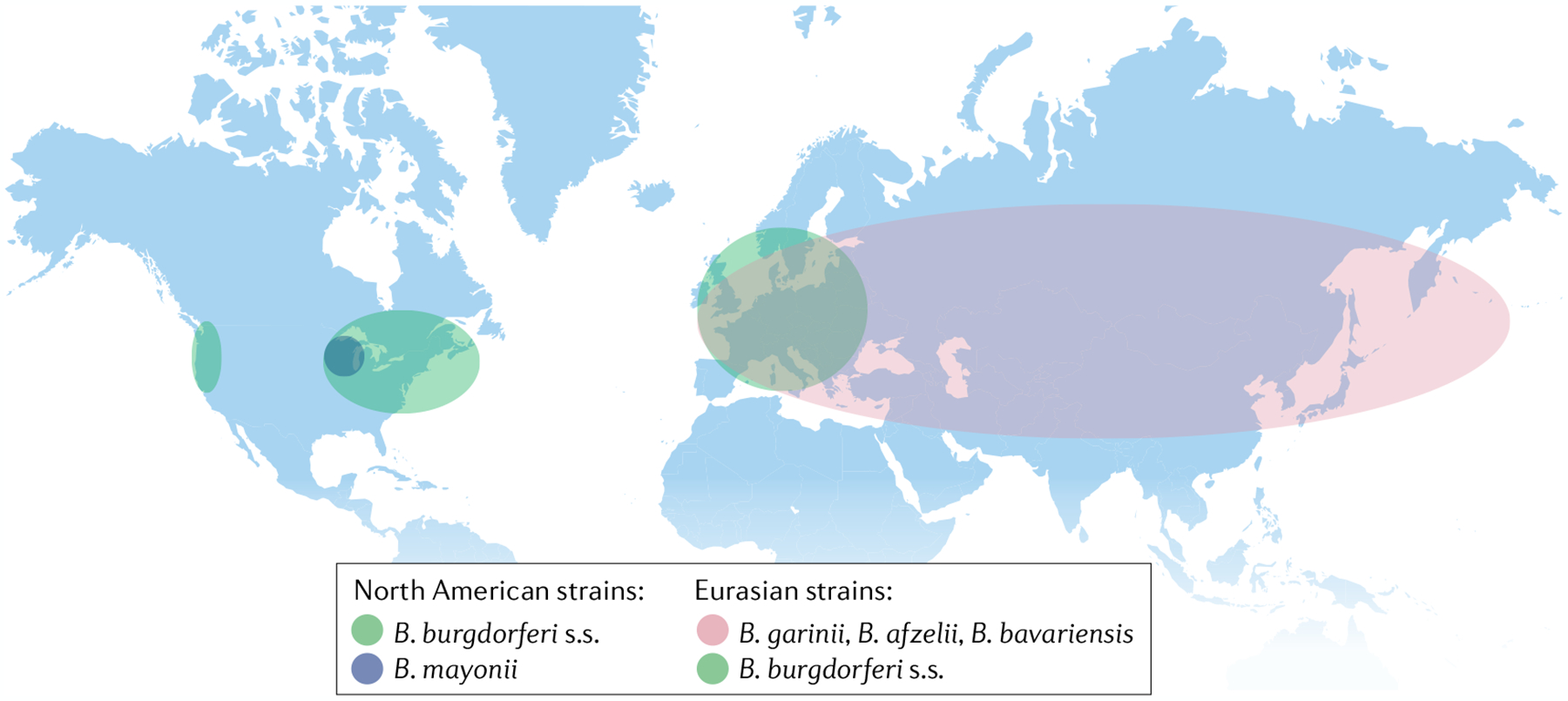
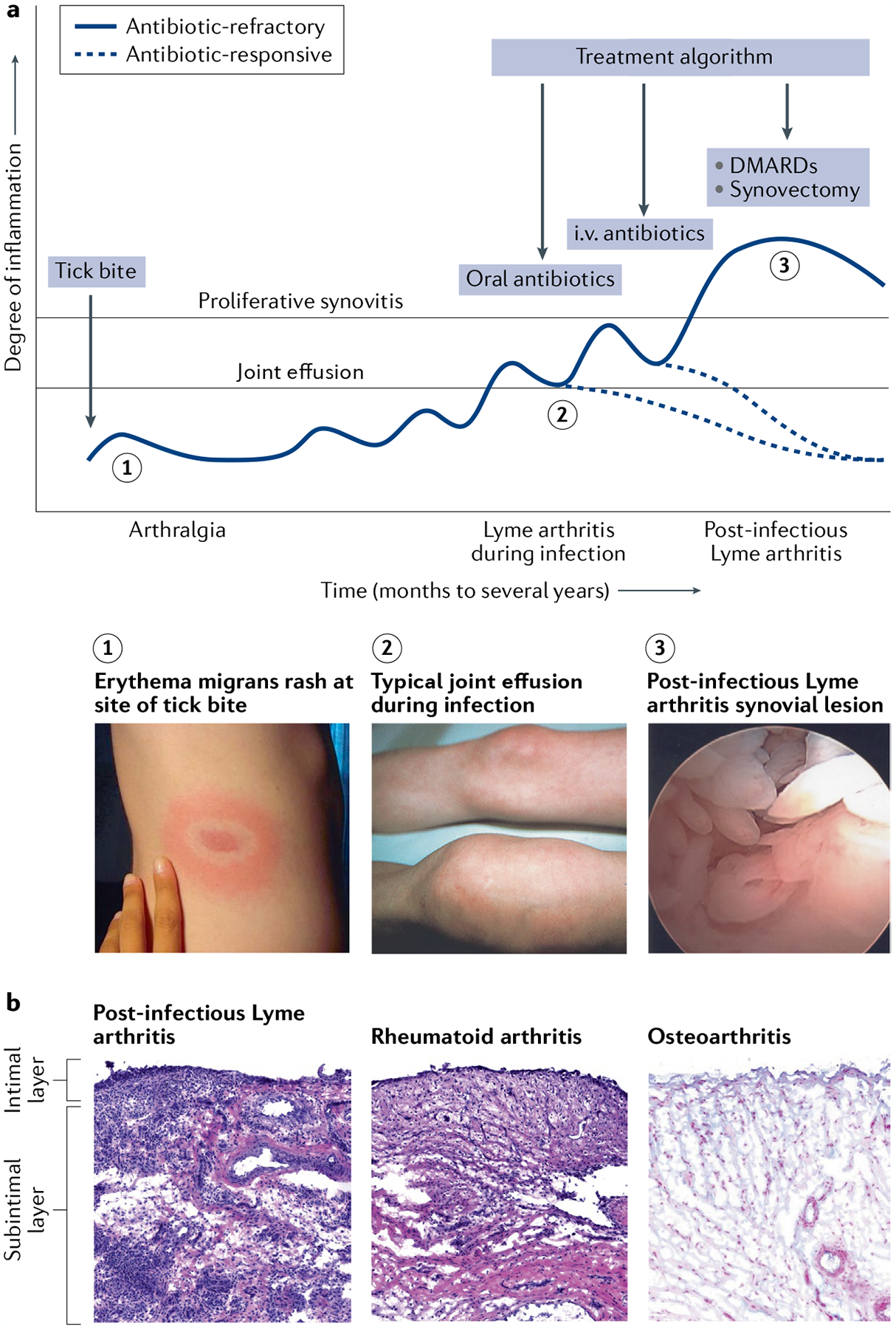
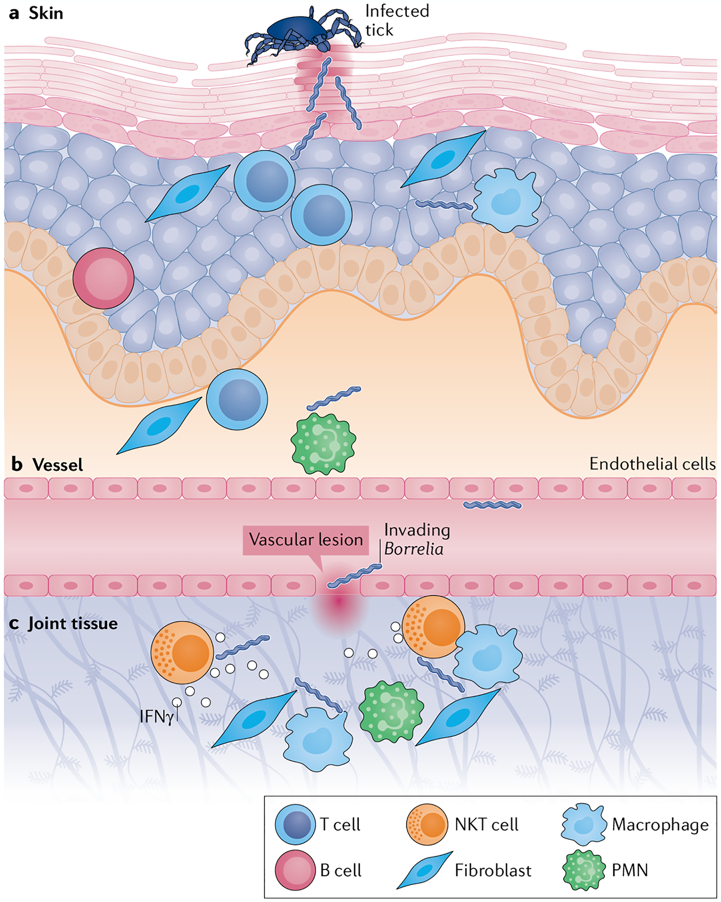
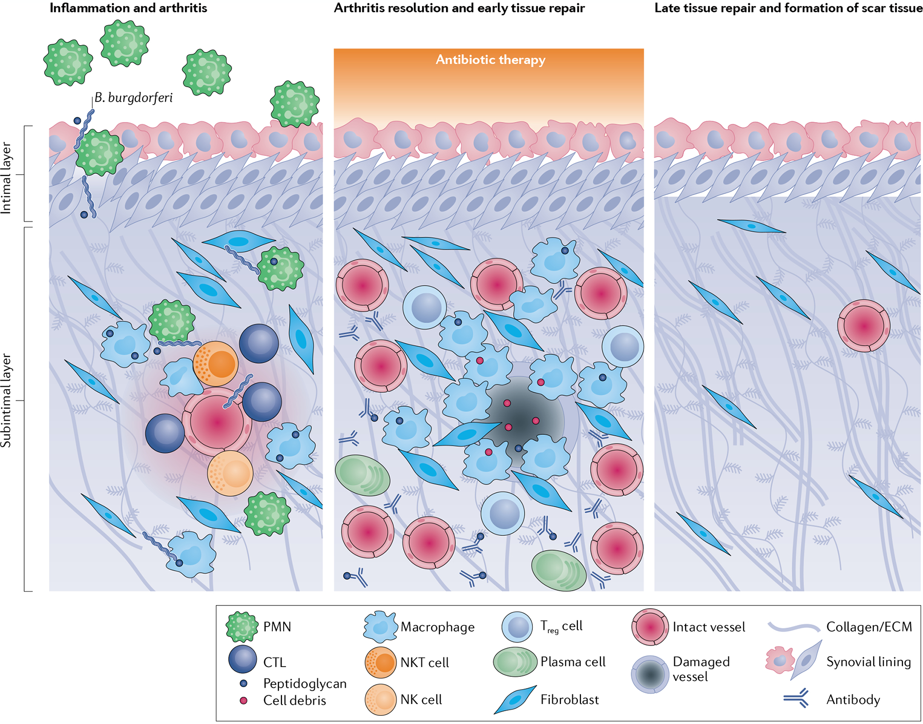
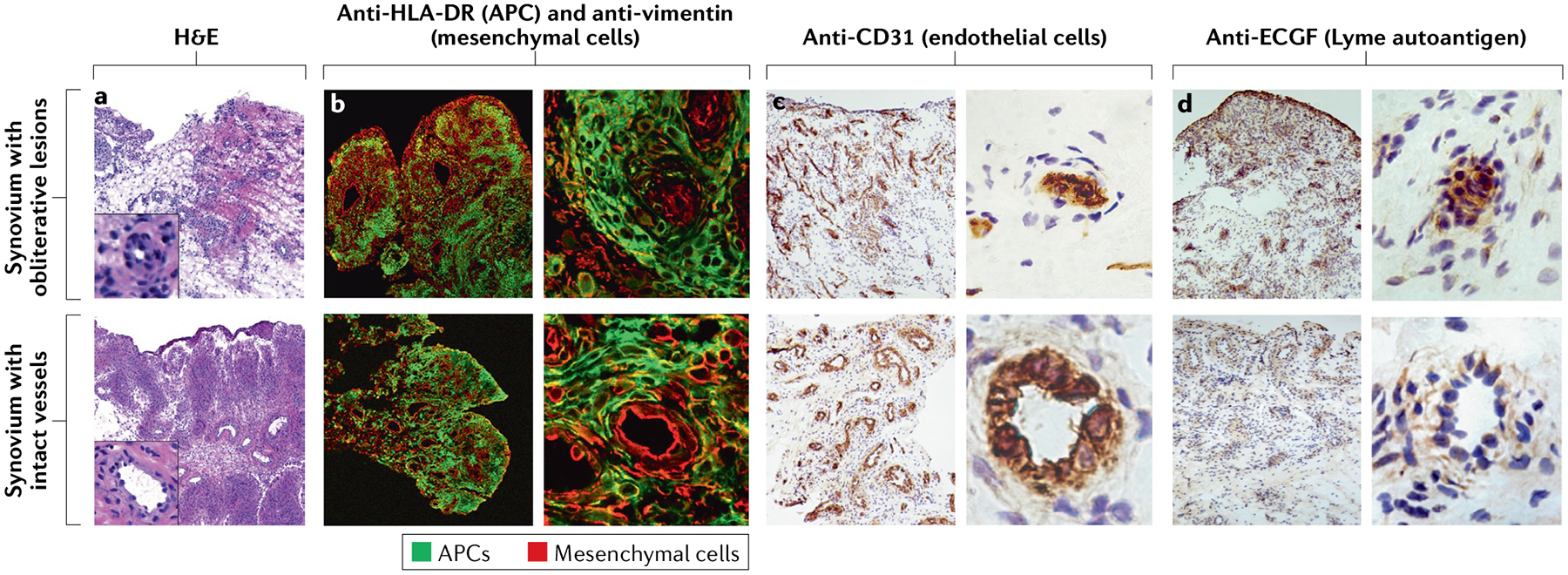
Infectious agents can trigger autoimmune responses in a number of chronic inflammatory diseases. Lyme arthritis, which is caused by the tick-transmitted spirochaete Borrelia burgdorferi, is effectively treated in most patients with antibiotic therapy; however, in a subset of patients, arthritis can persist and worsen after the spirochaete has been killed (known as post-infectious Lyme arthritis). This Review details the current understanding of the pathogenetic events in Lyme arthritis, from initial infection in the skin, through infection of the joints, to post-infectious chronic inflammatory arthritis. The central feature of post-infectious Lyme arthritis is an excessive, dysregulated pro-inflammatory immune response during the infection phase that persists into the post-infectious period. This response is characterized by high amounts of IFNγ and inadequate amounts of the anti-inflammatory cytokine IL-10. The consequences of this dysregulated pro-inflammatory response in the synovium include impaired tissue repair, vascular damage, autoimmune and cytotoxic processes, and fibroblast proliferation and fibrosis. These synovial characteristics are similar to those in other chronic inflammatory arthritides, including rheumatoid arthritis. Thus, post-infectious Lyme arthritis provides a model for other chronic autoimmune or autoinflammatory arthritides in which complex immune responses can be triggered and shaped by an infectious agent in concert with host genetic factors.


Conflict of interest statement

Competing interests

The authors declare no competing interests.

Figures

Fig. 1 |
Fig. 1 |. Geographic distribution of Borrelia burgdorferi species relevant to human disease.
Borrelia burgdorferi sensu stricto (B. burgdorferi s.s.) is the major species in North America and is primarily found in the USA in the north-eastern and mid-Atlantic states, the upper Midwest, in northern California and, to a lesser degree, in Oregon and Washington. B. burgdorferi s.s. also extends into Canada at each of the bordering USA locations. Borrelia mayonii is much less common than B. burgdorferi s.s. and is restricted to the upper midwestern states in the USA. European strains include Borrelia garinii and Borrelia afzelii and, to a lesser extent, B. burgdorferi s.s. and Borrelia bavariensis, which is closely related to B. garinii. In Asia, B. garinii is the predominant species, but B. bavariensis and B. afzelii are also found there.
Fig. 2 |
Fig. 2 |. Lyme arthritis stages and characteristics.
a | In untreated patients, Lyme disease occurs in stages, with different manifestations present at each stage. A slowly expanding erythema migrans rash commonly appears 3 to 32 days after a bite by a Borrelia burgdorferi-infected Ixodes tick (1), which can be accompanied by flu-like symptoms such as fever, headache, myalgias, arthralgias, malaise and fatigue. In the north-eastern states of the USA, Lyme arthritis typically causes large joint effusions, particularly affecting the knees (2), which develop a median of 6 months after the initial skin lesion. Arthritis usually resolves after 1–3 months of oral and, if necessary, intravenous (i.v.) antibiotic therapy (antibiotic-responsive Lyme arthritis). In a small subset of patients, arthritis persists or worsens despite 2–3 months of antibiotic therapy and apparent spirochaetal killing (post-infectious Lyme arthritis). These patients typically develop a highly proliferative synovial lesion (3) that does not respond to further courses of antibiotic therapy (antibiotic-refractory Lyme arthritis). Treatments such as DMARDs or arthroscopic synovectomy help to resolve their arthritis. b | The synovial lesion in post-infectious Lyme arthritis is similar to the lesion in rheumatoid arthritis and other inflammatory arthritides. By contrast, osteoarthritis synovium typically has minimal cellular infiltrate, the intimal layer is not inflamed or thickened, and the subintimal layer is composed of healthy, intact microvasculature and highly organized collagen fibres. In this figure, the synovial lesions from Lyme arthritis and rheumatoid arthritis are stained with haematoxylin and eosin (H&E), and osteoarthritis synovium is stained with H&E and Alcian blue to show acidic glycosaminoglycans on the outer surface of collagen fibre bundles and along the synovial lining. Image of the knee in part a reprinted with permission from REF., Elsevier.
Fig. 3 |
Fig. 3 |. Spirochaete invasion into joint tissue.
a | Spirochaetes invade the skin during the bloodmeal of an infected Ixodes tick. Upon entry, tissue-resident T cells, B cells, resident antigen-presenting cells (such as macrophages and dendritic cells), some polymorphonuclear cells (PMNs) and stromal cells (such as fibroblasts, keratinocytes, epithelial cells and endothelial cells) are responsible for early-acute immune responses to infection. b | A small number of spirochaetes escape the site of invasion and enter the vasculature, where Borrelia surface lipoproteins interact with vascular endothelial cells to induce the loosening of tight junctions. c | Spirochaetes enter the extracellular matrix of joint tissue through vascular lesions. Once in the joint tissue, they induce acute inflammatory responses by resident cells such as endothelial cells and synovial fibroblasts, which produce adhesion molecules, matrix metalloproteinases and innate immunity cytokines and chemokines. Natural killer T (NKT) cells produce IFNγ in response to CD1-presented immunogenic Borrelia glycolipids, thereby enhancing vascular barrier function and limiting spirochaetal invasion and chronic inflammation. The cytotoxic function of NKT cells might also directly contribute to spirochaetal killing. The nature and magnitude of these early immune responses in the skin and joint help to set the stage for subsequent arthritis development.
Fig. 4 |
Fig. 4 |. Stages of arthritis and proposed tissue repair in antibiotic-responsive lyme arthritis.
Immune responses to Borrelia burgdorferi and B. burgdorferi peptidoglycan by endothelial cells, fibroblasts, lymphocytes (such as natural killer (NK cells), natural killer T (NKT) cells and cytotoxic T lymphocytes (CTLs)) and myeloid cells (such as macrophages and polymorphonuclear cells (PMNs)) in the synovium trigger localized inflammation, tissue damage and arthritis. Antibiotic therapy is given during acute joint infection and inflammation, facilitating arthritis resolution and the initiation of early tissue repair responses. These responses are dominated by pro-angiogenic factors and the activation of tissue-repairing macrophages and fibroblasts, which remove bacterial debris and damaged cells from the damaged microvasculature, extracellular matrix (ECM) and fibrotic tissue. Other immune cells such as regulatory T (Treg) cells and plasma cells might also be present during arthritis resolution. Over several months, synovial fibroblasts differentiate into myofibroblasts and lay down collagen and form scar tissue, leading to full recovery.
Fig. 5 |
Fig. 5 |. Microvascular involvement in the synovial lesion of post-infectious lyme arthritis.
a | Haematoxylin and eosin (H&E)-stained sections of synovial tissue from representative patients with post-infectious Lyme arthritis with varying degrees of inflammation, fibrosis and vascular damage (insets show enlarged images of vessels). b | Synovial tissue sections stained with fluorescently labelled anti-HLA-DR (green) and anti-vimentin (red) antibodies, showing localization of antigen-presenting cells (APCs) and mesenchymal cells (such as fibroblasts and endothelial cells), respectively. c | Around half of patients with post-infectious Lyme arthritis have evidence of vascular damage, including obliterative microvascular lesions, as shown by staining with the endothelial cell marker CD31. d | Obliterative microvascular lesions are also enriched with the Lyme disease autoantigen endothelial cell growth factor (ECGF). In panels b–d, the panels on the left show the general architecture of the synovial tissue at low magnification, and the panels on the right show single blood vessels from the same sections at high magnification. Parts c and d adapted with permission from REF., Wiley. © 2014 by the American College of Rheumatology.
Fig. 6 |
Fig. 6 |. Cellular architecture of the post-infectious lyme arthritis synovial lesion.
a | The post-infectious Lyme arthritis synovial lesion is characterized by widespread fibrosis and areas of marked inflammation. Fibrotic areas contain large numbers of synovial fibroblasts, obliterative microvascular lesions, disordered collagen and other extracellular matrix (ECM) proteins. Areas of inflammation are found primarily in highly vascularized synovial intimal and subintimal layers that can contain obliterative microvascular lesions and/or intact vessels. Immune cells, such as macrophages, CD4+ T helper (TH) cells, cytotoxic T lymphocytes (CTLs; mostly CD8+ T cells with a few γδ T cells), natural killer (NK) cells, and large numbers of antibody-producing plasma cells, are found primarily in vascularized areas, but can be found throughout the tissue. Vascularized areas also contain many HLA-DR-expressing synovial fibroblasts; however, they tend to have less fibrotic tissue. Bacterial peptidoglycan is present in synovial fluid and might additionally be present in inflamed tissue, along with degraded cellular and ECM debris. Only a few polymorphonuclear cells (PMNs) are present in post-infectious Lyme arthritis synovial tissue, but more are present in synovial fluid. b | In this panel, a hypothesis is developed regarding the roles of important cells and immune mediators in autoimmune-mediated damage to the endothelium in inflamed synovia. Large amounts of IFNγ produced by TH cells, CTLs and NK cells induce potent responses by HLA-DR-expressing synovial fibroblasts and macrophages, which upregulate proteins associated with antigen presentation, T cell activation and inflammation. Borrelia burgdorferi peptidoglycan and cell debris might amplify these responses. Synovial fibroblasts and macrophages present MHC class II-restricted peptides derived from Lyme autoantigens, which are abundant in synovial tissue, to autoreactive TH cells, perpetuating IFNγ responses in the tissue. Endothelial cells, which were damaged during infection with B. burgdorferi, can be targeted for killing by CTLs, either through direct CTL-mediated killing or through autoantibody-dependent cell-mediated cytotoxicity (ADCC), or both. Further damage to the microvasculature releases more autoantigens and debris, leading to a feedback loop of chronic inflammation and tissue damage.

References

    1. von Herrath MG, Fujinami RS & Whitton JL Microorganisms and autoimmunity: making the barren field fertile? Nat. Rev. Microbiol 1, 151–157 (2003). - PubMed
    1. Steere AC et al. Lyme borreliosis. Nat. Rev. Dis. Prim 2, 16090 (2016). - PMC - PubMed
    1. Steere AC Lyme disease. N. Engl. J. Med 321, 586–596 (1989). - PubMed
    1. Radolf JD, Strle K, Lemieux JE & Strle F Lyme disease in humans. Curr. Issues Mol. Biol 42, 333–384 (2021). - PMC - PubMed
    1. Steere AC, Schoen RT & Taylor E The clinical evolution of Lyme arthritis. Ann. Intern. Med 107, 725–731 (1987). - PubMed

MeSH terms