. 2021 Jul 1;10(8):2.
doi: 10.1167/tvst.10.8.2.

Deep Learning Applied to Automated Segmentation of Geographic Atrophy in Fundus Autofluorescence Images

Affiliations

Deep Learning Applied to Automated Segmentation of Geographic Atrophy in Fundus Autofluorescence Images

Janan Arslan et al. Transl Vis Sci Technol. .

Abstract

Purpose: This study describes the development of a deep learning algorithm based on the U-Net architecture for automated segmentation of geographic atrophy (GA) lesions in fundus autofluorescence (FAF) images.

Methods: Image preprocessing and normalization by modified adaptive histogram equalization were used for image standardization to improve effectiveness of deep learning. A U-Net-based deep learning algorithm was developed and trained and tested by fivefold cross-validation using FAF images from clinical datasets. The following metrics were used for evaluating the performance for lesion segmentation in GA: dice similarity coefficient (DSC), DSC loss, sensitivity, specificity, mean absolute error (MAE), accuracy, recall, and precision.

Results: In total, 702 FAF images from 51 patients were analyzed. After fivefold cross-validation for lesion segmentation, the average training and validation scores were found for the most important metric, DSC (0.9874 and 0.9779), for accuracy (0.9912 and 0.9815), for sensitivity (0.9955 and 0.9928), and for specificity (0.8686 and 0.7261). Scores for testing were all similar to the validation scores. The algorithm segmented GA lesions six times more quickly than human performance.

Conclusions: The deep learning algorithm can be implemented using clinical data with a very high level of performance for lesion segmentation. Automation of diagnostics for GA assessment has the potential to provide savings with respect to patient visit duration, operational cost and measurement reliability in routine GA assessments.

Translational relevance: A deep learning algorithm based on the U-Net architecture and image preprocessing appears to be suitable for automated segmentation of GA lesions on clinical data, producing fast and accurate results.


Conflict of interest statement

Disclosure: J. Arslan, None; G. Samarasinghe, None; A. Sowmya, None; K.K. Benke, None; L.A.B. Hodgson, R.H. Guymer, Bayer (C), Novartis (C), Roche Genentech (C), Apellis (C); P.N. Baird, None

Figures

Figure 1.
Figure 1.
Image preprocessing using the CLAHE technique. (A) Original FAF image and (B) CLAHE-applied FAF image. The original image was very dark in contrast and illumination. Using the original image would have resulted in contrast-related errors, making it more difficult for the algorithm to distinguish the GA lesions. The CLAHE-applied image shows the dramatic improvement using this technique. CLAHE, Contrast Limited Adaptive Histogram Equalization; FAF, Fundus autofluorescence.
Figure 2.
Figure 2.
U-Net architecture. The U-Net architecture was created for biomedical image segmentation. The U-Net is aptly named because of the arrangements of the filters in a “U” shape. The contracting pathway (left) consists of two 3 × 3 convolutions, ReLU activation and 2 × 2 max pooling. The expansive pathway (right) consist of 2 × 2 convolution, a concatenation with the correspondingly cropped feature map from the contracting pathway, two 3 × 3 convolutions and ReLU activation (see Ronneberger et al.37).
Figure 3.
Figure 3.
Fivefold cross-validation. This cross-validation was chosen because it has been empirically shown to yield test error rates not excessively influenced by high bias and variances.
Figure 4.
Figure 4.
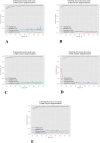
Learning curves with training/validation loss and accuracy across all fivefolds of cross-validation. (A) Cross-validation 1. (B) Cross-validation 2. (C) Cross-validation 3. (D) Cross-validation 4. (E) Cross-validation 5. The learning curve illustrates a consistent outcome of high accuracy and low loss throughout all fivefolds.
Figure 5.
Figure 5.
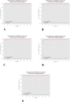
Learning curves for DSC and DSCloss across all fivefolds of cross-validation. (A) Cross-validation 1. (B) Cross-validation 2. (C) Cross-validation 3. (D) Cross-validation 4. (E) Cross-validation 5. The learning curve illustrates a consistent outcome of high DSC and low loss throughout all 5-folds. DSC, Dice similarity coefficient.
Figure 6.
Figure 6.
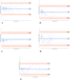
Bland-Altman plots and coefficient of repeatability across all fivefolds of cross-validation (in units of pixels).
Figure 7.
Figure 7.
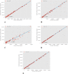
Spearman's correlations and regression lines across all fivefolds of cross-validation (in units of pixels).
Figure 8.
Figure 8.
Qualitative assessment of model prediction outcomes. Test cases A and B. In addition to assessing the performance of U-Net quantitatively, we evaluated the performance by visually assessing the degree of accuracy of U-Net-based lesion segmentation. The test cases presented demonstrate a good segmentation outcome.
Figure 9.
Figure 9.
Qualitative assessment of model prediction outcomes. Test cases A and B. In addition to assessing the performance of U-Net quantitatively, we evaluated the performance by visually assessing the degree of accuracy of U-Net-based lesion segmentation. The test cases presented demonstrate a good segmentation outcome.

References

    1. Resnikoff S, Pascolini D, Etya'ale D, et al. .. Global data on visual impairment in the year 2002. Bull World Health Organ. 2002; 82: 844–851. - PMC - PubMed
    1. Wong WL, Su X, Li X, et al. .. Global prevalence of age-related macular degeneration and disease burden projection for 2020 and 2040: a systematic review and meta-analysis. Lancet Global Health. 2014; 2: e106–e116. - PubMed
    1. Boyer DS, Schmidt-Erfurth U, van Lookeren Campagne M, Henry EC, Brittain C.. The pathophysiology of geographic atrophy secondary to age-related macular degeneration and the complement pathway as a therapeutic target. Retina. 2017; 37: 819–835. - PMC - PubMed
    1. Holz FG, Strauss EC, Schmitz-Valckenberg S, van Lookeren Campagne M.. Geographic atrophy: clinical features and potential therapeutic approaches. Ophthalmology. 2014; 121: 1079–1091. - PubMed
    1. Solomon SD, Lindsley K, Vedula SS, Krzystolik MG, Hawkins BS.. Anti-vascular endothelial growth factor for neovascular age-related macular degeneration. Cochrane Database Syst Rev. 2014; 8(8): CD005139. - PMC - PubMed

Publication types