. 2021 Sep 16;89(10):e0017821.
doi: 10.1128/IAI.00178-21. Epub 2021 Jul 6.

Trypanosoma cruzi Exploits E- and P-Selectins To Migrate Across Endothelial Cells and Extracellular Matrix Proteins

Affiliations

Trypanosoma cruzi Exploits E- and P-Selectins To Migrate Across Endothelial Cells and Extracellular Matrix Proteins

Smaro Panagiotidou et al. Infect Immun. .

Abstract

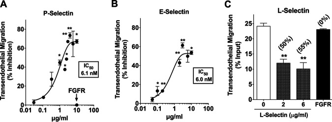
The Chagas disease parasite Trypanosoma cruzi must extravasate to home in on susceptible cells residing in most tissues. It remains unknown how T. cruzi undertakes this crucial step of its life cycle. We hypothesized that the pathogen exploits the endothelial cell programming leukocytes use to extravasate to sites of inflammation. Transendothelial migration (TEM) starts after inflammatory cytokines induce E-selectin expression and P-selectin translocation on endothelial cells (ECs), enabling recognition by leukocyte ligands that engender rolling cell adhesion. Here, we show that T. cruzi upregulates E- and P-selectins in cardiac ECs to which it binds in a ligand-receptor fashion, whether under static or shear flow conditions. Glycoproteins isolated from T. cruzi (TcEx) specifically recognize P-selectin in a ligand-receptor interaction. As with leukocytes, binding of P-selectin to T. cruzi or TcEx requires sialic acid and tyrosine sulfate, which are pivotal for downstream migration across ECs and extracellular matrix proteins. Additionally, soluble selectins, which bind T. cruzi, block transendothelial migration dose dependently, implying that the pathogen bears selectin-binding ligand(s) that start transmigration. Furthermore, function-blocking antibodies against E- and P-selectins, which act on endothelial cells and not T. cruzi, are exquisite in preventing TEM. Thus, our results show that selectins can function as mediators of T. cruzi transendothelial transmigration, suggesting a pathogenic mechanism that allows homing in of the parasite on targeted tissues. As selectin inhibitors are sought-after therapeutic targets for autoimmune diseases and cancer metastasis, they may similarly represent a novel strategy for Chagas disease therapy.

Keywords: Chagas disease; E-selectin; L-selectin; P-selectin; Trypanosoma cruzi; sialic acid; sulfate; transendothelial migration.


Figures

FIG 1
FIG 1
T. cruzi augments expression of P- and E-selectins and of inflammatory cytokines and Toll-like receptor 2 on cardiac endothelial cells and binds to P-, E-, and L-selectins dose dependently and saturable under static conditions. (A) T. cruzi selectively upregulates selectins on cardiac endothelial cells. Confluent mouse cardiac endothelial cells and human cardiomyocytes (myocytes) were incubated for 3 h with Colombiana strain trypomastigotes (MOI, 1) or control vehicle PBS; monolayers were processed for RNA-seq (in triplicates). Results are expressed as fold increase relative to uninfected (vehicle treated) cells;
FIG 2
FIG 2
T. cruzi subjected to shear flow reduces motility on P-selectin surface. (A) Visualization by video microscopy (20× objective) of T. cruzi Colombiana trypomastigotes flowing on BSA (vehicle) or human P-selectin (SELP) immobilized on glass. Lines depict the trajectory of individual trypomastigotes. (B) T. cruzi velocity quantified by Image J software. Each dot represents an individual pathogen. Data are derived from three independent experiments. Error bars indicate SD; Mann-Whitney U test was performed. ****, P < 0.001; ns, not significant. Figure S1 in the supplemental material displays T. cruzi trajectories for the duration of the video microscopy.
FIG 3
FIG 3
E-selectin, EDTA, plant lectins, neuraminidase, and sulfatase specifically inhibit the binding of P-selectin to T. cruzi. T. cruzi trypomastigotes (Tulahuen strain) were incubated without (vehicle) or with human SELE, ICAM-1, or VCAM-1 and then with SELP-Fc. Parasite-bound SELP-Fc was detected by chemiluminescent Western blotting using HRP-labeled anti-human Fc-specific antibodies. Band intensity was quantified using ImageJ and displayed as percent inhibition (bottom blot display). (B) Dose-response curve of T. cruzi-SELP binding inhibition by SELE. The protocol was the same as that described above. (C) T. cruzi trypomastigotes (Colombiana strain) were incubated with SELP-Fc with PBS (vehicle) or EDTA. The protocol was that described above except that SELP-Fc was detected by fluorescent Western blotting using the LI-COR Odyssey Western blotting system. (D) T. cruzi trypomastigotes (Colombiana strain) were incubated with five plant lectins having distinct sugar-binding specificities (WGA, wheat germ agglutinin; ConA, Concanavalin A; SNA, Sambuca nigra agglutinin; MAA, Maackia amurensis agglutinin; and UEA-1, Ulex europaeus agglutinin-1), followed by SELP-Fc. Results were obtained by the LI-COR Odyssey Western blotting system. (E) T. cruzi trypomastigotes (Colombiana strain) were incubated without (vehicle) or with Vibrio cholerae neuraminidase (NANAse) and P-selectin-Fc. Results were obtained by the LI-COR Odyssey Western blotting system. (F) T. cruzi trypomastigotes (Colombiana strain) were incubated without (vehicle) or with sulfatase (30 min) and P-selectin-Fc. Results were obtained by the LI-COR Odyssey Western blotting system. Panels A, B, C, E, and F are representative of three independent experiments. (D) Data shown represent the means from three independent experiments. Error bars indicate SD.
FIG 4
FIG 4
Binding of P-, E-, and L-selectins to glycoproteins/mucins isolated from T. cruzi reproduces molecular characteristics of selectin binding to live parasite. (A) Ninety-six-well plates were coated with TcEx or VeroEX and incubated with the indicated amount of SELP or FGFR. The plot was made using nonlinear regression. (B) Human PSGL-1 as a positive control or TcEx as experimental subject and incubated with SELP. (C) Human PSGL-1 incubated with SELP with or without treatment with V. cholerae neuraminidase (NANAse) or tyrosine sulfatase. (D) TcEx incubated with SELP with or without V. cholerae neuraminidase (SELP + NANAse) or sulfatase (SELP + Sulfatase). (E) TcEx treated with NANAse or sulfatase, washed to remove the enzymes, and then incubated with SELP. (F) TcEx with vehicle, SELE, or SELL and subsequently with SELP. In all experiments, plate-bound P-selectin was identified and quantified using biotinylated anti-human P-selectin antibody, followed by streptavidin-HRP and TMB substrate. Results are combinations of two, three, or four independent experiments, with each point representing one well. Data are presented as the means. Error bars indicate SD. One-way ANOVA was used. *, P ≤ 0.05; **, P ≤ 0.001; ****, P ≤ 0.0001.
FIG 5
FIG 5
T. cruzi enters confluent endothelial cells preferentially compared to multiplying subconfluent cells, and entry in confluent endothelial cells is specifically inhibited by soluble P- and E-selectins in a sialic acid-dependent manner. Confluent and approximately 60% subconfluent mouse cardiac endothelial cells were infected with trypomastigotes of T. cruzi Tulahuen or CL Brenner strain (expressing dtTomato fluorescent protein) at an MOI of 20. After 3 to 4 days, cells were fixed with methanol and stained with Diff-Quik. Percent infection was calculated by optical microscopy. Infection by the Colombiana strain gave similar results (data not shown). (B) Visualization of a representative field of a microtiter well infected with CL Brener strain T. cruzi amastigotes inside the cells, seen as red dots. (C) Confluent monolayers (in triplicates) of human umbilical vein endothelial cells (HUVECs) were exposed to T. cruzi trypomastigotes (Colombiana strain, MOI of 20) for 3 h in the presence of PBS (Veh) or the indicated concentrations of P-selectin (SELP) or fibroblast growth factor receptor (FGFR) (5 μg/ml). T. cruzi entry into HUVECs was assessed 3 days later by counting >300 endothelial cells/well containing intracellular amastigotes. (D) Experimental protocol is as described above except for the use of SELE. (E) T. cruzi was allowed to enter mcECs in the absence or presence of SELE (5 μg/ml) without (T. cruzi + SELE) or with treatment with V. cholerae neuraminidase (Tc + NANAse + SELE). Results are combinations of three independent experiments. Data are presented as the means. Error bars indicate SD. Unpaired t test (A) or one-way ANOVA (C, D, and E) were used. *, P ≤ 0.05; **, P ≤ 0.001; ***, P < 0.005; ns, not significant.
FIG 6
FIG 6
Sialic acid and sulfate epitopes are required for T. cruzi migration through endothelial cells and extracellular matrix proteins. (A) Boyden chamber transendothelial migration setup. The upper layer of sterile 3.0-μm pore inserts was coated with extracellular matrix (ECM) collagens (gelatin, 1 mg/ml) and fibronectin (0.5 μg/ml) and placed in a 24-well plate, to which primary human vein endothelial cells (HUVEC) were seeded at a density of 5 × 104 cells/transwell and grown overnight at 37°C in endothelial cell (EC) growth medium. T. cruzi trypomastigotes (Colombiana strain) were tagged with the fluorescent CellTracker red dye, resuspended in endothelial cell medium, and added (MOI, 10) to the monolayers without or with various treatments. After interaction with cell adhesion molecules (CAMS) such as selectins, parasites traverse the endothelial cell monolayer and extracellular matrix layer, penetrate the 3-μm pores, and then fall into the basolateral medium (10% FBS–DMEM) in the lower chamber of the transwell setup. T. cruzi that transmigrated to the lower chamber was measured using BioTek Synergy HT plate reader and quantified using a calibration curve constructed with predetermined doses of red-tagged trypomastigotes. Illustration was created using www.biorender.com. (B) Sialic acid and sulfate epitopes are critical for T. cruzi transendothelial migration. T. cruzi was treated with PBS (vehicle), V. cholerae neuraminidase (NANAse), sulfatase, or α-mannosidase (α-ManAse) and the mixtures added to the 24-well plate inserts (duplicates). Transendothelial migration was assessed at 2 h and 4 h. Numbers above bars represent percent inhibition relative to vehicle-treated cells set at 100% migration for two experiments with similar results. (C) Visualization by fluorescence microscopy of T. cruzi that transmigrated HUVEC after 2 h; before visualization, parasites were pelleted by centrifuging the 24-well plates at 2,000 × g for 10 min. Results are a combination of two independent experiments. Data are presented as the means. Error bars indicate SD. One-way ANOVA was used. *, P ≤ 0.05; **, P ≤ 0.001; ***, P < 0.005; ns, not significant.
FIG 7
FIG 7
Soluble selectins are competitive inhibitors of T. cruzi transendothelial migration. (A, B, and C) T. cruzi trypomastigotes (Colombiana strain) were incubated with the indicated concentrations of the soluble extracellular domain of recombinant human P-selectin (A), E-selectin (B), or L-selectin (C). The mixtures were added to confluent HUVEC monolayers (in duplicates), and transendothelial migration was allowed to proceed for 2 h. Transmigrating parasites were quantified using a BioTek Synergy HT plate reader (see Fig. 6A). The plots are a composite of seven (A), four (B), and two (C) distinct experiments. Numbers in parentheses in panel C represent percent inhibition. Data are presented as the means. Error bars indicate SD. One-way ANOVA; *, P ≤ 0.05; **, P ≤ 0.001; ***, P < 0.005; ns, not significant.
FIG 8
FIG 8
Selectin blockade prevents transendothelial migration of T. cruzi. (A) Red-fluorescent T. cruzi trypomastigotes (Colombiana or Tulahuen strain) were mixed with vehicle (Veh) or 1 μg/ml anti-E-selectin function-blocking antibody clone 9A9, and the mixtures were added to confluent HUVEC on transwells (see Fig. 6A). After 2 h, parasites that transmigrated through the human endothelial cells and extracellular matrix proteins were quantified on a BioTek plate reader. (B) Dose-response of the inhibition of T. cruzi trypomastigote (Colombiana strain) transendothelial migration by anti-E-selectin MAb, clone 9A9 MAb (see the legend to Fig. 6A for protocol). (C) Dose-response of the inhibition of T. cruzi trypomastigote (Colombiana strain) transendothelial migration by anti-P-selectin MAb, clone AK4. (D) Red-fluorescent T. cruzi trypomastigotes (Colombiana strain) was mixed with 3 μg/ml anti-P-selectin function-blocking antibody clone AK4 or anti-E-selectin antibody clone 9A9, or with a mix of both antibodies each at 3 μg/ml, and added to confluent HUVEC monolayers. After 2 h, transmigrating parasites were quantified in the BioTek fluorescence plate reader; percent inhibition was calculated relative to parasites without antibodies set at 0% inhibition. (E) Visualization by fluorescence microscopy of T. cruzi transendothelial migration through HUVEC monolayer without (0) or with 3 μg/ml of AK4 and 3 μg/ml of 9A9 antibodies (3 + 3). Each dot or stick represents a swimming trypanosome in the basolateral medium that traversed the endothelial cell and extracellular matrix protein barrier. The data presented in panel A are a composite of two of five independent experiments (B and C). Data are presented as the means. Error bars indicate SD. One-way ANOVA was used. *, P ≤ 0.05; **, P ≤ 0.001; ns, not significant.

Similar articles

Cited by

References

    1. Perez-Molina JA, Molina I. 2018. Chagas disease. Lancet 391:82–94. 10.1016/S0140-6736(17)31612-4. - DOI - PubMed
    1. RassiA, Jr, Rassi A, Marin-Neto JA. 2010. Chagas disease. Lancet 375:1388–1402. 10.1016/S0140-6736(10)60061-X. - DOI - PubMed
    1. Prata A. 2001. Clinical and epidemiological aspects of Chagas disease. Lancet Infect Dis 1:92–100. 10.1016/S1473-3099(01)00065-2. - DOI - PubMed
    1. Brener Z. 1973. Biology of Trypanosoma cruzii. Annu Rev Microbiol 27:347–382. 10.1146/annurev.mi.27.100173.002023. - DOI - PubMed
    1. Borsig L. 2018. Selectins in cancer immunity. Glycobiology 28:648–655. 10.1093/glycob/cwx105. - DOI - PMC - PubMed

Publication types

MeSH terms