Review
. 2021 Oct 6:37:199-232.
doi: 10.1146/annurev-cellbio-032321-035734. Epub 2021 Jul 6.

Spatial Organization of Chromatin: Emergence of Chromatin Structure During Development

Affiliations
Review

Spatial Organization of Chromatin: Emergence of Chromatin Structure During Development

Rajarshi P Ghosh et al. Annu Rev Cell Dev Biol. .

Abstract

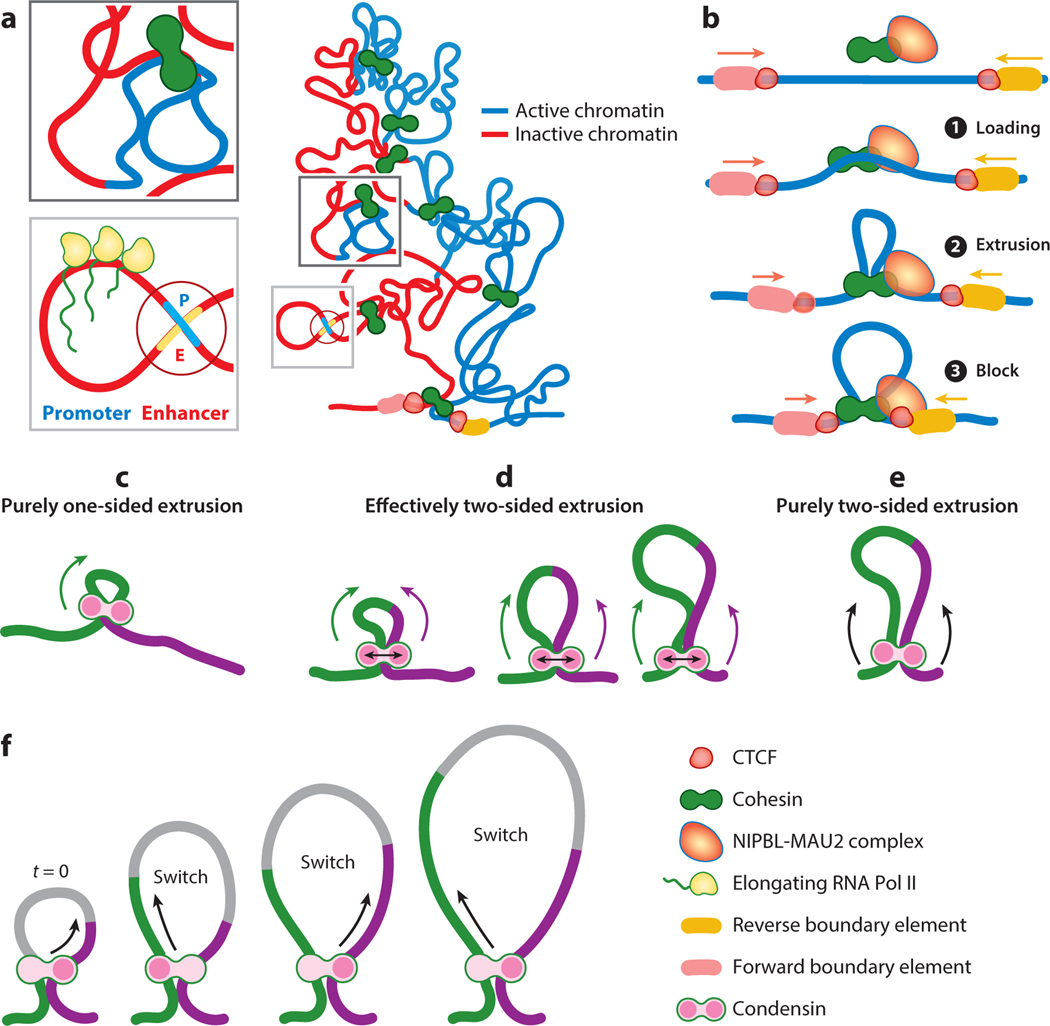
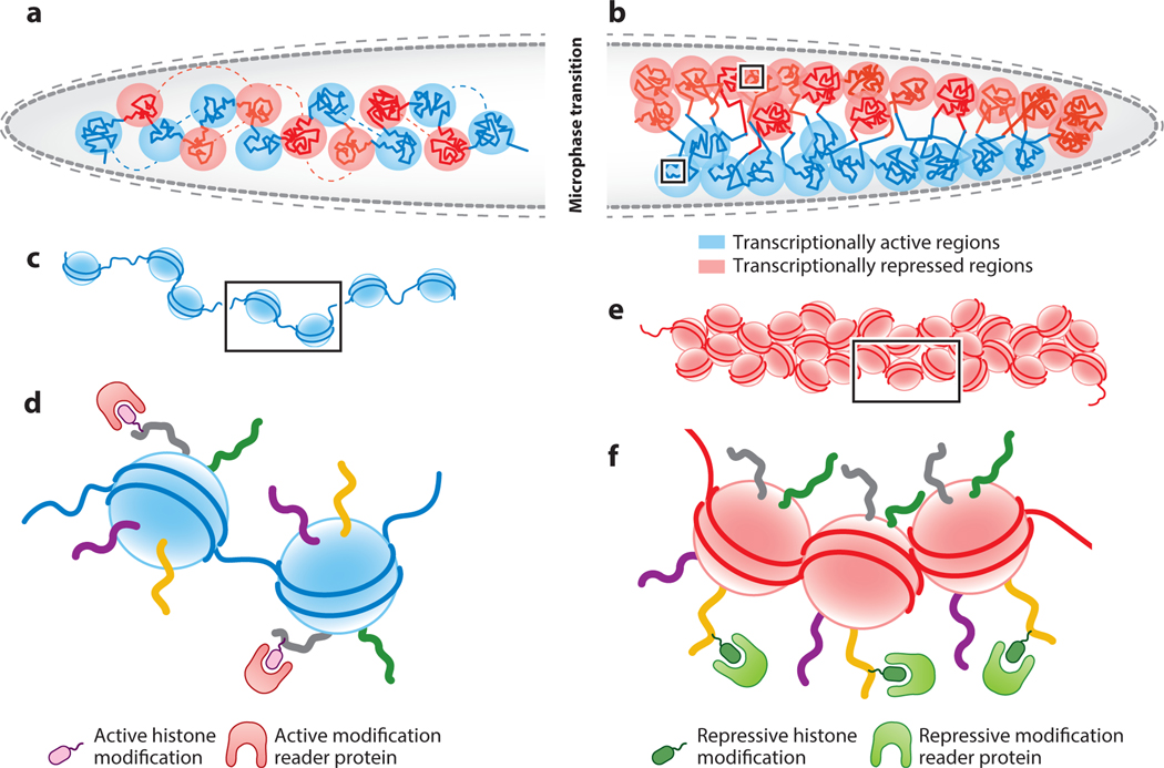
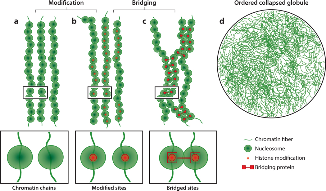
Nuclei are central hubs for information processing in eukaryotic cells. The need to fit large genomes into small nuclei imposes severe restrictions on genome organization and the mechanisms that drive genome-wide regulatory processes. How a disordered polymer such as chromatin, which has vast heterogeneity in its DNA and histone modification profiles, folds into discernibly consistent patterns is a fundamental question in biology. Outstanding questions include how genomes are spatially and temporally organized to regulate cellular processes with high precision and whether genome organization is causally linked to transcription regulation. The advent of next-generation sequencing, super-resolution imaging, multiplexed fluorescent in situ hybridization, and single-molecule imaging in individual living cells has caused a resurgence in efforts to understand the spatiotemporal organization of the genome. In this review, we discuss structural and mechanistic properties of genome organization at different length scales and examine changes in higher-order chromatin organization during important developmental transitions.

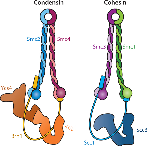
Keywords: chromosome topology; cohesin and condensin complexes; gametogenesis; loop extrusion; neural development; phase separation; zygotic genome activation.


Figures

Figure 1
Figure 1
Formation of topologically associated domains (TADs). (a, right) Shown is a TAD that includes both active (blue) and inactive (red) chromatin. TADs are flanked by forward- and reverse-oriented boundary elements facing each other in a convergent orientation. CCCTC-binding factor (CTCF) binds to the flanking boundaries. (Top left) This inset shows an enlargement of the dark gray boxed region that highlights a chromatin loop extruded by cohesin. The loop contains stretches of active and inactive chromatin. (Bottom left) This inset shows an enlargement of the light gray boxed region that highlights a chromatin loop extruded by cohesin. The loop joins an enhancer and a promoter to initiate transcription by RNA polymerase II (Pol II). (b) Loop extrusion of chromatin by cohesin occurs in a stepwise fashion. ❶ Cohesin is loaded onto chromatin by the nipped B-like protein (NIPBL)-MAU2 complex. The cohesin loader remains engaged with cohesin throughout the loop-extrusion process and plays a role in loop stability. ❷ After loading, cohesin extrudes chromatin loops in a two-sided manner until ❸ it is blocked by CTCF or any other barrier protein. Cohesin is then released with the aid of the cohesin-release factor WAPL (not shown). (c–e) Condensin-mediated loop extrusion is predominantly one sided. As shown in panel c, purely one-sided loop extrusion by condensin generates much smaller loops than purely two-sided loop extrusion (see panel e) and is unable to achieve the levels of compaction required for mitotic condensation. As shown in panel d, effectively two-sided loop extrusion is proposed as an alternate model for efficient loop extrusion. The effectively two-sided model matches the values of the extrusion parameters. In this model, condensin subunits stochastically switch between extruding from one side and then extruding from the other. In any one instant, condensin extrudes in a one-sided manner, but on average the switching causes the asymmetric one-sided extrusion to be effectively two sided. Panel d shows three different levels of loop extrusion, in which the size of the extruded loop scales with the switching frequency. (f) A detailed schematic representation of stepwise asymmetric strand extrusion by condensin. The chromatin is color coded (green and purple) to help visualize the chromatin segments that are being alternately extruded.
Figure 2
Figure 2
Subunit composition of Saccharomyces cerevisiae condensin and cohesin complexes. Figure adapted with permission from Yatskevich et al. (2019).
Figure 3
Figure 3
Microphase separation leads to the formation of active and inactive chromatin domains. (a) Chromatin can be envisioned as a block copolymer of alternative repeating units of transcriptionally active (blue) and repressed (red) regions. (b) Units of the same activity state interact with one another (blue with blue, red with red) to undergo microphase separation to form small domains of active and inactive chromatin. (c, d) Panel c shows an enlargement of the boxed area in the active block in panel b and represents an open conformation of nucleosomes (blue nucleosomes). These nucleosomes are marked with active histone modifications that are bound by proteins harboring recognition domains for active modifications shown in panel d. (e, f) Panel e shows an enlargement of the boxed area in the repressed block of panel b and represents compactly arranged nucleosomes (red nucleosomes). These nucleosomes are marked with repressive histone modifications that are bound by proteins harboring recognition domains for repressive modifications shown in panel f.
Figure 4
Figure 4
Polymer–polymer phase separation (PPPS) of chromatin. (a, b) Chromatin fibers are shown with nucleosomes and histone modifications. Enlargements of the black boxed areas are shown at the bottom. (c) Histone modifications can act as recruitment platforms for proteins that bridge chromatin fibers using multiple modular chromatin-binding domains (bridging proteins). An enlargement of the black boxed area is shown at the bottom. (d) Cross-linking of chromatin fibers by bridging proteins leads to PPPS and the formation of an ordered collapsed globule.
Figure 5
Figure 5
Liquid–liquid phase separation (LLPS) in chromatin organization. (a–c) Characteristics of droplets formed by LLPS. As shown in panel a, cells contain membrane-less bodies of varying sizes that form through LLPS. These liquid droplets have spherical shapes due to surface tension. As shown in panel b, the size of these liquid droplets scales with the concentration of the component molecules. As shown in panel c, droplets can undergo spontaneous fusion or fission. (d, e) During LLPS, a homogeneous mixture of molecules shown in panel d sorts into two liquid phases, a dense phase with a higher concentration of molecules and a dilute phase surrounding the dense phase shown in panel e. Panel e is an enlargement of the boxed area in panel a. Molecules in liquid phases can rearrange dynamically. (f–i) Chromatin mechanically regulates droplet growth. Droplet growth dynamics can be regulated by the stiffness of surrounding chromatin. Heterochromatin, shown in panel f, is stiffer than euchromatin, shown in panel h, and is less permissive of droplet growth. Growing droplets can, in turn, mechanically deform the surrounding chromatin. Panel g shows an enlargement of the purple boxed area in panel f, while panel i shows an enlargement of the purple boxed area in panel h. (j, k) Super-enhancer (SE) clusters can drive coactivator condensation. As shown in panel j, adjacent enhancers in an SE cluster can nucleate multiple coactivator droplets, where coactivators recruited by transcription factors can phase separate through homotypic polyvalent binding with other coactivators. As shown in panel k, these droplets can coalesce to generate a regulatory hub for transcriptional control of genes without the need for physical juxtaposition of distal enhancers and promoters.

References

    1. Agbleke AA, Amitai A, Buenrostro JD, Chakrabarti A, Chu L, et al. 2020. Advances in chromatin and chromosome research: perspectives from multiple fields. Mol. Cell 79(6):881–901 - PMC - PubMed
    1. Alberti S. 2017. Phase separation in biology. Curr. Biol 27(20):R1097–102 - PubMed
    1. Alipour E, Marko JF. 2012. Self-organization of domain structures by DNA-loop-extruding enzymes. Nucleic Acids Res. 40(22):11202–12 - PMC - PubMed
    1. Andergassen D, Smith ZD, Lewandowski JP, Gerhardinger C, Meissner A, Rinn JL. 2019. In vivo firre and Dxz4 deletion elucidates roles for autosomal gene regulation. eLife 8:e47214. - PMC - PubMed
    1. Anderson EC, Frankino PA, Higuchi-Sanabria R, Yang Q, Bian Q, et al. 2019. X chromosome domain architecture regulates Caenorhabditis elegans lifespan but not dosage compensation. Dev. Cell 51(2):192–207. e6 - PMC - PubMed

Publication types