. 2021 Jul 6;18(7):e1003656.
doi: 10.1371/journal.pmed.1003656. eCollection 2021 Jul.

SARS-CoV-2 neutralizing antibodies: Longevity, breadth, and evasion by emerging viral variants

Affiliations

SARS-CoV-2 neutralizing antibodies: Longevity, breadth, and evasion by emerging viral variants

Fiona Tea et al. PLoS Med. .

Abstract

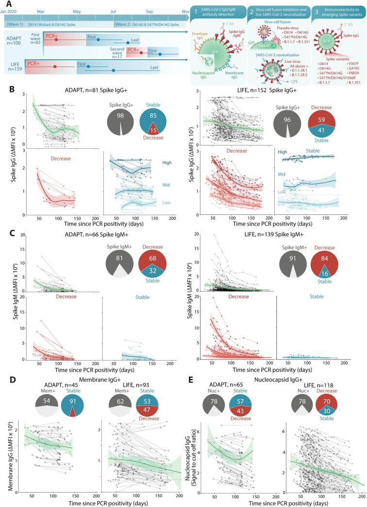
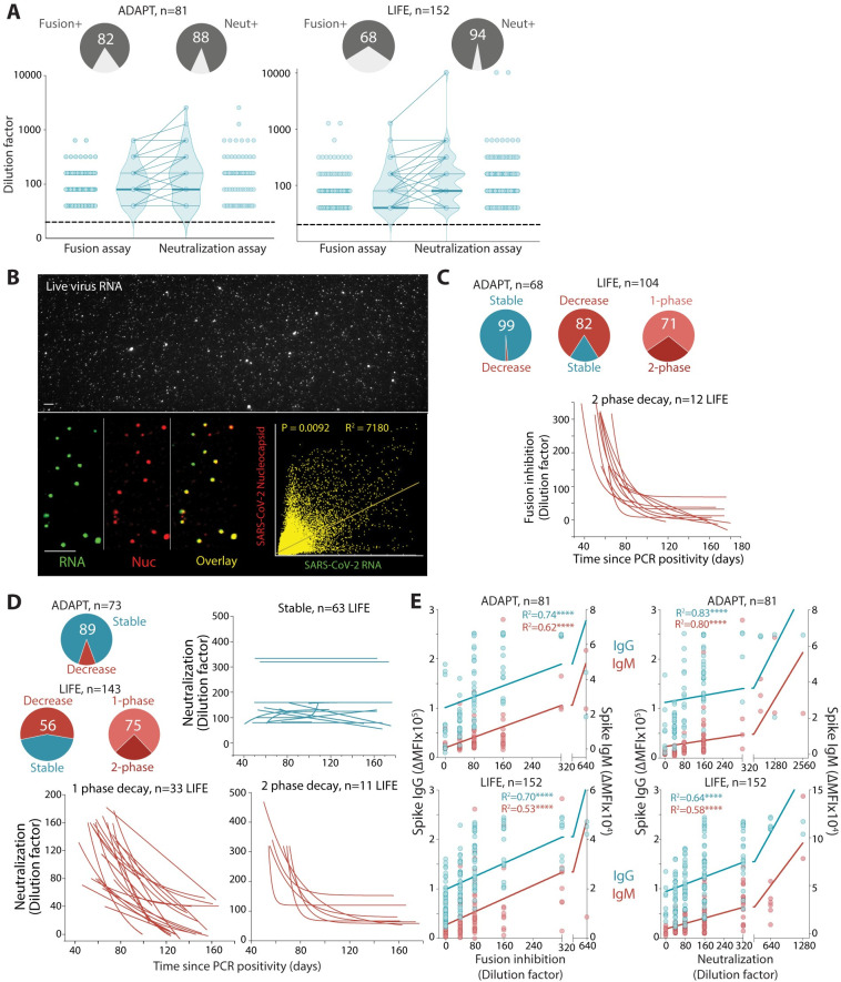
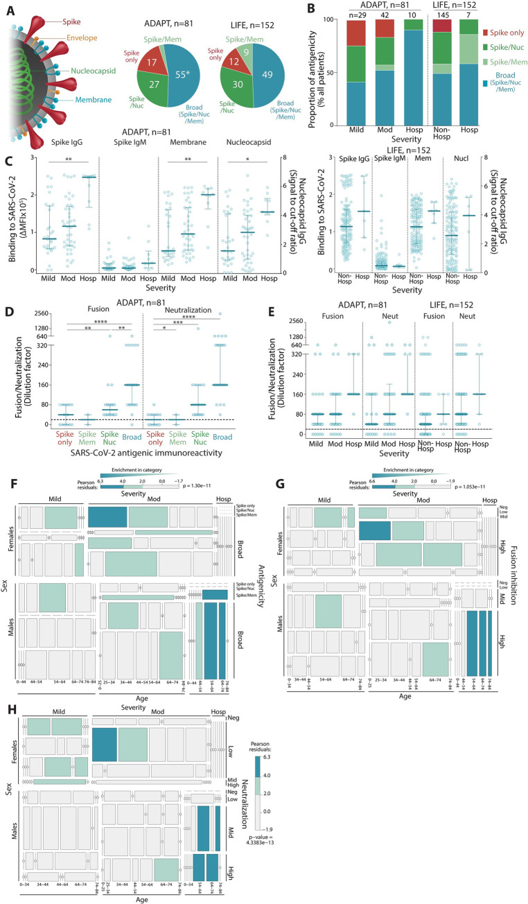
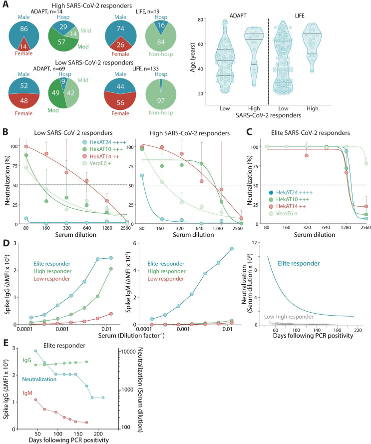
The Severe Acute Respiratory Syndrome Coronavirus 2 (SARS-CoV-2) antibody neutralization response and its evasion by emerging viral variants and variant of concern (VOC) are unknown, but critical to understand reinfection risk and breakthrough infection following vaccination. Antibody immunoreactivity against SARS-CoV-2 antigens and Spike variants, inhibition of Spike-driven virus-cell fusion, and infectious SARS-CoV-2 neutralization were characterized in 807 serial samples from 233 reverse transcription polymerase chain reaction (RT-PCR)-confirmed Coronavirus Disease 2019 (COVID-19) individuals with detailed demographics and followed up to 7 months. A broad and sustained polyantigenic immunoreactivity against SARS-CoV-2 Spike, Membrane, and Nucleocapsid proteins, along with high viral neutralization, was associated with COVID-19 severity. A subgroup of "high responders" maintained high neutralizing responses over time, representing ideal convalescent plasma donors. Antibodies generated against SARS-CoV-2 during the first COVID-19 wave had reduced immunoreactivity and neutralization potency to emerging Spike variants and VOC. Accurate monitoring of SARS-CoV-2 antibody responses would be essential for selection of optimal responders and vaccine monitoring and design.


Conflict of interest statement

I have read the journal’s policy and the authors of this manuscript have the following competing interests: FB has received honoraria from Biogen Idec and Merck Serono as invited speaker. The other authors have declared that no competing interests exist.

Figures

Fig 1
Fig 1. SARS-CoV-2 antibody responses are sustained and are predominantly focused on Spike.
(A) The first wave of Australian infections were from D614 and D614G Spike and the S477N/D614G Spike variant emerged during the second wave. Convalescent patient sera from ADAPT (first and second waves) and LIFE (first wave) were examined for SARS-CoV-2 antibodies. Mean time and range of PCR positivity (red) and dates of first and last sample collection (blue) are shown. Seropositive patients with at least 3 weeks between first and last samples were examined over time. Summary schematic of the current study that includes examination of (1) patient antibody responses toward various SARS-CoV-2 antigens; (2) functional virus–cell fusion and viral neutralization; and (3) immunoreactivity toward emerging Spike variants and VOC (S1 Table). (B) 96%–98% (gray) of patients were Spike IgG+. Most ADAPT patients had stable levels overtime, whereas most of LIFE Spike IgG levels decreased. No patients seroreverted. (C) 81%–91% (gray) were Spike IgM+, most had decreasing levels over time, and Spike IgM+ individuals started with and maintained low IgM levels. (D) 54%–57% (gray) of sera were Membrane IgG+, and most ADAPT had stable levels, whereas a larger proportion of LIFE had decreasing levels. (E) 78% of sera were Nucleocapsid IgG+, most were stable in ADAPT, whereas most decreased in LIFE. Loess curves with 95% confidence intervals are shown. PCR, polymerase chain reaction; SARS-CoV-2, Severe Acute Respiratory Syndrome Coronavirus 2.
Fig 2
Fig 2. Viral neutralization and inhibition of viral–cell fusion are strongly correlated with Spike antibody titers and sustained overtime.
(A) 68%–82% of convalescent sera inhibited virus–cell fusion, whereas 88%–94% sera neutralized live authentic SARS-CoV-2. Neutralization titers were higher than viral–cell fusion titers in ADAPT (ns, not significant) and LIFE (P < 0.0001). (B) Approximately 75% of virus particles were SARS-CoV-2 Nucleocapsid- and RNA positive (overlay, yellow). (C) All but one ADAPT patient had stable responses over time, whereas most LIFE donors (82%) had a decreased virus–cell fusion over time, with the majority (71%) exhibiting a single-phase decay. (D) In sera capable of viral neutralization, most ADAPT sera were stable (89%), whereas most LIFE sera (56%) had a decreased score over time, with the majority (75%) exhibiting a single-phase decay. Serum curves unable to be fitted were classified as undetermined. (E) Spike IgG and IgM levels were correlated with inhibition of virus–cell fusion and neutralization scores. R2 values are shown, and **** indicates significance (P < 0.0001). SARS-CoV-2, Severe Acute Respiratory Syndrome Coronavirus 2.
Fig 3
Fig 3. The antibody responses of patients with more severe COVID-19 disease have broader SARS-CoV-2 polyantigenicity.
(A) Approximately half of patients (49%–55%) had broad SARS-CoV-2 antibodies (blue, * includes n = 2 patient seropositive for Envelope IgG). Some had responses to 2 antigens (light and dark green), and a few reacted to Spike only (red). (B) Hospitalized patients were more likely to have broad SARS-CoV-2 polyantigenic immunoreactivity, whereas patients with only Spike reactivity exhibited mild-moderate symptoms. (C) Hospitalized patients exhibited higher Spike IgG, IgM, Membrane IgG, and Nucleocapsid IgG levels. High virus–cell fusion inhibition and neutralization titers were observed in patients with broad polyantigenic immunoreactivity (D) and in hospitalized patients (E). Older males were more likely to present with broader polyantigenic immunoreactivity (F), higher virus–cell fusion inhibition (G), and neutralization scores (H). Younger females were more enriched in mild to moderate disease severity, with narrow antigenicity (F), and lower virus–cell fusion inhibition (G) and neutralization scores (H). *: P < 0.05, **: P < 0.01, ***: P < 0.001, ****: P < 0.0001, *****: P < 0.00001. Not significant if significance is not indicated. P values in LIFE hospitalized patients (C) were not calculated due to low sample size. COVID-19, Coronavirus Disease 2019; SARS-CoV-2, Severe Acute Respiratory Syndrome Coronavirus 2.
Fig 4
Fig 4. High and elite responders are discriminated with SARS-CoV-2–permissive cells.
(A) Patients with high and robust SARS-CoV-2 responses were more likely male, hospitalized (left), and of older age (right). (B) Low and high responders to SARS-CoV-2 showed limited neutralization in hyperpermissive HekAT24 clonal cells. Permissiveness is indicated by +. (C) Only elite responders showed neutralization in HekAT24 cells. (D) Serum titration curves from an elite responder (blue) showed IgG and IgM levels greater than low (red) and high (green) responders and incredibly high neutralization titers (≥10,000) that decreased and stabilized at high levels (≥1280). (E) The elite donor demonstrated stable high Spike IgG, but the early decrease in viral neutralization was parallel to IgM decline before stabilization (at high titer). PCR, polymerase chain reaction; SARS-CoV-2, Severe Acute Respiratory Syndrome Coronavirus 2.
Fig 5
Fig 5. SARS-CoV-2 antibody responses show evasion by emerging Spike variants.
(A) Most patients had broad recognition of Spike variants (blue), whereas a smaller group had restricted Spike variant recognition and did not have a strong immunoreactivity to D614G Spike (red). Patients with reduced binding to D614G Spike had lower virus–cell fusion (ADAPT P < 0.01, LIFE P < 0.05) and neutralization scores (ADAPT P < 0.0001, LIFE P < 0.05) (B) and presented with less broad polyantigenic SARS-CoV-2 recognition (C). (D) D614G Spike-binding sera had greater inhibition of D614G Spike-pseudotyped virus–cell fusion (ns). (E) In Australia, D614G Spike was the predominant variant during the first wave and acquired additional mutations during the second wave (S477N, V1068F). VOCs, with high mutations within Spike, appeared in late December 2021. Pango lineages and Clades are shown in brackets. Graph adapted from Nextstrain [54]. (F) All patients had decreased immunoreactivity to S477N/D614G and S477N/D614G/V1068F Spike, while V1068F did not have an additive effect (ns, not significant). (G) Patients had reduced virus–cell fusion inhibition (first wave P < 0.0001, second wave P < 0.05) and neutralization (first wave P < 0.01, second wave ns) (H) to the S477N/D614G Spike variant compared to D614G. (I) Patients had reduced binding to VOC B.1.1.7 (UK) and B.1.351 (SA) Spike, with greater reduction toward B.1.351 (first wave P < 0.0001, second wave P < 0.0001). Virus–cell fusion inhibition (J) and neutralization (K) was also reduced against the VOC B.1.1.7 (UK, (J) first wave P < 0.0001, second wave P < 0.0001; (K) first wave P < 0.01, second wave ns) and B.1.351 (SA), but more so against the VOC B.1.351 (SA, (J) first wave P < 0.0001, second wave P < 0.05; (K) first wave P < 0.00001, second wave P < 0.0001). (L) Reduced neutralization was also observed against the authentic VOC B.1.1.28.1 (Brazil) (first wave P < 0.00001, second wave P < 0.01) and B.1.1.28.2 (Brazil) (first wave P < 0.00001, second wave P < 0.01). The level of decreased binding (F, I), virus–cell fusion inhibition (G, J), and neutralization (H, K) was irrespective of the virus that infected patients during the second wave. PCR, polymerase chain reaction; SARS-CoV-2, Severe Acute Respiratory Syndrome Coronavirus 2; VOC, variant of concern.

References

    1. Gudbjartsson DF, Norddahl GL, Melsted P, Gunnarsdottir K, Holm H, Eythorsson E, et al. Humoral Immune Response to SARS-CoV-2 in Iceland. N Engl J Med. 2020;383(18):1724–34. Epub 2020/09/02. doi: 10.1056/NEJMoa2026116 ; PubMed Central PMCID: PMC7494247. - DOI - PMC - PubMed
    1. Isho B, Abe KT, Zuo M, Jamal AJ, Rathod B, Wang JH, et al. Persistence of serum and saliva antibody responses to SARS-CoV-2 spike antigens in COVID-19 patients. Sci Immunol. 2020;5(52). Epub 2020/10/10. doi: 10.1126/sciimmunol.abe5511 . - DOI - PMC - PubMed
    1. Ibarrondo FJ, Fulcher JA, Goodman-Meza D, Elliott J, Hofmann C, Hausner MA, et al. Rapid Decay of Anti–SARS-CoV-2 Antibodies in Persons with Mild Covid-19. New England Journal of Medicine. 2020;383(11):1085–7. doi: 10.1056/NEJMc2025179 - DOI - PMC - PubMed
    1. Pollán M, Pérez-Gómez B, Pastor-Barriuso R, Oteo J, Hernán MA, Pérez-Olmeda M, et al. Prevalence of SARS-CoV-2 in Spain (ENE-COVID): a nationwide, population-based seroepidemiological study. Lancet. 2020;396(10250):535–44. Epub 2020/07/10. doi: 10.1016/S0140-6736(20)31483-5 ; PubMed Central PMCID: PMC7336131. - DOI - PMC - PubMed
    1. Rogers TF, Zhao F, Huang D, Beutler N, Burns A, He WT, et al. Isolation of potent SARS-CoV-2 neutralizing antibodies and protection from disease in a small animal model. Science. 2020;369(6506):956–63. Epub 2020/06/17. doi: 10.1126/science.abc7520 ; PubMed Central PMCID: PMC7299280. - DOI - PMC - PubMed

Publication types