

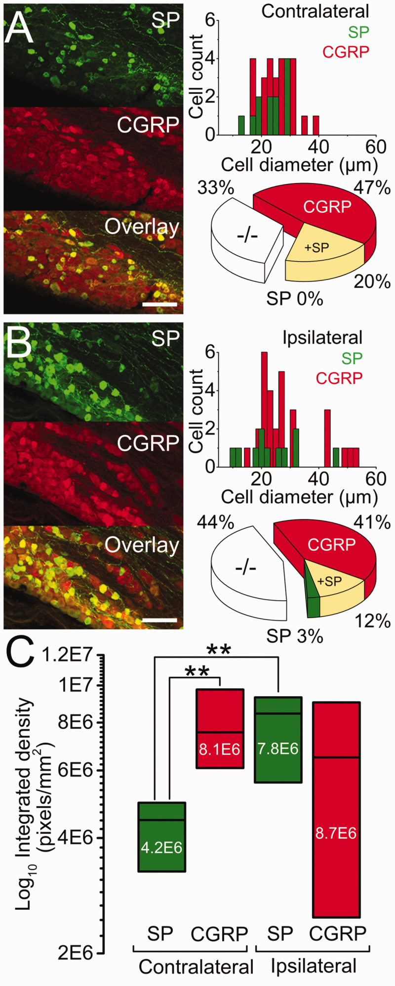
Seeding of breast cancer cell line (MDA-MB-231LUC+) to the mandible induces overexpression of substance P and CGRP throughout the trigeminal ganglion and widespread peripheral sensory neuropathy throughout all three of its divisions
- PMID: 34229504
- PMCID: PMC8267036
- DOI: 10.1177/17448069211024082
Seeding of breast cancer cell line (MDA-MB-231LUC+) to the mandible induces overexpression of substance P and CGRP throughout the trigeminal ganglion and widespread peripheral sensory neuropathy throughout all three of its divisions
Abstract
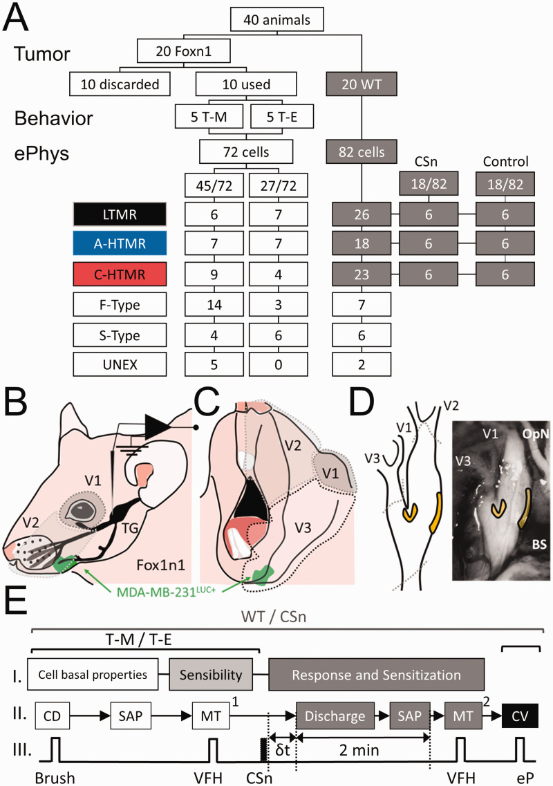
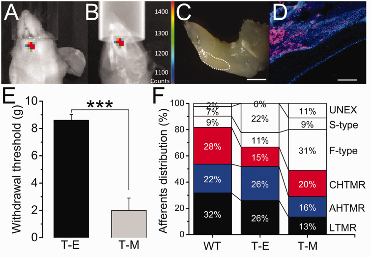
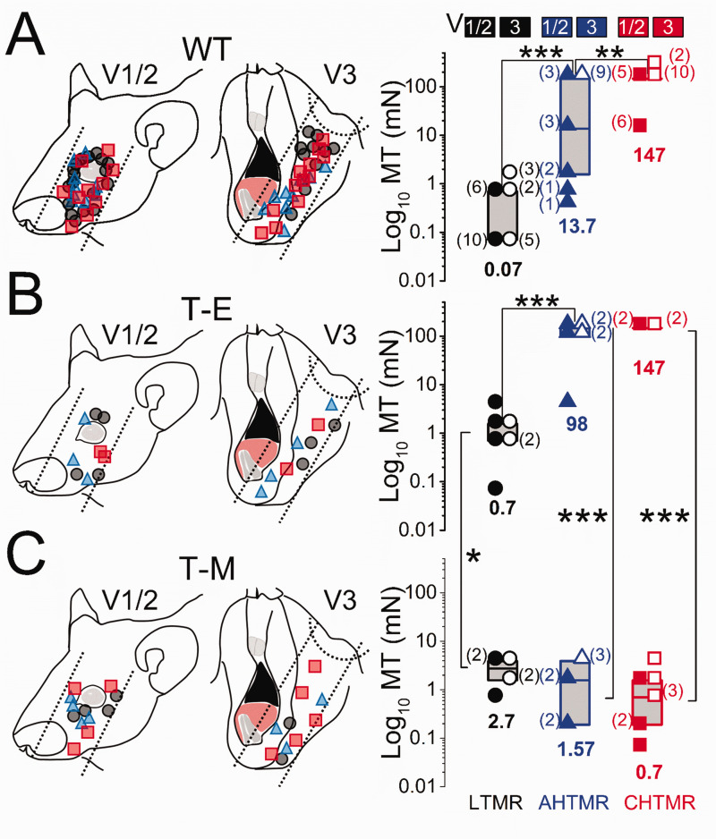
Some types of cancer are commonly associated with intense pain even at the early stages of the disease. The mandible is particularly vulnerable to metastasis from breast cancer, and this process has been studied using a bioluminescent human breast cancer cell line (MDA-MB-231LUC+). Using this cell line and anatomic and neurophysiologic methods in the trigeminal ganglion (TG), we examined the impact of cancer seeding in the mandible on behavioral evidence of hypersensitivity and on trigeminal sensory neurons. Growth of cancer cells seeded to the mandible after arterial injection of the breast cancer cell line in Foxn1 animals (allogeneic model) induced behavioral hypersensitivity to mechanical stimulation of the whisker pad and desensitization of tactile and sensitization of nociceptive mechanically sensitive afferents. These changes were not restricted to the site of metastasis but extended to sensory afferents in all three divisions of the TG, accompanied by widespread overexpression of substance P and CGRP in neurons through the ganglion. Subcutaneous injection of supernatant from the MDA-MB-231LUC+ cell culture in normal animals mimicked some of the changes in mechanically responsive afferents observed with mandibular metastasis. We conclude that released products from these cancer cells in the mandible are critical for the development of cancer-induced pain and that the overall response of the system greatly surpasses these local effects, consistent with the widespread distribution of pain in patients. The mechanisms of neuronal plasticity likely occur in the TG itself and are not restricted to afferents exposed to the metastatic cancer microenvironment.
Keywords: CGRP; Cancer; MDA-MB-231LUC+; SP; nociception; pain; sensitization.
Conflict of interest statement
Figures
Similar articles
-
Elevated levels of calcitonin gene-related peptide in upper spinal cord promotes sensitization of primary trigeminal nociceptive neurons.Neuroscience. 2016 Dec 17;339:491-501. doi: 10.1016/j.neuroscience.2016.10.013. Epub 2016 Oct 13. Neuroscience. 2016. PMID: 27746346 Free PMC article.
-
Distribution of a splice variant of choline acetyltransferase in the trigeminal ganglion and brainstem of the rat: comparison with calcitonin gene-related peptide and substance P.J Comp Neurol. 2008 Aug 1;509(4):436-48. doi: 10.1002/cne.21754. J Comp Neurol. 2008. PMID: 18521856
-
Neurochemical effects of photobiostimulation in the trigeminal ganglion after inferior alveolar nerve injury.J Biol Regul Homeost Agents. 2017 Jan-Mar;31(1):147-152. J Biol Regul Homeost Agents. 2017. PMID: 28337884
-
Neuropeptide effects in the trigeminal system: pathophysiology and clinical relevance in migraine.Keio J Med. 2011;60(3):82-9. doi: 10.2302/kjm.60.82. Keio J Med. 2011. PMID: 21979827 Review.
-
[Neuropeptide effects on the trigeminal system: pathophysiology and clinical significance for migraine].Schmerz. 2011 Aug;25(4):393-8, 400-1. doi: 10.1007/s00482-011-1069-5. Schmerz. 2011. PMID: 21818718 Review. German.
Cited by
-
Evaluation of pain related behaviors and disease related outcomes in an immunocompetent mouse model of prostate cancer induced bone pain.J Bone Oncol. 2023 Oct 30;43:100510. doi: 10.1016/j.jbo.2023.100510. eCollection 2023 Dec. J Bone Oncol. 2023. PMID: 38075938 Free PMC article.
-
Advances in Head and Neck Cancer Pain.J Dent Res. 2022 Aug;101(9):1025-1033. doi: 10.1177/00220345221088527. Epub 2022 Apr 13. J Dent Res. 2022. PMID: 35416080 Free PMC article.
-
Advanced cancer perineural invasion induces profound peripheral neuronal plasticity, pain, and somatosensory mechanical deactivation, unmitigated by the lack of TNFR1. Part 2. Biophysics and gene expression.Mol Pain. 2025 Jan-Dec;21:17448069251323666. doi: 10.1177/17448069251323666. Epub 2025 Feb 13. Mol Pain. 2025. PMID: 39945101 Free PMC article.
-
Effects of systemic oxytocin administration on ultraviolet B-induced nociceptive hypersensitivity and tactile hyposensitivity in mice.Mol Pain. 2024 Jan-Dec;20:17448069241226553. doi: 10.1177/17448069241226553. Mol Pain. 2024. PMID: 38172079 Free PMC article.
-
Advanced cancer perineural invasion induces profound peripheral neuronal plasticity, pain, and somatosensory mechanical deactivation, unmitigated by the lack of TNFR1. Part. 1: Behavior and single-cell in vivo electrophysiology.Mol Pain. 2025 Jan-Dec;21:17448069251314738. doi: 10.1177/17448069251314738. Epub 2025 Feb 8. Mol Pain. 2025. PMID: 39921540 Free PMC article.
References
Publication types
MeSH terms
Substances
LinkOut - more resources
Full Text Sources
Medical
Research Materials
Miscellaneous
