. 2021 Jul 6;18(1):152.
doi: 10.1186/s12974-021-02202-2.

Group 2 innate lymphoid cells are numerically and functionally deficient in the triple transgenic mouse model of Alzheimer's disease

Affiliations

Group 2 innate lymphoid cells are numerically and functionally deficient in the triple transgenic mouse model of Alzheimer's disease

Ivan Ting Hin Fung et al. J Neuroinflammation. .

Abstract

Background: The immune pathways in Alzheimer's disease (AD) remain incompletely understood. Our recent study indicates that tissue-resident group 2 innate lymphoid cells (ILC2) accumulate in the brain barriers of aged mice and that their activation alleviates aging-associated cognitive decline. The regulation and function of ILC2 in AD, however, remain unknown.

Methods: In this study, we examined the numbers and functional capability of ILC2 from the triple transgenic AD mice (3xTg-AD) and control wild-type mice. We investigated the effects of treatment with IL-5, a cytokine produced by ILC2, on the cognitive function of 3xTg-AD mice.

Results: We demonstrate that brain-associated ILC2 are numerically and functionally defective in the triple transgenic AD mouse model (3xTg-AD). The numbers of brain-associated ILC2 were greatly reduced in 7-month-old 3xTg-AD mice of both sexes, compared to those in age- and sex-matched control wild-type mice. The remaining ILC2 in 3xTg-AD mice failed to efficiently produce the type 2 cytokine IL-5 but gained the capability to express a number of proinflammatory genes. Administration of IL-5, a cytokine produced by ILC2, transiently improved spatial recognition and learning in 3xTg-AD mice.

Conclusion: Our results collectively indicate that numerical and functional deficiency of ILC2 might contribute to the cognitive impairment of 3xTg-AD mice.

Keywords: Alzheimer’s disease (AD); Cognitive function; Group 2 innate lymphoid cells (ILC2); IL-5; Innate lymphoid cells (ILC).


Conflict of interest statement

Q.Y. reports a patent HRFM Ref. No. 0410.054AWO. The other authors declare that they have no competing interests.

Figures
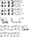
Fig. 1
Fig. 1
ILC2 are numerically and functionally deficient in 3xTg-AD mice. a Representative flow cytometric profiles of ILC2 in the whole-brain tissue of 7-month-old female and male 3xTg-AD mice and control wild-type (WT) mice. ILC2 were identified as LinCD45+Thy1+T1/ST2+ cells. Lin1 markers included B220, NK1.1, and CD11b. Total Lin markers included B220, NK1.1, CD11b, CD3, and TCRβ. The values shown represent the percentages of gated cells from total cells in the flow cytometry plot. b Numbers of ILC2 in the whole-brain tissue of 7-month-old male 3xTg-AD mice and control wild-type mice. Data are from 6 mice per group, two independent experiments. Sex effect: F [1, 20] = 15.88, p < 0.01; strain effect: F [1, 20] = 99.39, p < 0.01; interaction: F [1, 20] = 10.16, p < 0.01. c Numbers of ILC2 at different anatomic sites in 7-month-old female and male 3xTg-AD mice and sex and age-matched control wild-type mice. PFC, prefrontal context; CTX, cortex; HP, hippocampus; HT, hypothalamus; LP, leptomeninges; CP, choroid plexus; PVS, perivascular space. Data are from 6 mice per group, two independent experiments. t [10] = 4.49, p < 0.01. d Representative flow cytometric profiles depicting IL-5 expression by ILC2 in the whole-brain tissue of 7-month-old female and male 3xTg-AD and control wild-type mice. Isotype controls were used as the negative control. Plots were pre-gated on ILC2 (LinCD45+Thy1+T1/ST2+). e Numbers of IL-5-expressing ILC2 in the whole-brain tissue of 7-month-old female and male 3xTg-AD and control wild-type mice. Data are from 4 mice per group, two independent experiments. Sex effect: F [1, 12] = 12.06, p < 0.01; strain effect: F [1, 12] = 54.64, p < 0.01; interaction: F [1, 12] = 6.21, p < 0.05. f Representative flow cytometric profile of IL-5 expression in T cells (CD45+CD3+) and B cells (CD45+CD19+) in the whole-brain tissue of 7-month-female and male 3xTg-AD mice and control wild-type mice. Plots were pre-gated on T cells (CD45+CD3+) or B cells (CD45+CD19+). Data represent 4 female and 4 male mice per group. Error bars = mean ± SEM. **p < 0.01; n.s., not significant
Fig. 2
Fig. 2
ILC2 from 3xTg-AD mice exhibit intrinsic defects in producing IL-5. a 100 ILC2 were purified from the whole-brain tissue of 7-month-old female and male 3xTg-AD mice and control wild-type mice by fluorescence-activated cell sorting (FACS). Specifically, after liberase digestion, whole-brain tissue cells from the two mice of the group were pooled into one sample, and FACS were performed to sort ILC2. We pooled cells from two mice into one sample, in order to ensure that 100 ILC2 were obtained from each sample. One hundred ILC2 were plated into round-bottom 96-well plates and cultured with 20 ng/ml IL-2, IL-7, and IL-33 for 7 days. Growth of ILC2 was calculated as the number of cells that emerged after 7 days of culture per cell plated. Each culture well contained cells pooled from two different mice of the same group. Data are from 4 independent culture well per group, 2 independent experiments. b Expression of IL-5 was examined after 7 days of culture. Isotype was used as a negative control. c The mean fluorescence intensity (MFI) of IL-5 expression by cultured ILC2, after 7 days of culture. Data are from 5 independent culture well per group, 2 independent experiments. Sex effect: F [1, 16] = 0.57, p = 0.46; strain effect: F [1, 16] = 98.12, p < 0.01; interaction: F [1, 16] = 0.15, p = 0.71. d Concentrations of the indicated cytokines in the culture supernatant after 7 days of culture, measured by LegendPLex assays. Data are from 5 independent culture wells per group, 2 independent experiments. Sex effect: F [1, 16] = 0.19, p = 0.67; strain effect: F [1, 16] = 48.83, p < 0.01; interaction: F [1, 16] = 0.78, p = 0.39. U.D., undetected. Error bars = mean ± SEM. **p < 0.01; n.s., not significant
Fig. 3
Fig. 3
Single-cell transcriptomal profiles of cultured ILC2 from 3xTg-AD mice and control wild-type mice. a scRNA-seq was performed with cultured ILC2 from 7-month-old female and male 3xTg-AD mice and age and sex-matched wild-type control mice, after 7 days of culture. Feature plots deposit the expression of the indicated genes. The red color indicates positive expression. The gray color indicates negative expression. b Percentages of Il5+ ILC2, Gzma+ ILC2, Il5+ Gzma+ ILC2 (double-positive), and Il5 Gzma ILC2 (double-negative) in cultured ILC2 from 3xTg-AD and wild-type mice, by scRNA-seq. c Genes differentially expressed between ILC2 from 3xTg-AD mice and those from control wild-type mice were identified by scRNA-seq. Gene pathway analyses were performed. d List of effector molecules and lymphocyte signaling molecules that were differentially expressed between ILC2 from 3xTg-AD mice and those from control wild-type mice. e Violin plots depicting the expression of the indicated genes. Cells were pooled from 4 female and 4 male mice per group. *p < 0.05; **p < 0.01; n.s., not significant
Fig. 4
Fig. 4
IL-5 treatment did not significantly influence Aβ concentrations in 7-month-old 3xTg-AD mice. a Seven-month-old female and male 3xTg-AD mice and sex and age-matched control wild-type mice were treated with IL-5 daily for 2 days. Concentrations of IL-5 in the cerebrospinal fluid (CSF) were examined by LegendPlex assays. Data are from 4 to 6 mice per group, 2 independent experiments. t [8] = 2.61, p < 0.05. b Concentrations of soluble Aβ were examined by ELISA. Data are from 4 mice per group, 2 independent experiments. Sex effect: F [1, 12] = 36.05, p < 0.01; treatment effect: F [1, 12] = 0.029, p = 0.87; interaction: F [1, 12] = 0.026, p = 0.87. c Concentrations of insoluble Aβ were examined by ELISA. Data are from 4 mice per group, 2 independent experiments. Error bars = mean ± SEM. *p < 0.05; **p < 0.01; n.s., not significant
Fig. 5
Fig. 5
IL-5 treatment improved spatial recognition and learning of 7-month-old female 3xTg-AD mice. a Seven-month-old female 3xTg-AD mice were treated with IL-5 and PBS and underwent behavior tests. Experimental scheme for cytokine treatment and behavior tests. OF, open field; EPM, elevated plus maze. b Total distance traveled and the percentages of time spent in the indicated zones by open field test. c Percentages of time spent in the closed arms in the elevated plus maze test. d Percentages of time spent in the novel (N) and familiar (F) arms and percentages of entries into the novel and familiar arms in the Y-maze forced alternation test. Percentage of time: IL-5-treated t [18] = 2.42, p < 0.05. Percentage of entries: IL-5-treated t [18] = 3.7, p < 0.01. e Escape latency and escape distance to the hidden platform, the thigmotaxis rate, and swim speed during the 4 days of trials in the water maze test. Escape latency: F(1, 72) = 8.53, p < 0.01. Escape distance: F(1, 72) = 8.31, p < 0.01. Data are from 10 mice per group, 2 independent experiments. Error bars = mean ± SEM. *p < 0.05; **p < 0.01; n.s., not significant

References

    1. Bondi MW, Edmonds EC, Salmon DP. Alzheimer’s disease: past, present, and future. J Int Neuropsychol Soc. 2017;23(9-10):818–831. doi: 10.1017/S135561771700100X. - DOI - PMC - PubMed
    1. Wang J, Gu BJ, Masters CL, Wang YJ. A systemic view of Alzheimer disease - insights from amyloid-beta metabolism beyond the brain. Nat Rev Neurol. 2017;13(11):703. doi: 10.1038/nrneurol.2017.147. - DOI - PubMed
    1. Navarro V, Sanchez-Mejias E, Jimenez S, Munoz-Castro C, Sanchez-Varo R, Davila JC, Vizuete M, Gutierrez A, Vitorica J. Microglia in Alzheimer’s disease: activated, dysfunctional or degenerative. Front Aging Neurosci. 2018;10:140. doi: 10.3389/fnagi.2018.00140. - DOI - PMC - PubMed
    1. Gate D, Saligrama N, Leventhal O, Yang AC, Unger MS, Middeldorp J, Chen K, Lehallier B, Channappa D, De Los Santos MB, McBride A, Pluvinage J, Elahi F, Tam GK, Kim Y, Greicius M, Wagner AD, Aigner L, Galasko DR, Davis MM, Wyss-Coray T. Clonally expanded CD8 T cells patrol the cerebrospinal fluid in Alzheimer’s disease. Nature. 2020;577(7790):399–404. doi: 10.1038/s41586-019-1895-7. - DOI - PMC - PubMed
    1. Zhang Y, Fung ITH, Sankar P, Chen X, Robison LS, Ye L, D’Souza SS, Salinero AE, Kuentzel ML, Chittur SV, Zhang W, Zuloaga KL, Yang Q. Depletion of NK cells improves cognitive function in the Alzheimer disease mouse model. J Immunol. 2020;205(2):502–510. doi: 10.4049/jimmunol.2000037. - DOI - PMC - PubMed