Review
. 2021 Jul 6;25(1):236.
doi: 10.1186/s13054-021-03659-6.

Infectious disease-associated encephalopathies

Affiliations
Review

Infectious disease-associated encephalopathies

Maria C Barbosa-Silva et al. Crit Care. .

Abstract

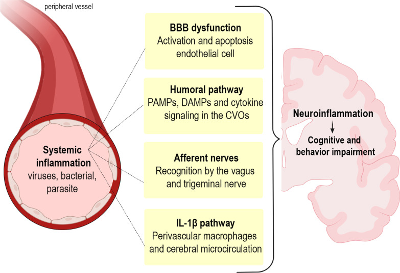
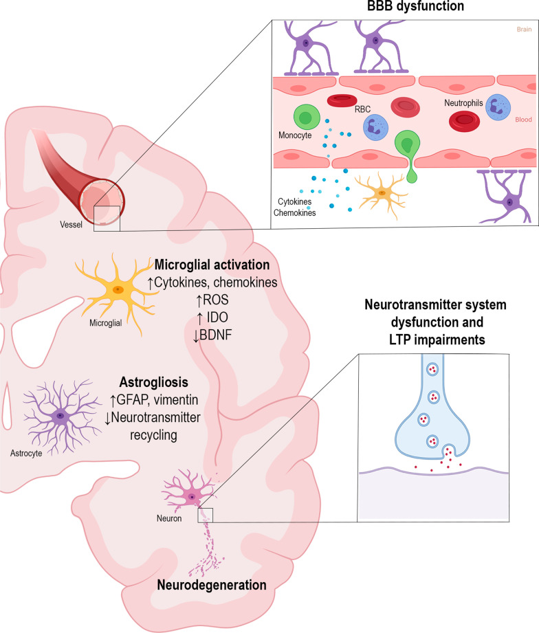
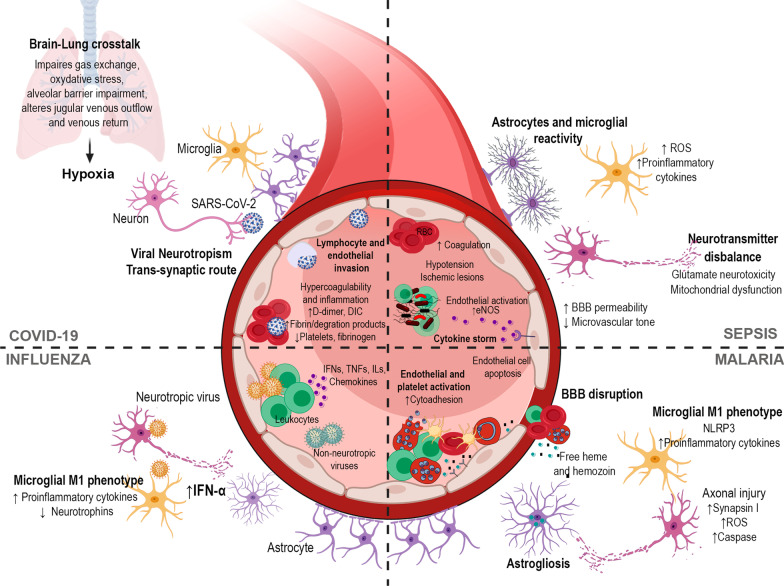
Infectious diseases may affect brain function and cause encephalopathy even when the pathogen does not directly infect the central nervous system, known as infectious disease-associated encephalopathy. The systemic inflammatory process may result in neuroinflammation, with glial cell activation and increased levels of cytokines, reduced neurotrophic factors, blood-brain barrier dysfunction, neurotransmitter metabolism imbalances, and neurotoxicity, and behavioral and cognitive impairments often occur in the late course. Even though infectious disease-associated encephalopathies may cause devastating neurologic and cognitive deficits, the concept of infectious disease-associated encephalopathies is still under-investigated; knowledge of the underlying mechanisms, which may be distinct from those of encephalopathies of non-infectious cause, is still limited. In this review, we focus on the pathophysiology of encephalopathies associated with peripheral (sepsis, malaria, influenza, and COVID-19), emerging therapeutic strategies, and the role of neuroinflammation.

Keywords: COVID-19; Cognition; Encephalopathy; Infection; Influenza; Malaria; Microglial priming; Neuroinflammation; SARS-CoV-2; Sepsis.


Conflict of interest statement

The authors declare that they have no competing interests.

Figures

Fig. 1
Fig. 1
Inflammatory signaling pathways to the brain. Systemic inflammation caused by pathogens, including viruses, bacteria, and parasites, leads to neuroinflammation with consequent cognitive and behavior impairments. The central nervous system is able to recognize systemic inflammation through (1) BBB dysfunction, with activation and apoptosis of endothelial cells, allowing cytokines and immune cells to invade the brain parenchyma; (2) the humoral pathway and saturable transport system in the blood–brain barrier (BBB), which involves the circumventricular organs (CVOs) and the choroid plexus, as local macrophage-like cells express innate immune receptors that recognize pathogen-associated molecular patterns (PAMPs), damage-associated molecular patterns (DAMPs), and cytokines, allowing inflammatory mediators to access the brain by volume diffusion and through cytokine-saturable transporters, since the CVOs do not have an intact BBB; (3) through activation of the afferent nerves (including the vagal nerves in abdominal/visceral infections and the trigeminal nerve in oro-lingual infections) by cytokines; and (4) IL-1β pathway signaling, through activation of IL-1 receptors expressed in perivascular macrophages and endothelial cells located in the brain microvasculature, initiating a local immune response
Fig. 2
Fig. 2
Molecular and cellular mechanisms of neuroinflammation. Blood–brain barrier (BBB) dysfunction contributes to the process of neuroinflammation. After losing its integrity, the BBB allows circulating leukocytes (e.g., monocytes and neutrophils) and proinflammatory mediators, such as cytokines, to enter the brain parenchyma. Microglia and astrocytes proliferate, become reactive, and undergo functional and morphological changes. Microglial cells increase the release of reactive oxygen species, cytokines, chemokines, and indoleamine 2,3-dioxygenase (IDO) expression/activity, as well as decrease brain-derived neurotrophic factor (BDNF) expression. Astrocytes increase the expression of glial fibrillary acidic protein (GFAP) and vimentin, which cause morphological changes, losing their function as supportive glial cells and developing impairment of neurotransmitter recycling. Neuroinflammation also impacts neurons and synaptic transmission, leading to impairments in long-term potentiation (LTP) and neurotransmitter system dysfunctions
Fig. 3
Fig. 3
Mechanisms implicated in neurological complications after infection. In COVID-19, SARS-CoV-2 can access the brain by a trans-synaptic route and also through endothelial and lymphocyte invasion, resulting in neuroinflammation. Lower thrombin, higher D-dimer, fibrin/fibrinogen degradation products, and fibrinogen levels are frequent in COVID-19, and activation of the coagulation cascade may contribute to the development of stroke and cerebrovascular accidents. Brain-lung crosstalk is an axis involved in brain hypoxia due to systemic oxygenation reduction and, subsequently, secondary brain oxygenation damage. In sepsis-associated encephalopathy, the cytokine storm leads to endothelial activation and increased eNOS activity, which results in nitric oxide (NO) production, leading to hypotension and ischemic lesions. Cytokines trigger glial reactivity, reactive oxygen species (ROS) production, mitochondrial dysfunction, and neurotransmitter imbalances, with consequent glutamate excitotoxicity. In malaria infection, there is an exacerbated inflammatory response to the parasite and activation of multiple cell death pathways leading to microcirculatory damage. Endothelial dysfunction, platelet activation, cytoadherence, and a downregulation of normal endogenous anticoagulant pathways are hallmarks. Dysregulation of the coagulation pathway leads to microvascular lesions; thrombin may be implicated. In the process of hemoglobin digestion, the malaria parasite releases heme and aggregates it into hemozoin, a highly toxic and proinflammatory signaling molecule. Hemozoin and free heme released into the bloodstream lead to exacerbated inflammation, tissue damage, apoptosis of microvascular brain endothelial cells through activation of STAT3, and loss of BBB integrity through binding to the metalloproteinase MMP3. The proinflammatory milieu leads to microglial M1 phenotype activation, release of proinflammatory cytokines, astrogliosis, axonal injury, and increase in synapsin I. In influenza infection, there is a peripheral inflammatory response and release of several proinflammatory mediators, including interferons (IFs), interleukins (ILs), tumor necrosis factor (TNF), and chemokines. Both neurotropic and non-neurotropic strains of influenza are able to induce neuroinflammation, with microglial activation, decrease in neurotrophin levels, and increase in IFN-α and other proinflammatory cytokines

References

    1. Erkkinen MG, Berkowitz AL. A clinical approach to diagnosing encephalopathy. Am J Med. 2019;132:1142–1147. - PubMed
    1. Turner L, Lavstsen T, Berger SS, Wang CW, Petersen JEV, Avril M, et al. Severe malaria is associated with parasite binding to endothelial protein C receptor. Nature. 2013;498(7455):502–505. - PMC - PubMed
    1. Gilden D, Nagel MA. Neurological complications of human herpesvirus infections. Int Neurol. 2016 doi: 10.1002/9781118777329.ch81. - DOI
    1. Ludlow M, Kortekaas J, Herden C, Hoffmann B, Tappe D, Trebst C, et al. Neurotropic virus infections as the cause of immediate and delayed neuropathology. Acta Neuropathol. 2016;131:159–184. - PMC - PubMed
    1. Koyuncu OO, Hogue IB, Enquist LW. Virus infections in the nervous system. Cell Host Microbe. 2013;13:379–393. - PMC - PubMed

Publication types