. 2021 Jul 6;11(1):13909.
doi: 10.1038/s41598-021-93158-z.

A bacteriocin-based treatment option for Staphylococcus haemolyticus biofilms

Affiliations

A bacteriocin-based treatment option for Staphylococcus haemolyticus biofilms

Christian Kranjec et al. Sci Rep. .

Abstract

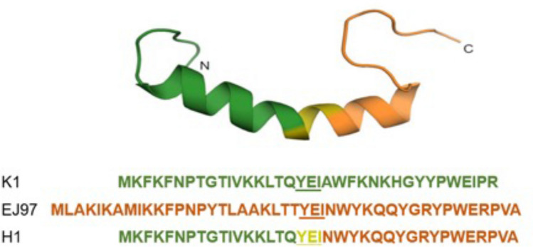
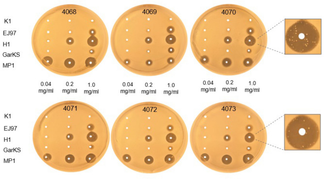
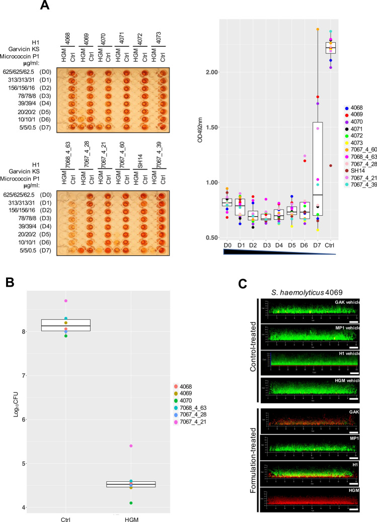
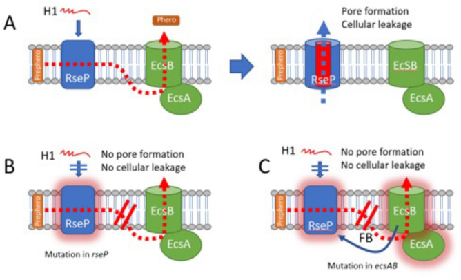
Bacteriocins are ribosomally-synthesized antimicrobial peptides, showing great potential as novel treatment options for multidrug-resistant pathogens. In this study, we designed a novel hybrid bacteriocin, Hybrid 1 (H1), by combing the N-terminal part and the C-terminal part of the related bacteriocins enterocin K1 (K1) and enterocin EJ97 (EJ97), respectively. Like the parental bacteriocins, H1 used the membrane-bound protease RseP as receptor, however, it differed from the others in the inhibition spectrum. Most notably, H1 showed a superior antimicrobial effect towards Staphylococcus haemolyticus-an important nosocomial pathogen. To avoid strain-dependency, we further evaluated H1 against 27 clinical and commensal S. haemolyticus strains, with H1 indeed showing high activity towards all strains. To curtail the rise of resistant mutants and further explore the potential of H1 as a therapeutic agent, we designed a bacteriocin-based formulation where H1 was used in combination with the broad-spectrum bacteriocins micrococcin P1 and garvicin KS. Unlike the individual bacteriocins, the three-component combination was highly effective against planktonic cells and completely eradicated biofilm-associated S. haemolyticus cells in vitro. Most importantly, the formulation efficiently prevented development of resistant mutants as well. These findings indicate the potential of a bacteriocins-based formulation as a treatment option for S. haemolyticus.


Conflict of interest statement

The authors declare no competing interests.

Figures

Figure 1
Figure 1
Predicted Structure and amino acid sequence of the hybrid bacteriocin and its parental bacteriocins. The predicted structure of the hybrid bacteriocin 1 (H1), formed by the N-terminal part of K1 (green) and the C-terminal part of EJ97 (orange). The central tripeptide sequence (YEI—underlined) in K1 and EJ97 serves as the joining segment in the fusion protein. The structure of H1 was modeled based on the NMR structural data of K1 using the structure prediction tool Swiss model.
Figure 2
Figure 2
Comparison of the antimicrobial activities of the bacteriocins against S. haemolyticus. The antimicrobial activity of the indicated bacteriocins was assessed by “spot-on-lawn” assay against six S. haemolyticus strains, 4068–4073. The bacteriocins were applied at three different concentrations (0.04, 0.2, and 1.0 mg/ml), and 5 µl of each was spotted on the lawns of the S. haemolyticus strains. All strains developed resistance to H1 after 24 h incubation, as illustrated for strains 4070 and 4073.
Figure 3
Figure 3
Boxplot of minimal inhibition concentrations (μg/ml) for the treatment with H1, garvicin KS, micrococcin P1 and their combinations against planktonic S. haemolyticus. The median distribution is indicated as thick line within boxes and the degree of variability (amplitude of the box or inter quartile region (IQR)) of the MIC50 values for the indicated strains obtained at three different time points: 5 h, 24 h and 48 h. Whiskers extending out the boxes mark the minimum and maximum observed values and the variability outside the middle 50% of values (whisker length). Outliers are represented as values that extend out of the whisker limit (1.5×IQR). Note that the treatment with the tricomponent combination (H1/GarKS/MP1) led to contained MIC values and inter-strain variability compared to the other treatments at all time-points. (*)Please note that for the treatment with H1 all strains were sensitive after a 5 h incubation (MIC50 = 0.78 μg/ml); (**) all strains were resistant after a 24 h incubation, except 4069 (MIC50 = 0.78 μg/ml); all strains were resistant after a 48 h incubation.
Figure 4
Figure 4
The tricomponent antimicrobial combination effectively inhibits the metabolic activity and viability of S. haemolyticus biofilm-associated cells in vitro. (A) The left panels show representative images of BOAT assays performed upon 24 h treatment with the tricomponent combination (HGM: H1, GarKS, MP1) for the indicated strains. The concentration (μg/ml) of the individual antimicrobials in the dilutions (dilution factors: D0 to D7) is indicated on the far left of the images. As controls, the assay was performed using the vehicles at their final concentrations (Ctrl). The development of red color indicates the retention of metabolic activity, and its quantification was performed by spectrophotometry at OD492. Metabolic activity values were plotted as a function of the dilution factor for the antimicrobial combination (right panel). Shown is the median distribution (thick line within boxes) and the degree of variability (amplitude of the box or inter quartile region (IQR)) of the metabolic activities for the indicated strains measured at increasing dilution factors (D0–D7). The boxplot components (whiskers and outliers) are displayed as specified in Fig. 3. Note that the treatment maintained the metabolic activitiy under detectable levels up to D6 for most strains. (B) The logarithmic colony formation unit (Log10CFU) was calculated after the BOAT assays were performed for the indicated strains. BOAT assays were performed as in (A), and the boxplot shows the median distribution of the Log10CFU values. The antimicrobial concentrations in the tricomponent combination were 625 μg/ml for H1 and garvicin KS and 62.5 μg/ml for micrococcin P1. The control was performed by using the antimicrobials vehicles at their final concentration. (C) The S. haemolyticus strain 4069 was allowed to form biofilms on glass-bottomed chambers for 24 h, prior to treatment with the indicated antimicrobials (lower set of panels) or the respective control-vehicles (upper set of panels) as detailed in (B). The biofilms were subsequently stained using the LIVE/DEAD biofilm staining kit and confocal microscope images were taken using a 63 × oil immersion objective. Scale bars correspond to 10 μm.
Figure 5
Figure 5
Proposed model for the EcsAB mediated H1-resistance in S.haemolyticus. (A) RseP is a inner-membrane protease working together the ABC-transporter EcsAB to cleave the hormone prepeptide (Prephero) and export the mature hormone peptide (Phero) to the external milieu. RseP is acting also as the receptor for the bacteriocin H1 which forms pores and causes lethal cellular leakage across the membrane. (B) When rseP is mutated resulting in a non-functional receptor (RseP with glow), cells become resistant to H1. (C) When the ecs system is mutated, the malfunctional ABC transporter can no longer export the hormone peptide. This jammed situation of hormone prepeptide/peptide within the membrane results in a feedback (FB) loop somehow causing RseP inactivity. An inactive RseP (RseP with glow) loses its function as a bacteriocin receptor, and cells therefore become resistant to H1.

References

    1. Becker K, Heilmann C, Peters G. Coagulase-negative staphylococci. Clin. Microbiol. Rev. 2014;27:870–926. doi: 10.1128/CMR.00109-13. - DOI - PMC - PubMed
    1. Czekaj T, Ciszewski M, Szewczyk EM. Staphylococcus haemolyticus—An emerging threat in the twilight of the antibiotics age. Microbiology (Reading) 2015;161:2061–2068. doi: 10.1099/mic.0.000178. - DOI - PubMed
    1. Takeuchi F, et al. Whole-genome sequencing of staphylococcus haemolyticus uncovers the extreme plasticity of its genome and the evolution of human-colonizing staphylococcal species. J. Bacteriol. 2005;187:7292–7308. doi: 10.1128/JB.187.21.7292-7308.2005. - DOI - PMC - PubMed
    1. Silva PV, et al. The antimicrobial susceptibility, biofilm formation and genotypic profiles of Staphylococcus haemolyticus from bloodstream infections. Mem. Inst. Oswaldo Cruz. 2013;108:812–813. doi: 10.1590/0074-0276108062013022. - DOI - PMC - PubMed
    1. Klingenberg C, et al. Persistent strains of coagulase-negative staphylococci in a neonatal intensive care unit: virulence factors and invasiveness. Clin. Microbiol. Infect. 2007;13:1100–1111. doi: 10.1111/j.1469-0691.2007.01818.x. - DOI - PubMed

Publication types

MeSH terms