Review
. 2022 Mar;22(3):188-199.
doi: 10.1038/s41577-021-00572-5. Epub 2021 Jul 6.

Phase separation in immune signalling

Affiliations
Review

Phase separation in immune signalling

Qian Xiao et al. Nat Rev Immunol. 2022 Mar.

Abstract

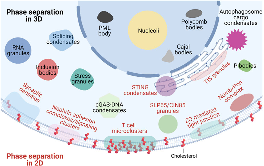
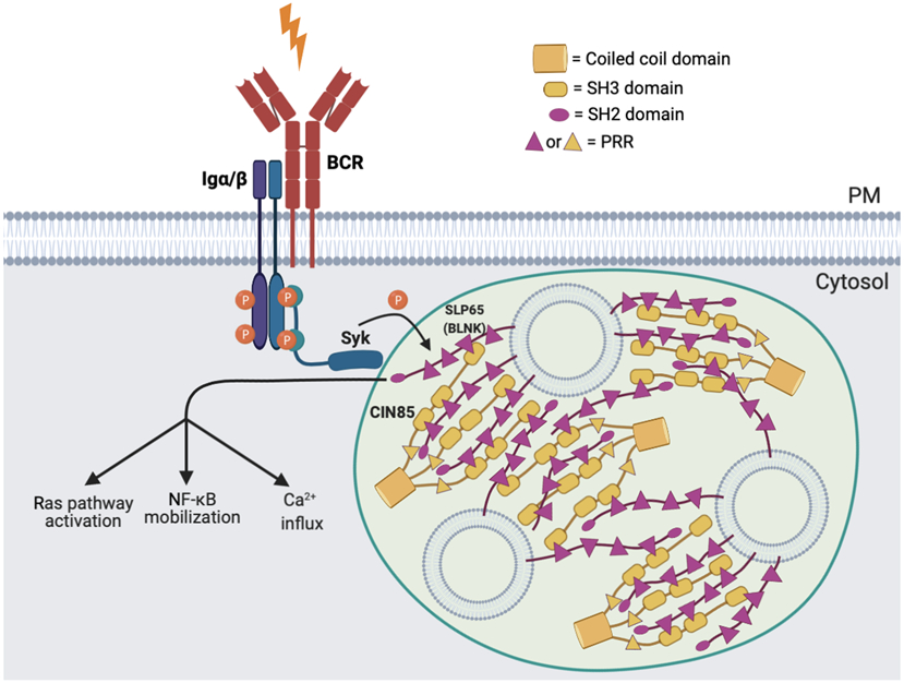
Immune signalling pathways convert pathogenic stimuli into cytosolic events that lead to the resolution of infection. Upon ligand engagement, immune receptors together with their downstream adaptors and effectors undergo substantial conformational changes and spatial reorganization. During this process, nanometre-to-micrometre-sized signalling clusters have been commonly observed that are believed to be hotspots for signal transduction. Because of their large size and heterogeneous composition, it remains a challenge to fully understand the mechanisms by which these signalling clusters form and their functional consequences. Recently, phase separation has emerged as a new biophysical principle for organizing biomolecules into large clusters with fluidic properties. Although the field is still in its infancy, studies of phase separation in immunology are expected to provide new perspectives for understanding immune responses. Here, we present an up-to-date view of how liquid-liquid phase separation drives the formation of signalling condensates and regulates immune signalling pathways, including those downstream of T cell receptor, B cell receptor and the innate immune receptors cyclic GMP-AMP synthase (cGAS)-stimulator of interferon genes (STING) and retinoic acid-inducible gene I protein (RIG-I). We conclude with a summary of the current challenges the field is facing and outstanding questions for future studies.


Figures

Figure 1 ∣
Figure 1 ∣. Phase separation in 3D and 2D.
The intracellular space is filled with biomolecular condensates formed through phase separation. These include 3D membrane-less organelles such as nucleoli, Cajal bodies, promyelocytic leukemia (PML) bodies and polycomb bodies in the nucleus; and stress granules, cGAS–DNA condensates, inclusion bodies, P bodies, splicing condensates, RNA granules, autophagosome cargo condensates, SLP65–CIN85 granules and endoplasmic reticulum-associated TIS granules and STING condensates in the cytoplasm. 2D condensates associated with the plasma membrane include T cell microclusters, nephrin-containing adhesion complexes, pre- or post-synaptic densities, NUMB–PON complexes and zonula occludens (ZO)-mediated tight junctions.
Figure 2 ∣
Figure 2 ∣. A microcluster view of T cell receptor signaling.
Upon antigen engagement, the T cell receptor (TCR) complex is phosphorylated by LCK on the immunoreceptor tyrosine-based activation motifs (ITAMs) of its CD3 chains. The phosphorylated TCR complex recruits the kinase ZAP70, which then phosphorylates LAT, resulting in the formation of liquid-like condensates of LAT. These LAT microclusters are enriched with adaptor proteins (such as GRB2, GADS, SLP76 and NCK1) and effector proteins (such as SOS1, PLCγ1, WASp and ARP2/3) to trigger the activation of downstream pathways, including RAS signaling, calcium influx (not shown) and actin remodeling. LAT microclusters exclude the phosphatase CD45 to protect phosphotyrosines, which are an activation marker of TCR signaling. CBL, an E3 ubiquitin ligase, is recruited to LAT microclusters to attenuate clustering and hence TCR signal transduction. The TCR co-receptors CD28 and PD1 overlap with LAT microclusters when engaging their own ligands.
Figure 3 ∣
Figure 3 ∣. Signaling condensates in the B cell receptor pathway.
The scaffold protein SLP65, its binding partner CIN85 and liposomes (spherical vesicles) form liquid-like signaling condensates in the cytosol of resting B cells. Condensate formation is mediated through multivalent interactions between the proline-rich motifs of SLP65 and the SH3 domains of CIN85, and between the amino-terminal lipid-binding domain of SLP65 and vesicles. CIN85 is trimerized by its coiled-coil domain, which further increases its interaction valency. Upon B cell receptor (BCR) stimulation, the kinase SYK is recruited and activated at the BCR, which phosphorylates SLP65 as the condensates approach the plasma membrane. Downstream pathways are further triggered including RAS activation, NF-κB mobilization and calcium influx.
Figure 4 ∣
Figure 4 ∣. Phase separation of intracellular innate immune signaling pathways.
a ∣ Cyclic GMP–AMP synthase (cGAS) forms liquid-like condensates with double-stranded DNA (dsDNA) to enhance the production of 2′3′-cyclic GMP–AMP (cGAMP) by protecting DNA from degradation by the exonuclease TREX1. Bacteria-derived streptavidin, free zinc ions, RNA and the stress granule protein G3BP1 regulate the formation of cGAS–dsDNA condensates and production of cGAMP. cGAMP, in turn, activates STING, leading to downstream signaling through TBK1 and IRF3 that induces the expression of type I interferons and other proinflammatory cytokines However, overproduction of cGAMP induces the formation of STING condensates on the endoplasmic reticulum (ER), which recruit TBK1 but exclude IRF3 and thereby prevent overactivation of the innate immune response by limiting interferon production. b ∣ The E3 ubiquitin ligase TRIM25 forms liquid-like condensates with RNA (preprint data; not yet peer reviewed), which recruit the RNA sensor retinoic acid-inducible gene I (RIG-I) and promote RIG-I activation through K63-linked ubiquitylation and downstream signaling through mitochondrial antiviral signaling protein (MAVS). In parallel, G3BP1 recruits the E3 ubiquitin ligase RNF125 into stress granules and destabilizes RNF125, which inhibits the K48-linked ubiquitylation of RIG-I that would otherwise lead to its proteasomal degradation. G3BP1 also promotes RNA binding to RIG-I to trigger RIG-I activation. c ∣ Nuclear factor-κB (NF-κB) functions downstream of RIG-I–MAVS signaling. The p65 subunit of NF-κB is trapped in the inclusion bodies that are formed by phase separation of the viral replication machinery. Trapped p65 is unable to translocate into the nucleus to induce the expression of pro-inflammatory cytokines.

References

    1. Zbinden A, Perez-Berlanga M, De Rossi P & Polymenidou M Phase Separation and Neurodegenerative Diseases: A Disturbance in the Force. Dev Cell 55, 45–68 (2020). - PubMed
    1. Su X et al. Phase separation of signaling molecules promotes T cell receptor signal transduction. Science 352, 595–9 (2016). - PMC - PubMed
    1. Huang WYC et al. A molecular assembly phase transition and kinetic proofreading modulate Ras activation by SOS. Science 363, 1098–1103 (2019). - PMC - PubMed
    1. Wong LE et al. Tripartite phase separation of two signal effectors with vesicles priming B cell responsiveness. Nat Commun 11, 848 (2020). - PMC - PubMed
    1. Stone MB, Shelby SA, Nunez MF, Wisser K & Veatch SL Protein sorting by lipid phase-like domains supports emergent signaling function in B lymphocyte plasma membranes. Elife 6 (2017). - PMC - PubMed

Publication types

Substances