Review
. 2021 Jun 1;25(1):2062.
doi: 10.4102/sajr.v25i1.2062. eCollection 2021.

PI-RADS v2.1: What has changed and how to report

Affiliations
Review

PI-RADS v2.1: What has changed and how to report

Robin Scott et al. SA J Radiol. .

Abstract

Multiparametric magnetic resonance imaging (MRI) of the prostate has become a vital imaging tool in daily radiological practice for the stratification of the risk of prostate cancer. There has been a recent update to the Prostate Imaging-Reporting and Data System (PI-RADS). The updated changes in PI-RADS, which is version 2.1, have been described with information pertaining to the recommended imaging protocols, the techniques on how to perform prostate MRI and a simplified approach to interpreting and reporting MRI of the prostate. Explanatory tables, schematic diagrams and key representative images have been used to provide the reader with a useful approach to interpreting and then stratifying lesions in the four anatomical zones of the prostate gland. The intention of this article is to address challenges of interpretation and reporting of prostate lesions in daily practice.

Keywords: PI-RADS; assessment categories to stratify risk; magnetic resonance imaging; prostate carcinoma; structured reporting; technical parameters for mpMRI of prostate.


Conflict of interest statement

The authors declare that they have no financial or personal relationships that may have inappropriately influenced them in writing this pictorial review.

Figures

FIGURE 1
FIGURE 1
Sector zone map demonstrating the named 41 zones of the prostate.
FIGURE 2
FIGURE 2
Workstation layout of four sequences used for multiparametric prostate magnetic resonance imaging evaluation of the prostate including (left to right) high b-value diffusion-weighted imaging, Apparent Diffusion Coefficient (ADC) map, axial and coronal T2-weighted imaging.
FIGURE 3
FIGURE 3
Recommended measurements for calculation of prostate volume at magnetic resonance imaging. Maximum longitudinal (L) diameter and maximum antero-posterior (AP) diameter measured on mid-sagittal T2-weighted imaging (T2WI) (a) and maximum transverse (T) diameter measured on axial T2WI (b).
FIGURE 4
FIGURE 4
Typical peripheral zone Prostate Imaging-Reporting and Data System (PI-RADS) v2.1 lesions. Peripheral zone (PZ) shows PI-RADS assessment categories with diffusion-weighted imaging (DWI) as the dominant sequence. (1) No abnormality (normal) on all sequences. (2) Iso-intense signal of the PZ on the high b-value DW image with an indistinct linear hypointense lesion on the Apparent Diffusion Coefficient (ADC) map (arrow) with corresponding T2W linear hypointense lesion. (3) Mildly hyperintense lesion of the PZ on the high b-value DW image with focal mildly hypointense lesion on the ADC map (arrow) and hyperintense non-circumscribed lesion on the T2W image. (4) Focal markedly hyperintense lesion (arrow) on the high b-value DW image with corresponding markedly hypointense signal intensity lesion on the ADC map. Lesion size < 1.5 cm in greatest dimension. T2W image demonstrates a circumscribed, homogeneous hyperintense mass confined to the prostate. (5) Same as (4) above but size > 1.5 cm in greatest dimension and could have extra-prostatic extension/invasive behaviour.
FIGURE 5
FIGURE 5
Typical Transition Zone Prostate Imaging-Reporting and Data System (PI-RADS) v2.1 lesions. Transition Zone (TZ) shows PI-RADS assessment categories with T2-weighted imaging as dominant sequence. (1) Homogeneous intermediate signal intensity OR round, completely encapsulated nodule(s), that is, ‘typical nodule’. (2) Mostly encapsulated nodule (arrow) OR homogeneous circumscribed nodule without encapsulation OR homogeneous mildly hypointense area between nodules. (3) Heterogeneous, hypointense signal intensity with obscured margins. On Apparent Diffusion coefficient (ADC) map, focal (discrete and different from background) hypointense and/or focal hyperintensity on high b-value diffusion-weighted image. (4) Non-circumscribed (arrow) homogeneous moderately hypointense lesion on T2-weighted image. Must be < 1.5 cm in its greatest dimension. On ADC map, markedly hypointense and markedly hyperintense on high b-value DW image. (5) Same as (4) above but >1.5 cm in greatest dimension or extra-prostatic extension/invasive behaviour.
FIGURE 6
FIGURE 6
Schematic diagram of transition zone nodules, the corresponding T2-weighted imaging scores and description of acceptable variants.
FIGURE 7
FIGURE 7
Schematic diagram detailing the multi-sequence evaluation of transition zone nodules including diffusion-weighted imaging. The dotted lines indicate a near-isointense lesion with indistinct borders.
FIGURE 8
FIGURE 8
Central zone (a, b, c and d). At the prostate base the central zone is homogeneously T2 hypointense.
FIGURE 9
FIGURE 9
The anterior fibromuscular stroma shows marked homogeneously T2-hypointense signal (a). It is ADC hypointense (b) and diffusion-weighted imaging iso-intense (c).
FIGURE 10
FIGURE 10
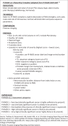
Sample reporting template devised using multiparametric prostate magnetic resonance imaging.
FIGURE 11
FIGURE 11
Flow diagram outlining the interplay of findings on relevant multiparametric prostate magnetic resonance imaging sequences for scoring of prostatic lesions in the peripheral zone and transition zone regions.,,

References

    1. Barret T, Rajesh A, Rosenkrantz AB, Choyke PL, Turkbey B. PI-RADS version 2.1: One small step for prostate MRI. Clin Radiol. 2019;74(11):841–852. 10.1016/j.crad.2019.05.019 - DOI - PubMed
    1. Steiger P, Thoeny HC. Prostate MRI based on PI-RADS version 2: How we review and report. Cancer Imaging. 2016;16:9. 10.1186/s40644-016-0068-2 - DOI - PMC - PubMed
    1. Weinreb JC, Barentz JO, Choyke PL, et al. . PI-RADS Prostate Imaging-Reporting and Data System, v2.1 [homepage on the Internet]. American College of Radiology; 2019. [cited 2021 Mar 15]. Available from: https://www.acr.org/-/media/ACR/Files/RADS/Pi-RADS/PIRADS-V2-1.pdf
    1. World Cancer Research Fund International/American Institute for Cancer Research . Continuous Update Project Report. Diet, nutrition, physical activity, and prostate cancer [homepage on the Internet]. 2014. [cited 2015 Dec 01]. Available from: www.wcrf.org/sites/default/files/Prostate-Cancer-2014-Report.pdf
    1. Singh E, Motsuku L, Sengayi-Muchengeti M, Chen W, Abraham N. Ekurhuleni population-based registry 2018 Annual report. Johannesburg, South Africa, March 2020. National Health Laboratory Service, National Cancer Registry, Department of Health. Johannesburg; 2020.

LinkOut - more resources