. 2021 Aug 25;95(18):e0079621.
doi: 10.1128/JVI.00796-21. Epub 2021 Aug 25.

Enhanced Ability of Plant-Derived PGT121 Glycovariants To Eliminate HIV-1-Infected Cells

Affiliations

Enhanced Ability of Plant-Derived PGT121 Glycovariants To Eliminate HIV-1-Infected Cells

Sai Priya Anand et al. J Virol. .

Abstract

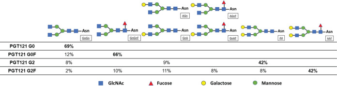
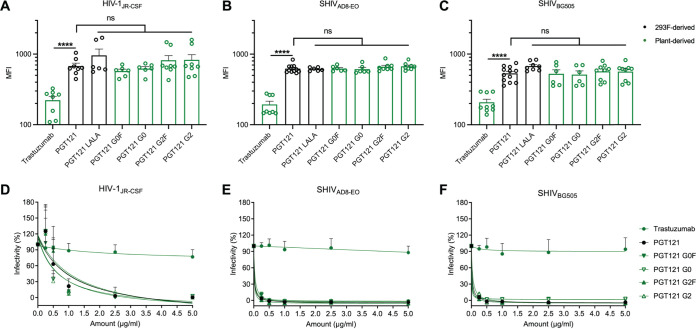
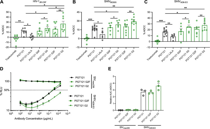
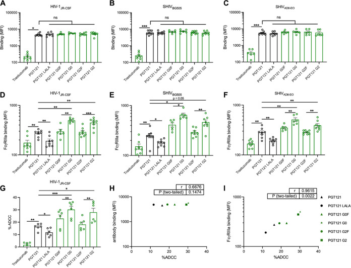
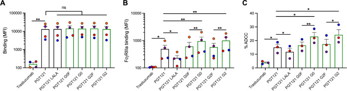
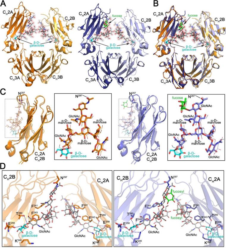
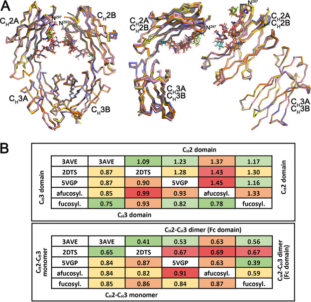
The activity of broadly neutralizing antibodies (bNAbs) targeting HIV-1 depends on pleiotropic functions, including viral neutralization and the elimination of HIV-1-infected cells. Several in vivo studies have suggested that passive administration of bNAbs represents a valuable strategy for the prevention or treatment of HIV-1. In addition, different strategies are currently being tested to scale up the production of bNAbs to obtain the large quantities of antibodies required for clinical trials. Production of antibodies in plants permits low-cost and large-scale production of valuable therapeutics; furthermore, pertinent to this work, it also includes an advanced glycoengineering platform. In this study, we used Nicotiana benthamiana to produce different Fc-glycovariants of a potent bNAb, PGT121, with near-homogeneous profiles and evaluated their antiviral activities. Structural analyses identified a close similarity in overall structure and glycosylation patterns of Fc regions for these plant-derived Abs and mammalian cell-derived Abs. When tested for Fc-effector activities, afucosylated PGT121 showed significantly enhanced FcγRIIIa interaction and antibody dependent cellular cytotoxicity (ADCC) against primary HIV-1-infected cells, both in vitro and ex vivo. However, the overall galactosylation profiles of plant PGT121 did not affect ADCC activities against infected primary CD4+ T cells. Our results suggest that the abrogation of the Fc N-linked glycan fucosylation of PGT121 is a worthwhile strategy to boost its Fc-effector functionality. IMPORTANCE PGT121 is a highly potent bNAb and its antiviral activities for HIV-1 prevention and therapy are currently being evaluated in clinical trials. The importance of its Fc-effector functions in clearing HIV-1-infected cells is also under investigation. Our results highlight enhanced Fc-effector activities of afucosylated PGT121 MAbs that could be important in a therapeutic context to accelerate infected cell clearance and slow disease progression. Future studies to evaluate the potential of plant-produced afucosylated PGT121 in controlling HIV-1 replication in vivo are warranted.

Keywords: ADCC; Env glycoproteins; Envelope glycoproteins; FcγRIIIa; HIV-1; Nicotiana benthamiana; PGT121; broadly neutralizing antibodies; fucose; galactose; glycosylation; neutralizing antibodies; plant antibodies.


Figures

FIG 1
FIG 1
N-linked glycans of N. benthamiana-produced PGT121 glycovariants. The percentages of predominant N-glycosylations on PGT121 G0, PGT121 G0F, PGT121 G2, and PGT121 G2F are presented in the table. Schematic diagrams are across the top, where Asn is asparagine 297, GnGn is diantennary N-acetylglucosamine, F is fucose, and A is galactose. Glycan species abundances are given as percentages, and minor glycoforms are not indicated. Glycan nomenclature is further described by ProGlycAn.
FIG 2
FIG 2
Fc glycosylation does not affect the ability of PGT121 to recognize infected cells or its neutralization capacity. Cell surface staining of CEM.NKr CCR5+ cells infected with HIV-1JRCSF (A), SHIVAD8-EO (B), and SHIVBG505 (C) was performed 48 h postinfection. Antibody binding was detected using Alexa Fluor 647-conjugated anti-human secondary Abs. Graphs represent the median fluorescence intensities (MFI) in the infected population (p24+ or p27+) determined from at least five independent experiments, with the error bars indicating means ± the standard errors of the mean (SEM). Statistical significance was tested using an unpaired t test or a Mann-Whitney U test based on statistical normality (****, P < 0.0001; ns, nonsignificant). (D to F) Lentiviral particles produced from HIV-1JRCSF (D), SHIVAD8-EO (E), and SHIVBG505 (F) IMCs. Viruses were incubated with serial dilutions of trastuzumab and PGT121 MAbs at 37°C for 1 h prior to infection of TZM-bl target cells. The infectivity at each Ab concentration tested is shown as the percentage of infection without Ab for each virus. Quadruplicate samples were analyzed in each experiment. The data shown are the means of results obtained in at least three independent experiments. Error bars indicate means ± the SEM. Black histogram/curves represent 293F cell-derived MAbs and green histogram/curves represent plant-derived MAbs.
FIG 3
FIG 3
Fc glycosylation profile of PGT121 regulates its ADCC capacity against infected cells. CEM.NKR-CCR5-sLTR-Luc cells infected with HIV-1JRCSF (A), SHIVBG505 (B), and SHIVAD8-EO (C) were used as target cells. PBMCs from uninfected donors were used as effector cells in a FACS-based ADCC assay. The graphs shown represent the percentages of ADCC obtained in the presence of the respective antibodies. (D and E) For the luciferase assay, CEM.NKr-CCR5-sLTR-Luc cells infected with SHIVAD8-EO, or SIVmac239 as a negative control. ADCC responses were measured as the dose-dependent loss of luciferase activity in RLU after incubation of infected CEM.NKR-CCR5-sLTR-Luc cells with CD16+ KHYG-1 effector cells in the presence of antibody. Values are the means ± standard deviations (error bars) for triplicate wells, and the dotted line indicates half-maximal lysis of infected cells. (E) Area under the curve (AUC) values were calculated using from curves of increasing MAb concentrations shown in panel D. Error bars indicate means ± the SEM. Statistical significance was tested using a paired t test or Wilcoxon matched-pairs signed-rank test based on statistical normality (*, P < 0.05; **, P < 0.01; ***, P < 0.001). Black histogram bars represent 293F cell-derived MAbs and green histogram bars represent plant-derived MAbs.
FIG 4
FIG 4
Fc glycosylation profile of PGT121 modulates FcγRIIIa interaction and ADCC against infected primary CD4+ T cells. Cell surface staining of primary CD4+ T cells infected with (A and D) HIV-1JRCSF, (B and E) SHIVAD8-EO, and (C and F) SHIVBG505 was performed 48 h postinfection. Antibody binding was detected either by using Alexa Fluor 647-conjugated anti-human secondary Abs (A to C) or by using biotin-tagged dimeric rsFcγRIIIa (0.2 μg/ml) followed by the addition of Alexa Fluor 647-conjugated streptavidin (D to F). (A to F) Graphs represent MFI values in the infected population (p24+ or p27+) determined from at least five independent experiments, with the error bars indicating means ± the SEM. (G) Primary CD4+ T cells infected with HIV-1JRCSF were used as target cells. Autologous PBMCs were used as effector cells in a FACS-based ADCC assay. The graph represents the percentages of ADCC obtained in the presence of the respective antibodies. Statistical significance was tested using a paired t test or Wilcoxon matched-pairs signed-rank test based on statistical normality (*, P < 0.05; **, P < 0.01; ***, P < 0.001; ns, nonsignificant). Black histogram bars represent 293F cell-derived MAbs, and green histogram bars represent plant-derived MAbs. (H and I) Correlations between the levels of ADCC and levels of antibody binding (H) or FcγRIIIa binding (I), as measured on primary CD4+ T cells infected with HIV-1JRCSF. Statistical significance was tested using a Pearson correlation test. Black points represent 293F cell-derived MAbs, and green points represent plant-derived MAbs.
FIG 5
FIG 5
Susceptibility of ex vivo-expanded endogenously infected primary CD4+ T cells from HIV-1-infected individuals to PGT121-mediated ADCC. Primary CD4+ T cells from four different HIV-1-infected individuals were isolated and reactivated with PHA-L for 48 h, followed by incubation with IL-2 to expand the endogenous virus. Cell surface staining of infected primary CD4+ T cells was performed upon reactivation. Antibody binding was detected either by using Alexa Fluor 647-conjugated anti-human secondary Abs (A) or biotin-tagged dimeric rsFcγRIIIa (0.2 μg/ml) followed by the addition of Alexa Fluor 647-conjugated streptavidin (B). (A and B) Graphs represent the MFI values in the infected population (p24+ or p27+) determined from at four different donors, with the error bars indicating means ± the SEM. (C) Ex vivo-expanded infected primary CD4+ T cells from three HIV-1-infected individuals were used as target cells. Autologous PBMCs were used as effector cells in a FACS-based ADCC assay. The graphs represent the percentages of ADCC obtained in the presence of the respective antibodies. ADCC susceptibility was only measured when the percentage of infection (p24+ cells) was higher than 10%. Statistical significance was tested using a paired t test or Wilcoxon matched-pairs signed-rank test based on statistical normality (*, P < 0.05; **, P < 0.01; ***, P < 0.001; ns, nonsignificant). Black histogram bars represent 293F cell-derived MAbs, and green histogram bars represent plant-derived MAbs.
FIG 6
FIG 6
Structural characterization of the Fc regions of N. benthamiana-produced PGT121. (A) Crystal structures of afucosylated (right) and fucosylated (left) Fc. The overall structure is shown in a ribbon diagram with the two heavy chains (CH2-CH3 domains) in lighter (chain B) and darker (chain A) shades of orange and blue for afucosylated and fucosylated variants, respectively. The sugars attached to asparagine 297 are shown as sticks and spheres colored by atom type (gray for carbon, red for oxygen, and blue for nitrogen). The fucose in the fucosylated Fc is colored green and the terminal galactose visible on the α6 arm of the glycan in both structures cyan. (B) Superposition of the afucosylated and fucosylated CH2-CH3 dimer (Fc domain) colored as in panel A. (C) Superposition of the CH2 domains from the afucosylated (left) and fucosylated (right) Fc dimer. Blow-up views to the right show the superposition of the glycan only with chain A shown as sticks and chain B as lines. Atom types are colored as in panel A. (D) Details of the glycan-glycan and glycan-protein contacts in the afucosylated (left) and fucosylated (right) Fc dimers. The glycan and interacting residues are shown as sticks and the protein backbone as a ribbon. Hydrogen bonds are shown with dashed lines. Atom types are colored as in panel A.
FIG 7
FIG 7
Comparison of the overall structures of N. benthamiana expressed and mammalian expressed human Fc domains. Structural alignment of CH2-CH3 dimers (Fc), CH2-CH3 monomers, CH2 domains, and CH3 domains of N. benthamiana and mammalian expressed human Fcs including the following: a human fucosylated Fc lacking the terminal galactose (PDB ID 3AVE, yellow), an afucosylated Fc (2DTS, pink), and a fucosylated Fc containing a terminal galactose (5VGP, gray). N. benthamiana expressed Fcs are colored as in Fig. 6, with fucose colored green and galactose cyan. (B) Average RMSD values for main chain atom pairwise comparisons of CH2 domains, CH3 domains, CH2-CH3 monomers, and CH2-CH3 dimers (Fc domain) shown in tabular format color coded with smaller RMSD values green and larger RMSD values red.

References

    1. Barouch DH, Whitney JB, Moldt B, Klein F, Oliveira TY, Liu J, Stephenson KE, Chang HW, Shekhar K, Gupta S, Nkolola JP, Seaman MS, Smith KM, Borducchi EN, Cabral C, Smith JY, Blackmore S, Sanisetty S, Perry JR, Beck M, Lewis MG, Rinaldi W, Chakraborty AK, Poignard P, Nussenzweig MC, Burton DR. 2013. Therapeutic efficacy of potent neutralizing HIV-1-specific monoclonal antibodies in SHIV-infected rhesus monkeys. Nature 503:224–228. 10.1038/nature12744. - DOI - PMC - PubMed
    1. Bournazos S, Klein F, Pietzsch J, Seaman MS, Nussenzweig MC, Ravetch JV. 2014. Broadly neutralizing anti-HIV-1 antibodies require Fc effector functions for in vivo activity. Cell 158:1243–1253. 10.1016/j.cell.2014.08.023. - DOI - PMC - PubMed
    1. Horwitz JA, Halper-Stromberg A, Mouquet H, Gitlin AD, Tretiakova A, Eisenreich TR, Malbec M, Gravemann S, Billerbeck E, Dorner M, Buning H, Schwartz O, Knops E, Kaiser R, Seaman MS, Wilson JM, Rice CM, Ploss A, Bjorkman PJ, Klein F, Nussenzweig MC. 2013. HIV-1 suppression and durable control by combining single broadly neutralizing antibodies and antiretroviral drugs in humanized mice. Proc Natl Acad Sci U S A 110:16538–16543. 10.1073/pnas.1315295110. - DOI - PMC - PubMed
    1. Klein F, Halper-Stromberg A, Horwitz JA, Gruell H, Scheid JF, Bournazos S, Mouquet H, Spatz LA, Diskin R, Abadir A, Zang T, Dorner M, Billerbeck E, Labitt RN, Gaebler C, Marcovecchio P, Incesu RB, Eisenreich TR, Bieniasz PD, Seaman MS, Bjorkman PJ, Ravetch JV, Ploss A, Nussenzweig MC. 2012. HIV therapy by a combination of broadly neutralizing antibodies in humanized mice. Nature 492:118–122. 10.1038/nature11604. - DOI - PMC - PubMed
    1. Hessell AJ, Poignard P, Hunter M, Hangartner L, Tehrani DM, Bleeker WK, Parren PW, Marx PA, Burton DR. 2009. Effective, low-titer antibody protection against low-dose repeated mucosal SHIV challenge in macaques. Nat Med 15:951–954. 10.1038/nm.1974. - DOI - PMC - PubMed

Publication types

MeSH terms