

Whole-body uptake classification and prostate cancer staging in 68Ga-PSMA-11 PET/CT using dual-tracer learning
- PMID: 34232350
- PMCID: PMC8803695
- DOI: 10.1007/s00259-021-05473-2
Whole-body uptake classification and prostate cancer staging in 68Ga-PSMA-11 PET/CT using dual-tracer learning
Abstract
Purpose: In PSMA-ligand PET/CT imaging, standardized evaluation frameworks and image-derived parameters are increasingly used to support prostate cancer staging. Clinical applicability remains challenging wherever manual measurements of numerous suspected lesions are required. Deep learning methods are promising for automated image analysis, typically requiring extensive expert-annotated image datasets to reach sufficient accuracy. We developed a deep learning method to support image-based staging, investigating the use of training information from two radiotracers.
Methods: In 173 subjects imaged with 68Ga-PSMA-11 PET/CT, divided into development (121) and test (52) sets, we trained and evaluated a convolutional neural network to both classify sites of elevated tracer uptake as nonsuspicious or suspicious for cancer and assign them an anatomical location. We evaluated training strategies to leverage information from a larger dataset of 18F-FDG PET/CT images and expert annotations, including transfer learning and combined training encoding the tracer type as input to the network. We assessed the agreement between the N and M stage assigned based on the network annotations and expert annotations, according to the PROMISE miTNM framework.
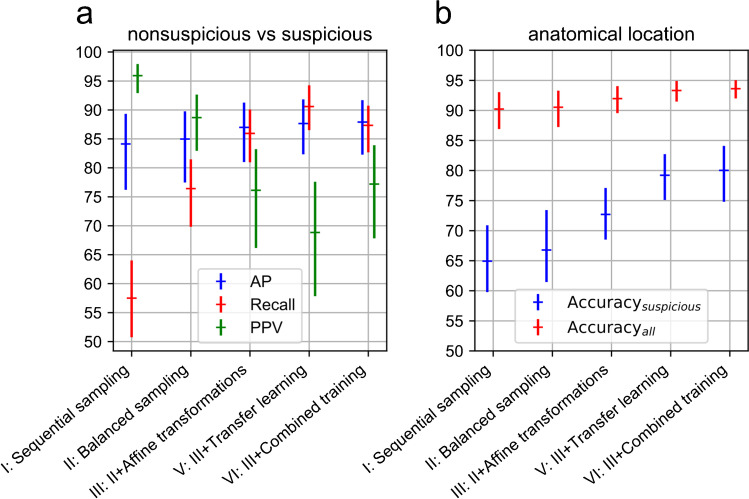
Results: In the development set, including 18F-FDG training data improved classification performance in four-fold cross validation. In the test set, compared to expert assessment, training with 18F-FDG data and the development set yielded 80.4% average precision [confidence interval (CI): 71.1-87.8] for identification of suspicious uptake sites, 77% (CI: 70.0-83.4) accuracy for anatomical location classification of suspicious findings, 81% agreement for identification of regional lymph node involvement, and 77% agreement for identification of metastatic stage.
Conclusion: The evaluated algorithm showed good agreement with expert assessment for identification and anatomical location classification of suspicious uptake sites in whole-body 68Ga-PSMA-11 PET/CT. With restricted PSMA-ligand data available, the use of training examples from a different radiotracer improved performance. The investigated methods are promising for enabling efficient assessment of cancer stage and tumor burden.
Keywords: Deep learning; PET/CT; PSMA; Prostate cancer; Staging; miTNM.
© 2021. The Author(s).
Conflict of interest statement
N.C. is a full-time employee at Siemens Healthcare GmbH receiving funding under the European Union’s Horizon 2020 Marie Skłodowska–Curie grant agreement (no. 764458). G.P. is a full-time employee at Siemens Healthcare GmbH. L.S., V.S., and B.S. are full-time employees at Siemens Medical Solutions USA, Inc. All the other authors declare to have no conflict of interest.
Figures


References
-
- Hofman MS, Lawrentschuk N, Francis RJ, Tang C, Vela I, Thomas P, et al. Prostate-specific membrane antigen PET-CT in patients with high-risk prostate cancer before curative-intent surgery or radiotherapy (proPSMA): a prospective, randomised, multicentre study. Lancet. 2020;395:1208–1216. doi: 10.1016/S0140-6736(20)30314-7. - DOI - PubMed
-
- Maurer T, Gschwend JE, Rauscher I, Souvatzoglou M, Haller B, Weirich G, et al. Diagnostic efficacy of 68 gallium-PSMA positron emission tomography compared to conventional imaging for lymph node Staging of 130 consecutive patients with intermediate to high risk prostate cancer. J Urol. 2016;195:1436–1443. doi: 10.1016/j.juro.2015.12.025. - DOI - PubMed
MeSH terms
Substances
LinkOut - more resources
Full Text Sources
Other Literature Sources
Medical
Miscellaneous
