Review
. 2022 Apr;141(3-4):363-382.
doi: 10.1007/s00439-021-02304-0. Epub 2021 Jul 7.

Human deafness-associated variants alter the dynamics of key molecules in hair cell stereocilia F-actin cores

Affiliations
Review

Human deafness-associated variants alter the dynamics of key molecules in hair cell stereocilia F-actin cores

Takushi Miyoshi et al. Hum Genet. 2022 Apr.

Abstract

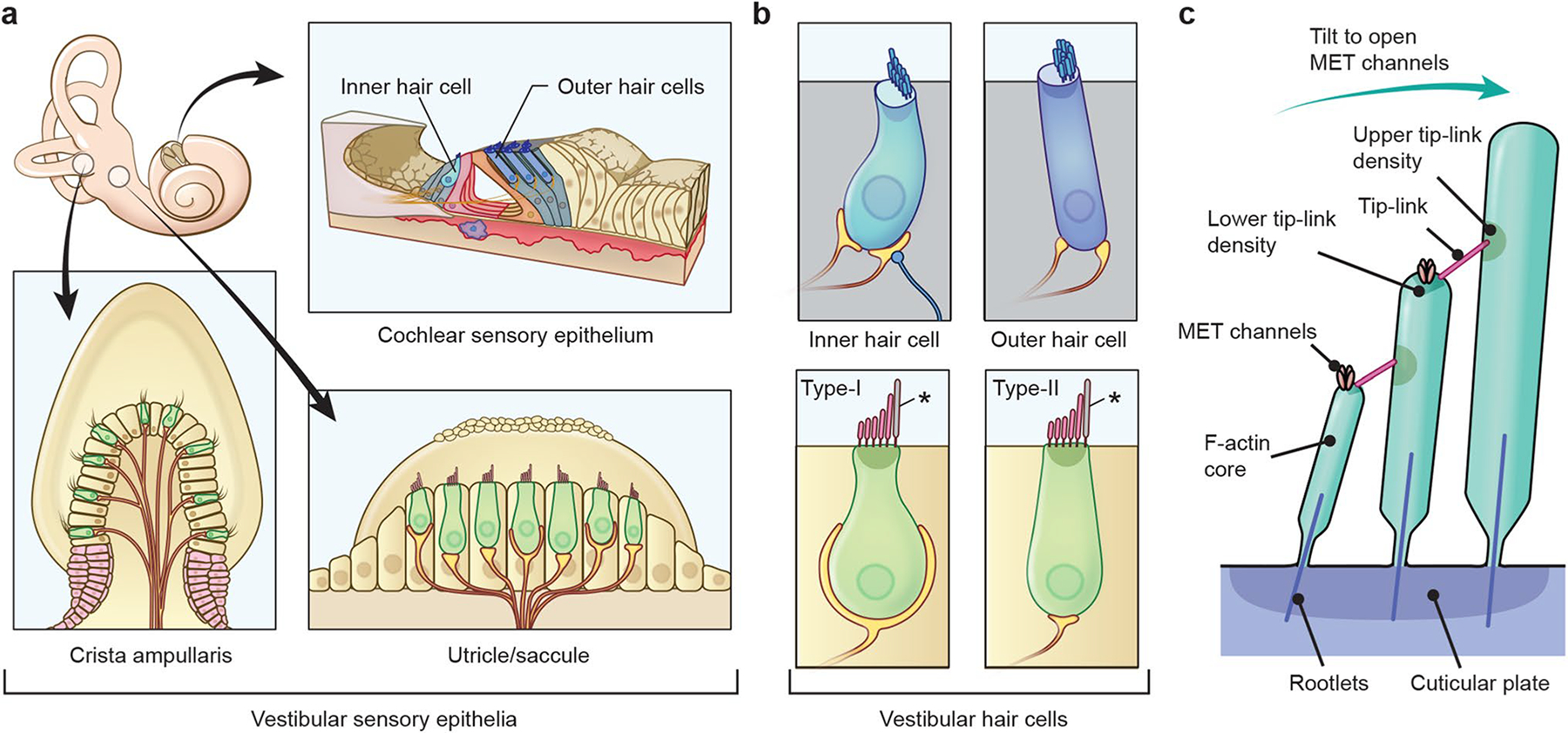
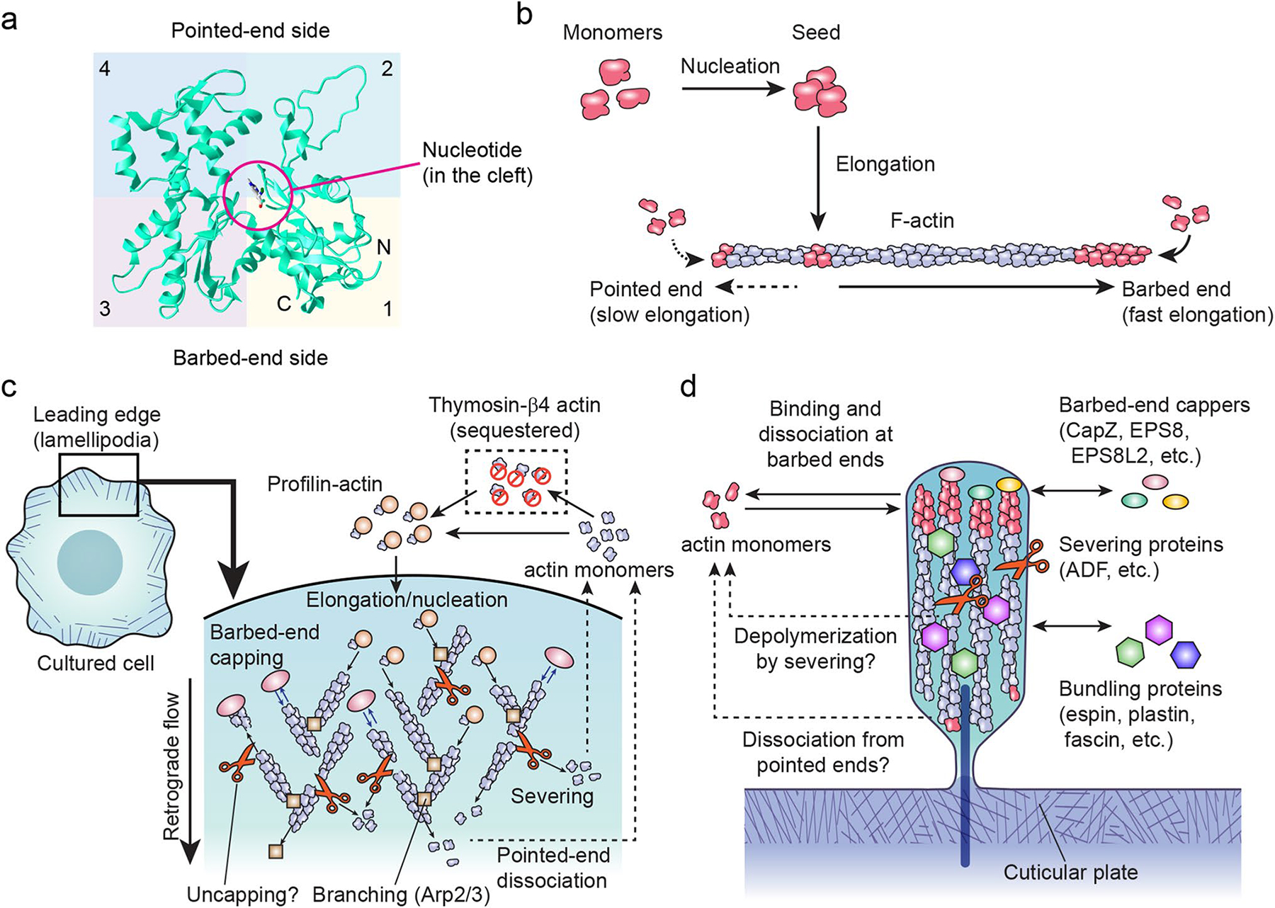
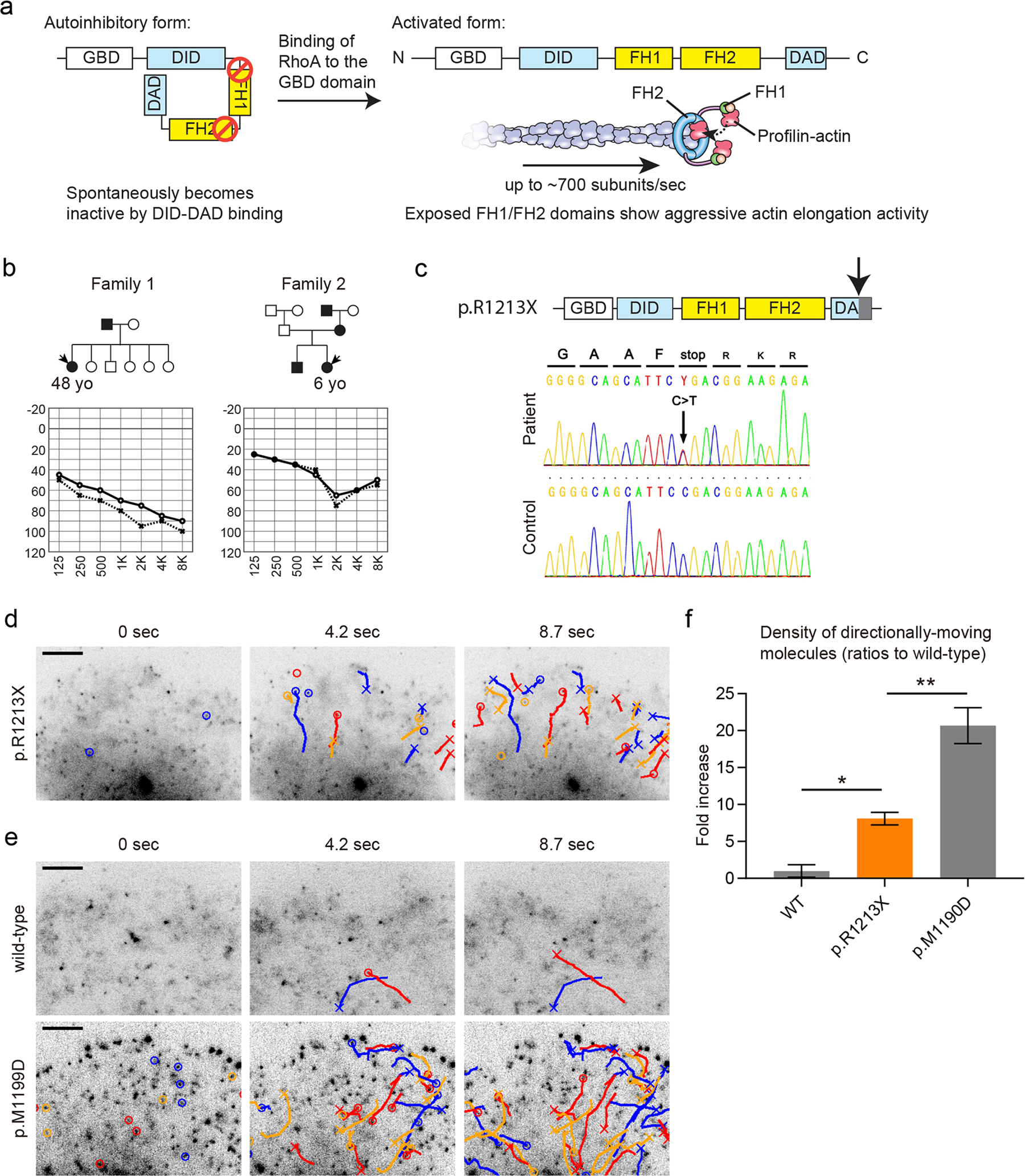
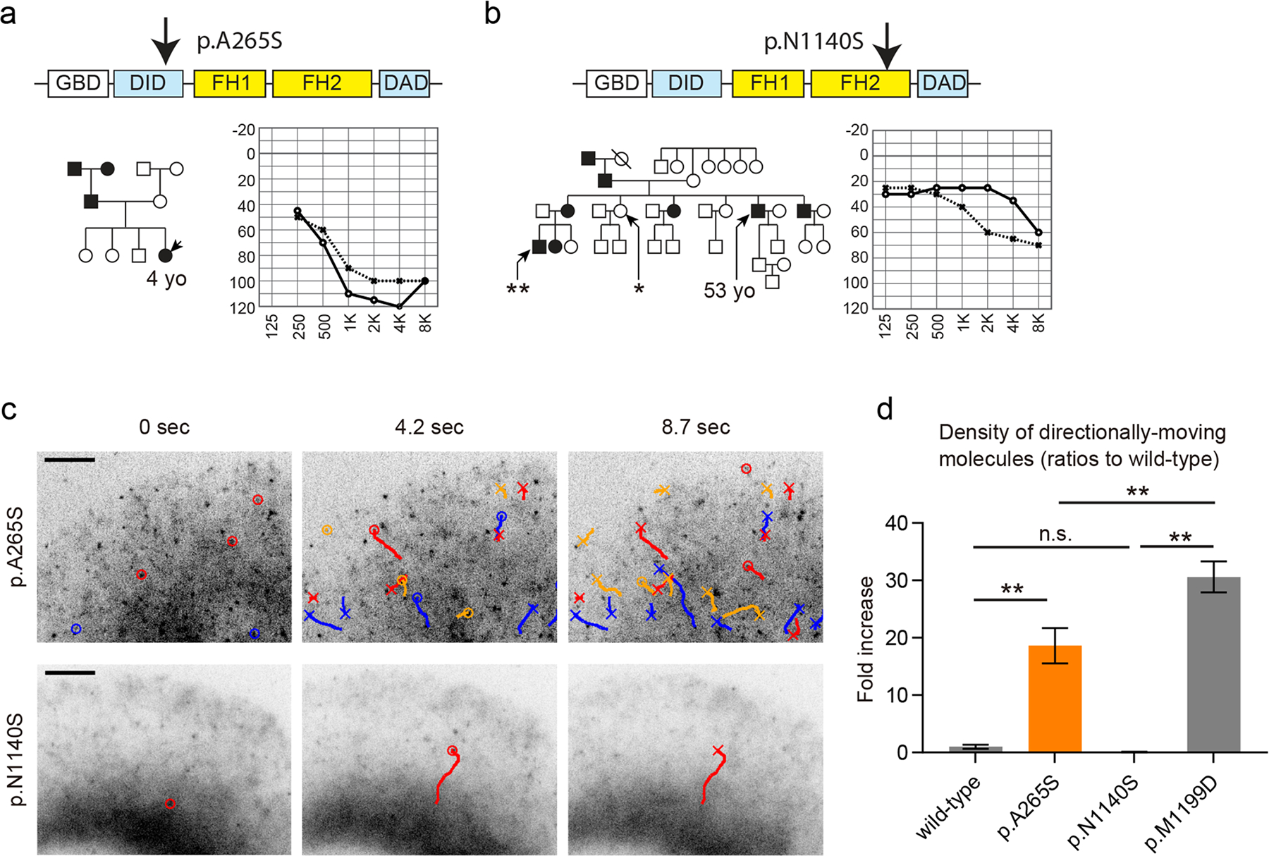
Stereocilia protrude up to 100 µm from the apical surface of vertebrate inner ear hair cells and are packed with cross-linked filamentous actin (F-actin). They function as mechanical switches to convert sound vibration into electrochemical neuronal signals transmitted to the brain. Several genes encode molecular components of stereocilia including actin monomers, actin regulatory and bundling proteins, motor proteins and the proteins of the mechanotransduction complex. A stereocilium F-actin core is a dynamic system, which is continuously being remodeled while maintaining an outwardly stable architecture under the regulation of F-actin barbed-end cappers, severing proteins and crosslinkers. The F-actin cores of stereocilia also provide a pathway for motor proteins to transport cargos including components of tip-link densities, scaffolding proteins and actin regulatory proteins. Deficiencies and mutations of stereocilia components that disturb this "dynamic equilibrium" in stereocilia can induce morphological changes and disrupt mechanotransduction causing sensorineural hearing loss, best studied in mouse and zebrafish models. Currently, at least 23 genes, associated with human syndromic and nonsyndromic hearing loss, encode proteins involved in the development and maintenance of stereocilia F-actin cores. However, it is challenging to predict how variants associated with sensorineural hearing loss segregating in families affect protein function. Here, we review the functions of several molecular components of stereocilia F-actin cores and provide new data from our experimental approach to directly evaluate the pathogenicity and functional impact of reported and novel variants of DIAPH1 in autosomal-dominant DFNA1 hearing loss using single-molecule fluorescence microscopy.


Conflict of interest statement

Declarations

Conflict of interests The authors declare no conflict of interest associated with this manuscript.

Figures

Fig. 1
Fig. 1
Sensory epithelia of the inner ear, hair cells and their stereocilia in mammals. a In the inner ear, there are three distinct types of sensory epithelia. The cochlear sensory epithelium detects and attenuates sound vibration using one row of inner hair cells and three rows of outer hair cells, which are a part of the organ of Corti. Vestibular sensory epithelia of utricle and saccule detect horizontal and vertical accelerations. The three cristae ampullaris detect angular accelerations of the head. b Alignment of stereocilia on the apical surface of hair cells. Each outer hair cell has three rows of V-shaped stereocilia while stereocilia of inner hair cell are organized in a straighter line. Vestibular hair cell stereocilia (type I and type II) are long and also align in a staircase manner although the alignment is not as organized as inner and outer hair cells. Asterisks indicate kinocilia, which are composed of microtubules. c Architecture of stereocilia F-actin cores and tip-links. Stereocilia shafts consist of F-actin, which are bundled more tightly near their bases to form rootlets and anchor stereocilia in the cuticular plate. Tip-links are tethered to F-actin cores via protein complexes in upper and lower tip-link densities. Tension caused by unidirectional deflection of all stereocilia within a bundle opens the mechanotransduction channels (MET channels) near the tip-links
Fig. 2
Fig. 2
Sructure and dynamics of actin monomers and filaments (F-actin). a Tertiary structure of an ACTB actin monomer (PDB number: 3j82) (Hanc et al. 2015). A large cleft divides subdomains 1 and 2 from subdomains 3 and 4. ATP or ADP and a cation are held in a pocket deep in the cleft (ADP shown). b Assembly of F-actin from actin monomers in vitro. Formation of F-actin seeds from monomers is a slow nucleation step. F-actin has two ends, a barbed end that can rapidly elongate and a pointed end that can elongate but more slowly. c Actin dynamics in the lamellipodia, a thin veil-like structure at the leading edge of cells. Quasi-two-dimensional actin mesh is dynamically remodeled and tracked toward the center of cells by the retrograde flow. Lamellipodia can be used to study the dynamics of actin monomers, barbed-end cappers, Arp2/3 complex and severing proteins, many of which play crucial roles in stereocilia. Exchange of ATP and ADP in actin monomers is not shown to simplify the diagram. d Dynamics of stereocilia components. Proteins, such as actin, barbed-end cappers and bundling proteins, turn over through processes involving binding and dissociation that are hypothesized to be mechanisms to maintain the entire architecture of stereocilia but are not well understood, especially those that balance polymerization and depolymerization of the F-actin and replace damaged components in stereocilia F-actin cores
Fig. 3
Fig. 3
Functional analysis of the DIAPH1 (p.R1213X) variant using single-molecule fluorescence microscopy. a Autoinhibitory mechanism regulating DIAPH1 actin elongation activity. The DID and DAD domains spontaneously interact with each other. Binding of RhoA to the GBD domain of DIAPH1 releases inhibition and allows for aggressive actin elongation activity by the FH1-FH2 domains of DIAPH1. The FH1 domain recruits profilin-actin while the FH2 domain holds onto the barbed end of F-actin to cause processive and high-speed actin elongation, which reaches an average speed of ~ 700 actin subunits/sec. b Two Japanese families segregating hearing loss analyzed to identify the DIAPH1 (p.R1213X) variant. Audiograms of probands are also shown. c Diagrams and Sanger sequencing results of DIAPH1 (p.R1213X) variant found in families shown in (b). The p.R1213X variant truncates the C-terminus of the DAD domain (downward pointing arrow). The DNA sequence is from the proband in Family 1. d Detection of abnormal, constitutive activation of DIAPH1 (p.R1213X) using single-molecule fluorescence microscopy. Time-lapse images of Xenopus XTC cells expressing GFP-DIAPH1 (p.R1213X) are shown. GFP-DIAPH1 (p.R1213X) molecules show frequent directional movements driven by actin elongation activity of FH1-FH2 domains, which were abnormally exposed by the disrupted DID-DAD interaction. Molecules moving directionally for more than two frames are indicated by circles and trajectories. Crosses indicate disappearances. Bar, 5 μm. e Time-lapse images of negative and positive controls, wild-type GFP-DIAPH1 and GFP-DIAPH1 (p.M1199D). Only a few molecules show directional movements in XTC cells expressing wild-type GFP-DIAPH1. Moving molecules were frequently observed in XTC cells expressing GFP-DIAPH1 (p.M1199D), which is a variant lacking the DID-DAD autoinhibitory interaction (Lammers et al. 2005). Bar, 5 μm. f Semi-quantitative comparison of directionally moving molecules showing moderate activation of DIAPH1 (p.R1213X). The densities of moving molecules were determined and normalized to the expression levels of GFP-DIAPH1 as described in our previous study (Ueyama et al. 2016). Directional movements of DIAPH1 (p.R1213X) were significantly more frequent than wild-type DIAPH1 and less frequent than DIAPH1 (p.M1199D). One-way ANOVA showed p < 0.0001. Post hoc Tukey’s multiple comparison is indicated by asterisk (p < 0.05) and double asterisks (p < 0.01). Number of replicates: n = 5 for wild-type, n = 6 for p.R1213X and p.M1199D. Error bars, SEMs
Fig. 4
Fig. 4
Pathogenicity screening of DIAPH1 variants using single-molecule fluorescence microscopy. a A Korean family segregating hearing loss. The p.A265S variant was found in this family and considered to be pathogenic using single-molecule fluorescence microscopy analyses in (c). An audiogram of the proband is shown. b A Japanese family segregating hearing loss. The p.N1140S variant was found in the proband of this family and predicted to be a benign polymorphism based upon our single-molecule fluorescence microscopy assay in (c). The p.N1140S variant was subsequently found in a female with normal hearing (asterisk), and another male with hearing loss in this family did not have the p.N1140S variant (double asterisks). An audiogram of the proband is shown. c Functional analyses of the p.A265S and p.N1140S variants using single-molecule fluorescence microscopy and Xenopus XTC cells expressing GFP-fused variants. GFP-DIAPH1 (p.A265S) showed frequent directional movements similar to p.R1213X indicating the pathogenicity of p.A265S variant. In contrast, the p.N1140S variant is likely a benign polymorphism since GFP-DIAPH1 (p.N1140S) did now show frequent directional movements. The movement distances were also similar to wild-type DIAPH1 despite the amino-acid substitution in the FH2 domain. Indications of markers are similar to Fig. 3d. Bar, 5 μm. d Semi-quantitative comparison of directionally moving molecules showing the constitutive activation of DIAPH1 (p.A265S) and the intact autoinhibition of DIAPH1 (p.N1140S). The densities of moving molecules were normalized by the expression levels as described in our previous study (Ueyama et al. 2016). Directional movements of DIAPH1 (p.A265S) were significantly more frequent than wild-type DIAPH1 and less frequent than DIAPH1 (p.M1199D) while DIAPH1 (p.N1140S) did not show an increase of moving molecules. New XTC cells were prepared to obtain data for the wild-type and p.M1199D in this graph. One-way ANOVA showed p < 0.0001. Post-hoc Tukey’s multiple comparison is indicated by double asterisks (p < 0.01). Number of replicates: n = 5 for p.N1140S, n = 6 for wild-type, p.A265S and p.M1199D. Error bars, SEMs

References

    1. Ahmed ZM, Smith TN, Riazuddin S, Makishima T, Ghosh M, Bokhari S, Menon PS, Deshmukh D, Griffith AJ, Riazuddin S, Friedman TB, Wilcox ER (2002) Nonsyndromic recessive deafness DFNB18 and Usher syndrome type IC are allelic mutations of USHIC. Hum Genet 110:527–531. 10.1007/s00439-002-0732-4 - DOI - PubMed
    1. Ahmed ZM, Morell RJ, Riazuddin S, Gropman A, Shaukat S, Ahmad MM, Mohiddin SA, Fananapazir L, Caruso RC, Husnain T, Khan SN, Riazuddin S, Griffith AJ, Friedman TB, Wilcox ER (2003a) Mutations of MYO6 are associated with recessive deafness, DFNB37. Am J Hum Genet 72:1315–1322. 10.1086/375122 - DOI - PMC - PubMed
    1. Ahmed ZM, Riazuddin S, Ahmad J, Bernstein SL, Guo Y, Sabar MF, Sieving P, Riazuddin S, Griffith AJ, Friedman TB, Belyantseva IA, Wilcox ER (2003b) PCDH15 is expressed in the neurosensory epithelium of the eye and ear and mutant alleles are responsible for both USH1F and DFNB23. Hum Mol Genet 12:3215–3223. 10.1093/hmg/ddg358 - DOI - PubMed
    1. Alderson-Day B, Fernyhough C (2015) Inner speech: development, cognitive functions, phenomenology, and neurobiology. Psychol Bull 141:931–965. 10.1037/bul0000021 - DOI - PMC - PubMed
    1. Al-Maawali A, Barry BJ, Rajab A, El-Quessny M, Seman A, Coury SN, Barkovich AJ, Yang E, Walsh CA, Mochida GH, Stoler JM (2016) Novel loss-of-function variants in DIAPH1 associated with syndromic microcephaly, blindness, and early onset seizures. Am J Med Genet A 170A:435–440. 10.1002/ajmg.a.37422 - DOI - PMC - PubMed

LinkOut - more resources