Review
. 2021 Jul 7;12(1):95.
doi: 10.1186/s13244-021-01046-x.

Identifying the deceiver: the non-neoplastic mimickers of genital system neoplasms

Affiliations
Review

Identifying the deceiver: the non-neoplastic mimickers of genital system neoplasms

Omer Onder et al. Insights Imaging. .

Abstract

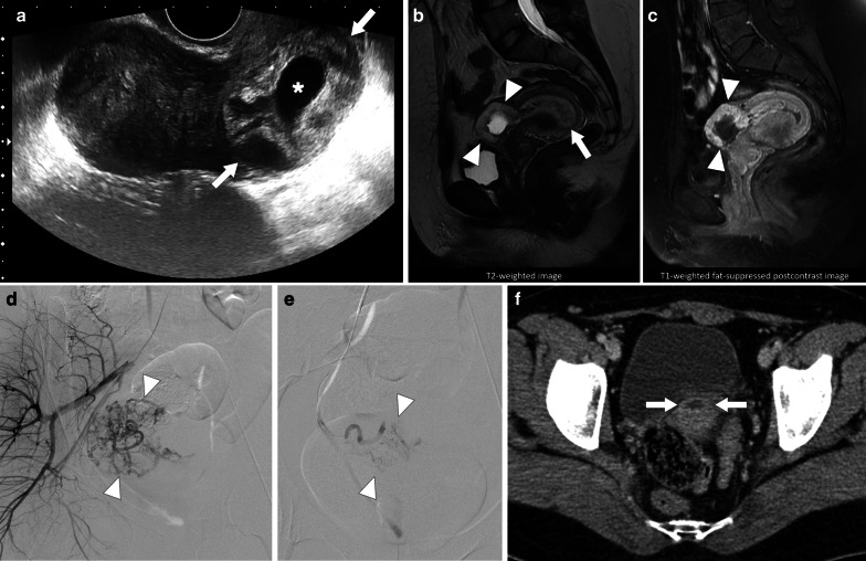
Tumors of the genital system are common and imaging is of crucial importance for their detection and diagnosis. Several non-neoplastic diseases may mimic these tumors and differential diagnosis may be difficult in certain cases. Misdiagnosing non-neoplastic diseases as tumor may prompt unnecessary medical treatment or surgical interventions. In this article, we aimed to present the imaging characteristics of non-neoplastic diseases of the male and female genital systems that may mimic neoplastic processes. Increasing awareness of the imaging specialists to these entities may have a severe positive impact on the management of these patients.

Keywords: Genital system; Infection; Inflammation; Mimicker; Neoplasm.


Conflict of interest statement

The authors declare that they have no competing interests.

Figures

Fig. 1
Fig. 1
Granulomatous prostatitis after BCG therapy. A 62-year-old man with a history of bladder cancer treated with cystoscopic surgery and intravesical BCG therapy now presenting with deep pelvic pain and difficulty with urination. Axial T2-weighted image a of the prostate demonstrates a large hypointense lesion (arrowheads) on the right lateral and posterior peripheral zone of the prostatic apex. Axial diffusion-weighted image b (b = 2000s/mm2; 3 T) demonstrates pronounced diffusion restriction (arrowheads) concerning for prostate cancer. Fusion-guided transrectal biopsy confirmed tuberculous prostatitis with no evidence of malignancy
Fig. 2
Fig. 2
Prostatic involvement by granulomatosis with polyangiitis. A 31-year-old man with no known systemic disease presented with severe dysuria and hematuria. Serum PSA levels were also increased. Axial T2-weighted MR image a of the prostate demonstrates heterogeneous ill-defined, mostly hypointense lesion (arrowheads) in the prostate apex. A small cystic cavity appears in the central part of the lesion (asterisk). Axial ADC map (b = 800 s/mm2; 1.5 T) b shows diffusion restriction in the peripheral part of the lesion, suggestive of increased cellularity (arrowheads). Fusion guided transrectal biopsy revealed granulomatous prostatitis consistent with granulomatosis with polyangiitis
Fig. 3
Fig. 3
IgG4-related prostatitis. A 32-year-old man with well-established IgG4-RD underwent PET/CT for evaluation of disease activity. a Axial plane PET/CT images revealed an FDG-avid nodular lesion (arrows) in the prostate gland. Because of the patient’s medical history, prostatic involvement of IgG4-RD was considered primarily. Patient declined histopathological evaluation, and preferred close follow-up. b Follow-up multiparametric prostate MRI 4 years after the initial PET/CT examination showed no significant change in size in the 4-year interval. Axial T2-weighted image and ADC map (b = 1000 s/mm2; 1.5 T), respectively, demonstrate T2-hypointense lesion with low ADC values (arrowheads)
Fig. 4
Fig. 4
Mumps orchitis. A 32-year-old man referred from infertility clinic for general evaluation and mildly enlarged left testis. US exam demonstrated severely heterogeneous left testis with no evident vascularity within the testis parenchyma. The right testis was completely normal (not shown). Orchiectomy revealed severe and diffuse testicular fibrosis consistent with chronic mumps orchitis
Fig. 5
Fig. 5
Tuberculous orchitis. A 13-year-old male with known inherited interleukin-12 receptor deficiency and recently diagnosed disseminated tuberculosis. Patient also complained of mild left testicular pain and asymmetric enlargement at his initial presentation. a Gray scale US image shows ill-defined heterogeneous hypoechoic areas (arrows) almost completely replacing the parenchyma. b Color flow Doppler US demonstrated mild-to-moderate vascularity within these hypoechoic areas. c Axial T2-weighted MR image shows enlarged left testis with irregular borders and low T2 signal intensity (arrowheads). Based on the clinical, microbiological and radiological findings, the putative diagnosis was tuberculous orchitis. Serial follow-up US examinations after the start of antituberculosis treatment demonstrated gradual testicular atrophy
Fig. 6
Fig. 6
Xanthogranulomatous orchitis. A 19-year-old male presenting with left testicular mass and scrotal swelling. a Gray scale US image shows large ill-defined, heterogeneous mass within the testis (asterisk). Also noted was focal discontinuity in tunica albuginea (arrowheads). b Power Doppler US did not demonstrate any prominent vascularity within the lesion (asterisk) with marked peripherally increased blood flow. Radical orchiectomy was performed, and histopathological examination confirmed xanthogranulomatous orchitis. No causative microorganism was detected in tissue culture and other microbiological studies
Fig. 7
Fig. 7
Testicular abscess. A 18-year-old male with known neurogenic bladder due to myelomeningocele presenting with high fever, right testicular swelling, and severe scrotal pain. Color Doppler US image shows heterogeneous, hypoechoic lesion (arrows) with no significant internal vascularity. Note was also made of scrotal skin thickening and edema. As the patient was almost septic, an emergent right orchiectomy was performed. Histopathological examination confirmed severe testicular inflammation and abscess formation
Fig. 8
Fig. 8
Testicular abscess. A 30-year-old healthy male with testicular pain and palpable scrotal mass. Clinical history and physical exam, apart from palpable left testicular mass, were unremarkable. a Color Doppler ultrasound image demonstrates a complex cystic mass with increased peripheral and mild central vascularity (arrows). b Postcontrast fat suppressed T1W axial plane image shows predominantly peripherally enhancing left testicular mass (arrows). Polymicrobial abscess was pathologically confirmed after orchiectomy. No obvious underlying risk factor could be discerned for testicular abscess
Fig. 9
Fig. 9
Mass-forming testicular abscess. A 69-year-old male who presented with recent onset of right testicular pain and palpable mass. Gray scale US image shows a heterogenous mass with ill-defined borders and central cystic areas. The imaging appearance was concerning for a paratesticular sarcoma (arrows) infiltrating testicular parenchyma (asterisk). Orchiectomy revealed no evidence of malignant disease but organized testicular abscess with paratesticular extension
Fig. 10
Fig. 10
Testicular choriocarcinoma. A 25-year-old male with an unremarkable past medical history presenting with right testicular palpable mass and mild tenderness. Color Doppler US shows a large heterogeneous solid mass (asterisk) with no apparent internal vascularity on color Doppler study. Differential diagnoses based on US findings were necrotic testicular tumor, testicular hematoma or testicular abscess. As the concomitant serum beta-hCG levels were extremely high (150.000 mlU/mL), a testicular choriocarcinoma was considered as the leading diagnosis. Histopathological examination confirmed testicular choriocarcinoma
Fig. 11
Fig. 11
Testicular vasculitis. A 37-year-old male with known Churg-Strauss disease now presenting with new testicular mass. The patient was afebrile and did not report any pain. Ultrasound image demonstrates focal well-defined hypoechoic, centrally cystic mass, almost completely replacing the left testis (arrows). Orchiectomy revealed necrotizing vasculitis with no malignant cells
Fig. 12
Fig. 12
Isolated testicular vasculitis. A 35-year-old healthy male with progressive severe testicular pain and palpable mass on physical exam. Power Doppler ultrasound image demonstrates a hypoechoic peripherally located testicular mass (arrows) with mild increase in perilesional blood flow. Orchiectomy revealed necrotizing vasculitis and hemorrhagic infarct. There was no evidence of systemic vasculitis elsewhere in the body in the post-operative medical evaluation
Fig. 13
Fig. 13
Testicular IgG4-related disease (IgG4-RD). A 35-year-old male with known systemic IgG4-RD presenting with new onset of blunt testicular pain and bilaterally enlarged testes. Ultrasound image demonstrates multiple irregular shaped hyperechoic lesions (arrowheads) scattered within both testes. As the process was bilateral and the patient had known IgG4-RD, a non-operative approach was elected. Intense medical treatment provided immediate relief and follow-up US study 6 months after the first US revealed stable findings
Fig. 14
Fig. 14
Testicular sarcoidosis. A 46-year-old male with known sarcoidosis presenting with mild testicular pain. Longitudinal plane gray-scale US image shows several subcentimeter hypoechoic nodules scattered throughout the testis parenchyma (arrowheads). These lesions were presumed to represent testicular involvement of the disease and were found to be stable on follow-up US studies (not shown)
Fig. 15
Fig. 15
Testicular hematoma. A 30-year-old male presenting after a fall from height for which he did not seek any medical care. Ten days after the initial event, he presented with a palpable left testicular mass. a Gray scale US shows a heterogeneous hypoechoic lesion in the left testis (arrowheads). b There was mild internal vascularity within this lesion on color Doppler US exam. Hematoma was considered as the initial diagnosis but as testicular tumor could not be ruled out an MRI order was placed. c-d Axial plane T2W fat-suppressed and T1W images show high T1 and T2 signal intensities within the central part of the lesion (arrows) which was thought to represent hemorrhage in subacute phase. The peripheral low T2 signal intensity was considered hemosiderin deposition. e Axial plane postcontrast MR image shows no internal enhancement within the central part (arrow). During the follow-up, the lesion completely regressed, leaving mild testicular atrophy (not shown)
Fig. 16
Fig. 16
Segmental testicular infarction. A 32-year-old male with known Behcet’s disease presenting to the ER with left testicular pain. There was no history of trauma. a Gray scale US shows a triangular heterogeneous hyperechoic lesion (arrows) within the left testis. b On Power mode Doppler US, there was no vascularity within this focal area (asterisk). c Coronal plane T2W MR image shows triangular heterogeneous hyperintense lesion with a T2 hypointense rim. d Coronal plane postcontrast MR image demonstrates a hypoenhancing, wedge-shaped parenchymal area with sharp borders. Imaging findings were considered to be consistent with segmental testicular infarction, likely related to Behcet’s disease. Patient was placed on supportive treatment, to which he responded well. Serial follow-up US examinations revealed almost-complete resolution within 3 weeks after the initial event
Fig. 17
Fig. 17
Leydig’s cell hyperplasia. A 34-year-old male presenting with infertility underwent testicular US exam for primary evaluation. a Gray-scale US image shows several hypoechoic lesions scattered throughout the parenchyma in both testes. The largest lesion was in the right testis (arrows). Both testes were deemed to be small in size when patient’s age was considered. b Color Doppler US image demonstrates intense vascularity within the largest lesion. Provisional diagnosis of Leydig’s cell hyperplasia was made. Genetic analysis confirmed Klinefelter’s syndrome
Fig. 18
Fig. 18
Two different patients with known history of congenital adrenal hyperplasia. a Gray scale and color Doppler US images show round lesions in both testes in 13-year old male. The most prominent lesion appears as a focal hypoechoic lesion within the mediastinum of the right testis (asterisk). There was no significant internal vascularity on color Doppler US. Based on the patient’s history and imaging findings, testicular adrenal rests was considered. Serial US exams over 2-years confirmed stability of testicular lesions. b-c Gray scale US b shows bilateral lobulated, large-sized hypoechoic lesions within the mediastinum testis in 15 years-old male. The largest lesion was detected within the right testis as a predominantly hypoechoic, heterogenous lesion (arrows). c On color Doppler US, there was intense internal vascularity within the lesion. These lesions were also considered to be consistent with testicular adrenal rests
Fig. 19
Fig. 19
Tubular ectasia of rete testis. A 57-year-old male with a history of bilateral vasectomy now presenting with a right testicular mass. Physical exam revealed bilateral small testes with a questionable palpable mass on the right side. Gray scale US image demonstrates multiple cystic-tubular anechoic structures completely replacing the mediastinum testis (asterisk) in the right. There were also similar findings in the left testis, to a lesser extent (not shown). Based on patient’s history the findings are consistent with extensive rete testis ectasia in the right testis. Follow-up imaging studies demonstrated stability of the findings over 2 years (not shown)
Fig. 20
Fig. 20
Cystic dysplasia of the rete testis in two different patients with known ipsilateral multicystic dysplastic kidney. a A 3-year-old boy with palpable right testicular mass. Gray scale testis US showed well-defined multicystic lesions within the right mediastinum testis. The lesion contains serpiginous tubular anechoic areas with intervening echogenic septations (arrowheads). No internal vascularity was detected on color flow Doppler US exam (not shown). Cystic dysplasia of rete testis was considered due to patient demographics and history. The lesion was stable on his last follow-up US exam 2 years after initial presentation. b A 7-year-old boy presenting with left testicular swelling. Gray scale US image shows a large multicystic lesion almost completely replacing the left testis parenchyma (arrows). Again the patient demographics, history and imaging findings were consistent with cystic dysplasia of the rete testis. Findings were stable on serial follow-up US exams
Fig. 21
Fig. 21
Torsion of the appendix testis. A 9-year-old boy who presented to ER with left-sided scrotal pain which started 3 days before presentation. Physical examination revealed left testicular tenderness and a small, firm scrotal mass. a Gray scale US image shows a round mildly heterogeneous solid-appearing extratesticular lesion adjacent to the upper pole of left testis (arrowheads). Minimal hydrocele and scrotal wall thickening were also noticed on US. b Color flow Doppler US shows the absence of vascularity within the lesion (asterisk). As clinical history was more suggestive for torsion of the appendix testis, the patient was placed on supportive treatment. He responded well to treatment, and his symptoms completely resolved within a week
Fig. 22
Fig. 22
Paratesticular inflammatory pseudotumor. A 11-year-old boy who presented to ER with painless left scrotal swelling. a Gray scale US image demonstrates a heterogeneous, predominantly hypoechoic extratesticular lesion (arrowheads), adjacent to the lower pole of the left testis (asterisk). Also noted were a few scattered intralesional calcifications. b Color Doppler US exam shows widespread internal vascularity. The findings suggest an intrascrotal-extratesticular tumor, such as rhabdomyosarcoma. Surgical removal of the tumor, with preservation of the left testis, instead found paratesticular inflammatory pseudotumor
Fig. 23
Fig. 23
Bilateral paratesticular tuberculomas. A 63-year-old male with known metastatic renal cell carcinoma being treated with chemotherapy and bladder cancer treated with intravesical BCG therapy now presenting with bilateral scrotal masses. a Gray scale US image of the right hemiscrotum shows an extratesticular hypoechoic mass (arrows) adjacent to the lower pole of the right testis (asterisk). Intralesional calcifications were also noted (arrowheads). On color Doppler US, there was a mild vascularity within this lesion. b Gray scale US image of the left hemiscrotum shows a large heterogeneous hypoechoic mass (arrows) adjacent to the lower pole of the left testis (asterisk), compressing the testicular parenchyma and extending into the testis and scrotal wall (arrowheads). c Color Doppler US of the left hemiscrotal lesion reveals widespread internal vascularity. Imaging findings were considered suspicious for malignant neoplastic processes, and bilateral inguinal orchiectomy performed. Extensive inflammation with conglomerate caseating granulomas and dystrophic calcifications were seen on the pathologic specimens. Histopathological examination confirmed paratesticular tuberculoma on the right side and paratesticular, intratesticular and scrotal tuberculomas on the left side
Fig. 24
Fig. 24
Pelvic inflammatory disease with tubo-ovarian abscess. A 23-year-old female with high fever, vaginal discharge and pain in her left lower quadrant and deep pelvis. Her medical history included chronic renal failure treated with transplantation. a Transabdominal sonography reveals large, complex-looking, mostly cystic left lower quadrant mass (arrows). b Axial T2W MR image shows a complex-looking mass with internal cystic components (arrows). c Axial postcontrast T1W image demonstrates intense contrast enhancement within the walls of the mass (arrows). Percutaneous aspiration produced pus, and biopsy showed intense inflammatory changes. Cultures grew Escherichia coli, and patient was placed on IV antibiotic treatment after aspiration
Fig. 25
Fig. 25
Pelvic actinomycosis. A 32-year-old woman with intrauterine contraceptive device now presenting with diffuse abdominal pain and serous vaginal discharge. a Coronally reformatted post contrast CT image shows the IUD (arrow). Pelvic fat planes are obliterated. b Coronally reformatted postcontrast CT image reveals bilateral severe hydroureteronephrosis (asterisks). Endometrial biopsy showed actinomycotic bacteria on both Gram and silver stains
Fig. 26
Fig. 26
Bilateral adnexal inflammatory pseudotumor. A 25-year-old female with progressively increasing dull pain in lower abdominal quadrants which started 4–6 weeks before her initial presentation. Physical examination revealed mild abdominal tenderness and suspicious palpable masses in the pelvis without fever. Serum CA-125 level was 250 U/mL (N: 0–35 U/mL), and an ovarian malignancy was suspected based on the clinical and laboratory findings. a Gray scale US showed bilateral adnexal complex cystic masses with thick irregular walls (arrowheads) and layering echogenic internal debris (asterisk). b Color Doppler US demonstrates internal vascularity within the irregularly thickened walls (arrows). Based on sonographic findings, malignancy could not be excluded, and MRI was performed for further characterization. c-d Axial plane T2-weighted and T1-weighted postcontrast MR images, respectively, better demonstrated the enhancing, irregularly thickened walls of the cystic lesions in both adnexa (arrows). Histopathological examination revealed chronic inflammatory changes secondary to plasma cell and macrophage infiltration, and found consistent with inflammatory pseudotumors. Cultures from the surgical specimen did not grow any microorganism
Fig. 27
Fig. 27
Extrapelvic endometriosis mimicking metastatic implants. A 35-year-old woman with a history of gastric adenocarcinoma now presenting with palpable mass in the abdominal wall. a Gray scale US image shows solid mass with lobulated contours (arrows) within the left rectus abdomens muscle. b Axial postcontrast fat suppressed T1W MR image shows intense enhancement of the mass (arrows). The lesion was isointense to rectus abdominis muscle on T1W image, and hypointense on T2W images with internal hyperintense foci (not shown). The lesion was resected with a preliminary diagnosis of metastatic implant but histopathologic study confirmed endometrioma within rectus abdominis muscle
Fig. 28
Fig. 28
Pelvic endometriosis. A 47-year-old woman with recent onset of pelvic pain and fullness. Gray scale US image a showed a large cystic mass (arrows) with solid-appearing mural nodules (arrowheads). There was no apparent blood flow on color Doppler imaging. Axial plane postcontrast CT image b shows the same cystic lesion (arrows) with asymmetrically thickened enhancing wall (approximately 45 HU) (arrowheads). Surgical resection confirmed endometrioma with no evidence of malignant component
Fig. 29
Fig. 29
Deep endometriosis. A 40-year-old woman with no known medical history presenting with pelvic pain and hematuria. Sagittal precontrast CT image shows a nodular soft tissue mass located in the bladder dome inseparable from the anterior uterine wall. A neoplastic uterine mass invading the bladder was suspected. Surgical resection confirmed endometriosis
Fig. 30
Fig. 30
Chronic ovarian torsion. A 42-year-old female patient with no significant past medical history presenting with progressively increasing blunt pelvic pain in the last two months. a US study demonstrated 6 cm mostly solid appearing mass (with central and peripheral cystic areas) (arrows) within the right adnexa. Axial plane postcontrast CT scan b confirmed the presence of the sonographically detected mass (arrows). Postsurgical pathological examination showed no evidence of malignant disease but confirmed chronic ovarian torsion
Fig. 31
Fig. 31
Intrauterine ovarian torsion. 10-day-old newborn girl presented with a questionable abdominal mass detected by her parents. She was otherwise asymptomatic. Physical examination was unremarkable except for palpable pelvic mass. a, b Gray scale abdominal US images showed a right adnexal complex cystic mass containing internal echogenic debris (arrowheads). The lesion was mostly cystic with irregularly thickened walls, internal septations (arrow), and a solid component with scattered calcifications (asterisk). Surgical resection and subsequent histopathological examination revealed cystic lesions with internal hemorrhage. The thick walls of the lesion were found to be non-neoplastic and composed of widespread fibrosis, necrosis and dystrophic calcifications. Findings were considered consistent with in utero ovarian torsion. Her recovery was uneventful and she was discharged 6 days after the surgery
Fig. 32
Fig. 32
Tubal ectopic pregnancy. A 24-year-old female with abdominal pain. Gray scale transabdominal US shows a complex-appearing, mostly solid left adnexal mass with mild internal vascularization on color Doppler US examination (arrows). Serum beta-hCG level was found to be mildly elevated (1120 mIU/mL). Imaging findings suggestive of ectopic pregnancy and she was placed on methotrexate treatment. Serial follow-up US exams did not demonstrate any decrease in size and her serum beta-hCG level remained persistently elevated between 1000 and 2000 mIU/mL. The lack of response to medical treatment prompted surgical removal of the mass which finally confirmed ectopic pregnancy with no histopathological evidence of neoplastic or inflammatory disease
Fig. 33
Fig. 33
Ectopic pregnancy in the cesarean scar. A 29-year-old female patient presenting with recent onset post-coital bleeding. Gynecological examination was suspicious for a cervical mass. Serum beta-hCG level was mildly elevated (2100 mIU/mL). a Transvaginal gray scale US examination clearly outlined a mass (arrows) with central cystic component (asterisk) and thick walls. There were no detectable fetal elements nor fetal cardiac activity within the cystic component of the lesion. b, c Sagittal plane T2W and T1W postcontrast images, respectively, better demonstrate the same lesion with central cystic component and intense enhancement in its thick walls (arrowheads). Also note was made of thick endometrial lining suggestive of decidual reaction (arrow). The imaging and laboratory findings were inconclusive but a conservative approach was elected as the patient expressed her desire to preserve fertility. Oral methotrexate was started and a simultaneous endoarterial embolization was performed. d Intense vascularity was noted at the site of the lesion in pre-embolization angiography (arrowheads). e The lesion becomes completely devascularized after embolization (arrowheads). Her recovery was uneventful and serum beta-hCG levels precipitously dropped after these treatments. f Axial plane postcontrast abdominal CT image 2 year after the initial presentation showed a small residual cystic lesion (arrows), and confirmed almost complete regression of this mass
Fig. 34
Fig. 34
Decidualized endometriomas. A 24-year-old pregnant patient with previous history of menstrual irregularity presenting for routine obstetric US. a US showed a normal intrauterine pregnancy with biometry compatible with 12 weeks and 5 days gestation (not shown). However, there were bilateral (left not shown) adnexal complex cystic lesions (arrowheads) with solid components (asterisk). About 10 days later, axial T1W fat-suppressed (b) and coronal T2W (c) non-contrast pelvic MR images showed bilateral T1 hyperintense endometriomas (arrows) with T2 hypointense nodules (asterisk) in the lesions bilaterally (right not shown). She was managed expectantly. She was induced at 41 weeks and the baby was healthy. Follow-up MRI at 12 and 30 months post-partum (not shown) showed diminished size of the lesions with some complexity remaining on the left. Two years after the first delivery she delivered a second child uneventfully

Similar articles

References

    1. Kitzing YX, Prando A, Varol C, Karczmar GS, Maclean F, Oto A. Benign conditions that mimic prostate carcinoma: MR imaging features with histopathologic correlation. Radiographics. 2016;36(1):162–175. doi: 10.1148/rg.2016150030. - DOI - PMC - PubMed
    1. Rosenkrantz AB, Taneja SS. Radiologist, be aware: ten pitfalls that confound the interpretation of multiparametric prostate MRI. AJR Am J Roentgenol. 2014;202(1):109–120. doi: 10.2214/AJR.13.10699. - DOI - PubMed
    1. Thomas S, Oto A. Multiparametric MR imaging of the prostate: pitfalls in interpretation. Radiol Clin N Am. 2018;56(2):277–287. doi: 10.1016/j.rcl.2017.10.009. - DOI - PubMed
    1. Cheng Y, Huang L, Zhang X, Ji Q, Shen W. Multiparametric magnetic resonance imaging characteristics of prostate tuberculosis. Korean J Radiol. 2015;16(4):846–852. doi: 10.3348/kjr.2015.16.4.846. - DOI - PMC - PubMed
    1. Stillwell TJ, DeRemee RA, McDonald TJ, Weiland LH, Engen DE. Prostatic involvement in Wegener's granulomatosis. J Urol. 1987;138(5):1251–1253. doi: 10.1016/S0022-5347(17)43569-5. - DOI - PubMed

LinkOut - more resources