. 2021 Jul 21;13(28):32823-32836.
doi: 10.1021/acsami.1c09294. Epub 2021 Jul 7.

Surface Engineering of FLT4-Targeted Nanocarriers Enhances Cell-Softening Glaucoma Therapy

Affiliations

Surface Engineering of FLT4-Targeted Nanocarriers Enhances Cell-Softening Glaucoma Therapy

Michael P Vincent et al. ACS Appl Mater Interfaces. .

Abstract

Primary open-angle glaucoma is associated with elevated intraocular pressure (IOP) that damages the optic nerve and leads to gradual vision loss. Several agents that reduce the stiffness of pressure-regulating Schlemm's canal (SC) endothelial cells, in the conventional outflow pathway of the eye, lower IOP in glaucoma patients and are approved for clinical use. However, poor drug penetration and uncontrolled biodistribution limit their efficacy and produce local adverse effects. Compared to other ocular endothelia, FLT4/VEGFR3 is expressed at elevated levels by SC endothelial cells and can be exploited for targeted drug delivery. Here, we validate FLT4 receptors as clinically relevant targets on SC cells from glaucomatous human donors and engineer polymeric self-assembled nanocarriers displaying lipid-anchored targeting ligands that optimally engage this receptor. Targeting constructs were synthesized as lipid-PEGx-peptide, differing in the number of PEG spacer units (x), and were embedded in micelles. We present a novel proteolysis assay for quantifying ligand accessibility that we employ to design and optimize our FLT4-targeting strategy for glaucoma nanotherapy. Peptide accessibility to proteases correlated with receptor-mediated targeting enhancements. Increasing the accessibility of FLT4-binding peptides enhanced nanocarrier uptake by SC cells while simultaneously decreasing the uptake by off-target vascular endothelial cells. Using a paired longitudinal IOP study in vivo, we show that this enhanced targeting of SC cells translates to IOP reductions that are sustained for a significantly longer time as compared to controls. Confocal microscopy of murine anterior segment tissue confirmed nanocarrier localization to SC within 1 h after intracameral administration. This work demonstrates that steric effects between surface-displayed ligands and PEG coronas significantly impact the targeting performance of synthetic nanocarriers across multiple biological scales. Minimizing the obstruction of modular targeting ligands by PEG measurably improved the efficacy of glaucoma nanotherapy and is an important consideration for engineering PEGylated nanocarriers for targeted drug delivery.

Keywords: FLT4; IOP; VEGFR3; drug delivery; nanoparticle; rational design; targeting ligand.


Figures

Figure 1.
Figure 1.
Normal and glaucomatous SC endothelial cells express FLT4/VEGFR3. (a) Illustration of the FLT4/VEGFR3 expression test in two normal SC endothelial cell strains (SC 78 and SC 84) and two SCg cell strains (SC 57g and SC 90g). (b) Flow cytometric analysis of FLT4 expression in SC and SCg cell strains. HUVECs are included as a control endothelial cell line that does not express FLT4 at high levels. FLT4 expression was detected by staining cells with antihuman FLT4-APC antibodies. Data are presented as mean ± s.e.m. (n = 3). Significant differences between SC and HUVEC FLT4 MFI were determined by ANOVA with Dunnett’s multiple comparison test (5% significance level). *p < 0.05. (c) FLT4 expression observed by widefield fluorescence microscopy (40×). Brightfield images are shown together with the DAPI channel (cell nuclei) and Cy5 channel (anti-FLT4-APC). “Merge” denotes the merged DAPI and Cy5 channels. Scale bar = 50 μm.
Figure 2.
Figure 2.
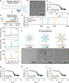
PEG-b-PPS MCs displaying lipid-anchored FLT4-/VEGFR3-binding peptides that differ in the length of their PEG spacers. (a,b) Illustration of PEG-b-PPS MCs (a) and cryo-TEM of blank MCs (i.e., without peptide) (b). (c) MC characterization by SAXS. (d) Illustration of the designed FLT4-binding peptide constructs and LC–MS performed on the synthesized peptide products. (e) Deconvoluted mass spectra of purified peptides. Peaks are 2151.1, 3046.7, and 4071.4 Da for PG6, PG24, and PG48, respectively. (f) Cryo-TEM of MCs displaying the specified targeting peptides at a 5% molar ratio (peptide/polymer). (g–i) Characterization of MCs displaying FLT4-binding peptides at a 5% molar ratio. The magnification is 10,000×, and the scale bar is 100 nm for all cryo-TEM micrographs. In all cases, SAXS was performed using synchrotron radiation at the Argonne National Laboratory and a core–shell model was fit to the data. χ2 ≪ 1.0 was obtained for all model fits (a good fit is indicated by χ2 < 1.0).
Figure 3.
Figure 3.
Differences in ligand biochemical accessibility modulate the rate of MC uptake by SC endothelial cells and vascular endothelial cells in vitro. (a,b) Determination of biochemical access to lipid-anchored targeting peptides displayed on polymeric MCs. (a) Illustration of the protease protection assay to evaluate peptide accessibility. (b) Trypsin proteolysis kinetics (n = 5). Concentrations: [peptide] = 40 nM and [trypsin] = 800 nM. Pseudo-first-order association model fits are displayed for comparison, y = y0 + (ymaxy0)*(1 – ekx), where k is the proteolysis rate (hours−1). In all cases, r2 > 0.94. (c–f) The surface-displayed PG48 FLT4-targeting peptide significantly increases MC uptake by human SC cells and decreases uptake by HUVECs in vitro. (c) Illustration of PEG-b-PPS MC formulations and the cellular uptake study. (d,e) Cellular uptake by normal SC cells (d) or HUVECs (e). MFI determined by flow cytometry. The mean ± s.e.m. is displayed (n = 3). (f) SC-targeting specificity defined here as SCMFI/HUVECMFI. For (d–f), statistical significance was determined by ANOVA with post hoc Tukey’s multiple comparison test and a 5% significance level. ****p < 0.0001, **p < 0.01, and *p < 0.05.
Figure 4.
Figure 4.
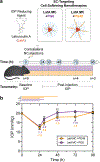
Increasing the biochemical accessibility of the FLT4-targeting peptide on SC-targeting nanocarriers enhances the efficacy of a model IOP-reducing agent in vivo. (a) Experimental overview. The performance of LatA-loaded MCs (18 μM LatA) displaying either the PG6 or PG48 FLT4-binding peptide was evaluated in a paired IOP study in vivo. Nanocarriers were injected intracamerally into the contralateral eyes of mice. IOP was measured prior to injection (baseline) and at 24, 30, 48, 72, and 96 h after injection. Nine mice (n = 9) were evaluated through 48 h. Measurements from four (n = 4) and three (n = 3) mice were obtained at 72 and 96 h timepoints, respectively. (b) IOP time course at the baseline and after the intracameral injection of the specified nanocarrier formulations. Statistically significant differences between the PG6 and PG48 treatment groups were determined using a paired, two-tailed t-test and a 5% significance level. ‡‡p < 0.01. The bars above the plot show statistically significant differences in IOP from the baseline value within the specified treatment group, assessed using a paired, two-tailed t-test. ****p < 0.0001; ***p ≤ 0.001; **p < 0.005; and *p < 0.05.
Figure 5.
Figure 5.
Confocal microscopy of MC + PG48 localization within the murine conventional outflow pathway in vivo. (a) Experimental illustration. DiI dye-loaded MC + PG48 (5%) nanocarriers (20 mg/mL polymer concentration) were administered intracamerally in C57BL/6J mice. After 45 min, eyes were enucleated and fixed for confocal microscopy analysis. (b,c) Images of conventional outflow pathway tissues obtained from the eyes of (b) the negative control group (no injection) and (c) the MC + PG48 treatment group. Images were acquired at 20× magnification. DAPI and DiI signals are overlaid to present the cell nuclei and nanocarrier localization, respectively. Scale bar = 50 μm. Tissue abbreviations: SC: Schlemm’s canal and TM: trabecular meshwork.

References

    1. Tham Y-C; Li X; Wong TY; Quigley HA; Aung T; Cheng C-Y Global Prevalence of Glaucoma and Projections of Glaucoma Burden through 2040. Ophthalmology 2014, 121, 2081–2090. - PubMed
    1. Mao LK; Stewart WC; Shields MB Correlation Between Intraocular Pressure Control and Progressive Glaucomatous Damage in Primary Open-Angle Glaucoma. Am. J. Ophthalmol 1991, 111, 51–55. - PubMed
    1. Quigley HA; Nickells RW; Kerrigan LA; Pease ME; Thibault DJ; Zack DJ Retinal Ganglion Cell Death in Experimental Glaucoma and after Axotomy Occurs by Apoptosis. Invest. Ophthalmol. Visual Sci 1995, 36, 774–786. - PubMed
    1. Allingham RR; de Kater AW; Ethier CR; Anderson PJ; Hertzmark E; Epstein DL The Relationship between Pore Density and Outflow Facility in Human Eyes. Invest. Ophthalmol. Visual Sci 1992, 33, 1661–1669. - PubMed
    1. Johnson M; Shapiro A; Ethier CR; Kamm RD Modulation of Outflow Resistance by the Pores of the Inner Wall Endothelium. Invest. Ophthalmol. Visual Sci 1992, 33, 1670–1675. - PubMed

MeSH terms