. 2021 Sep 9;95(19):e0068621.
doi: 10.1128/JVI.00686-21. Epub 2021 Sep 9.

Regulation of Human Papillomavirus 18 Genome Replication, Establishment, and Persistence by Sequences in the Viral Upstream Regulatory Region

Affiliations

Regulation of Human Papillomavirus 18 Genome Replication, Establishment, and Persistence by Sequences in the Viral Upstream Regulatory Region

Tami L Coursey et al. J Virol. .

Abstract

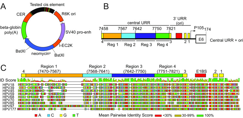
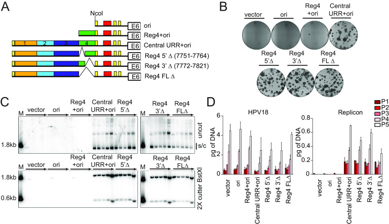
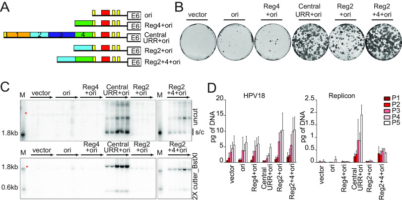
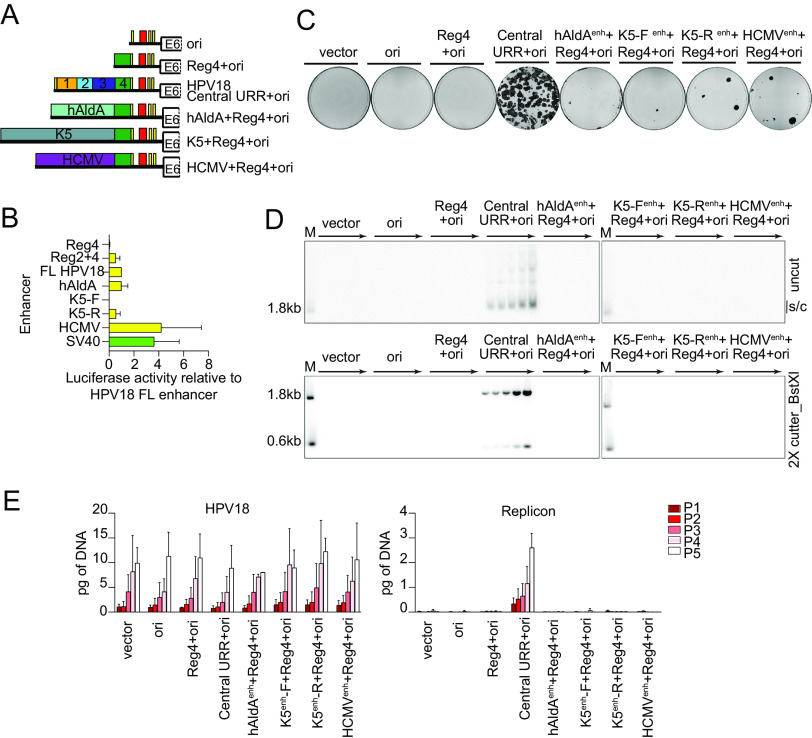
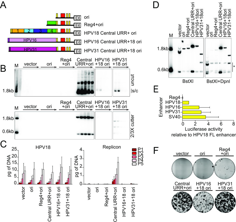
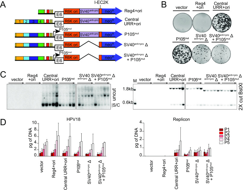
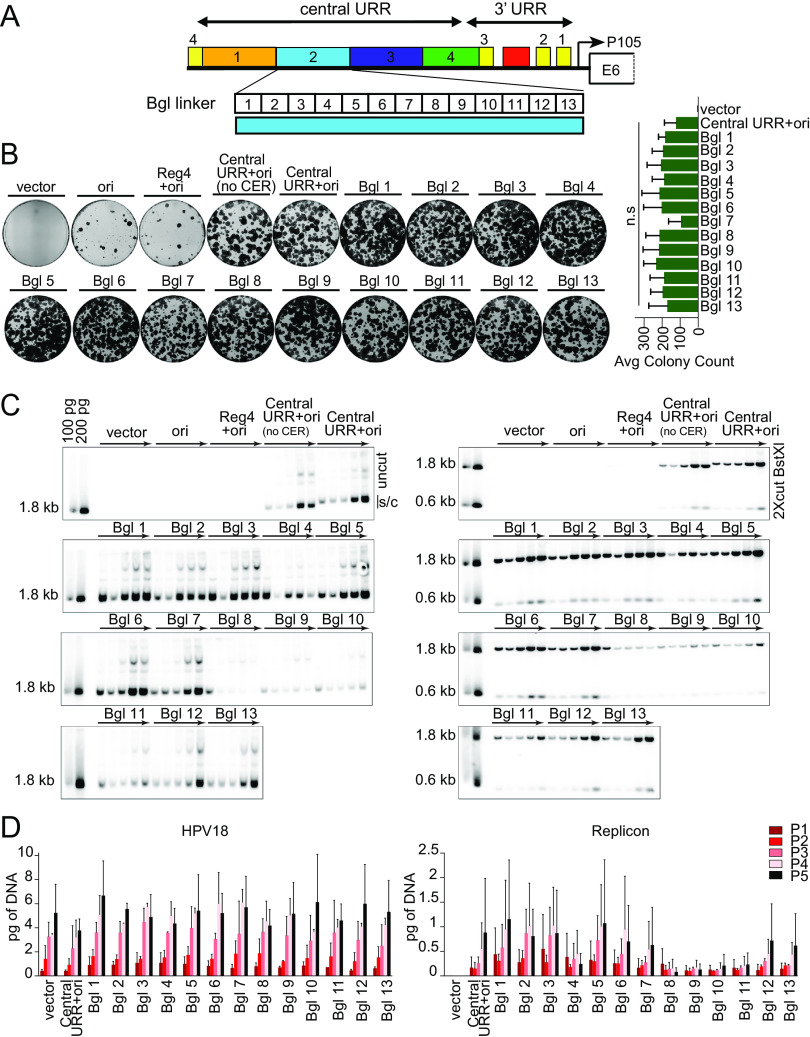
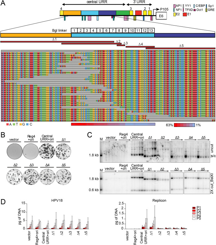
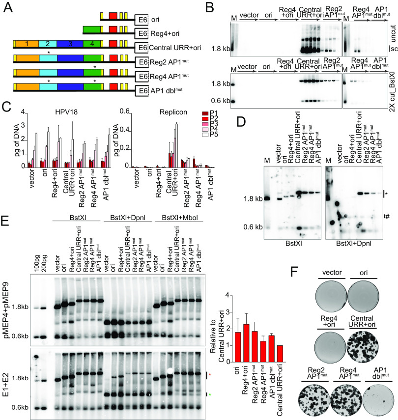
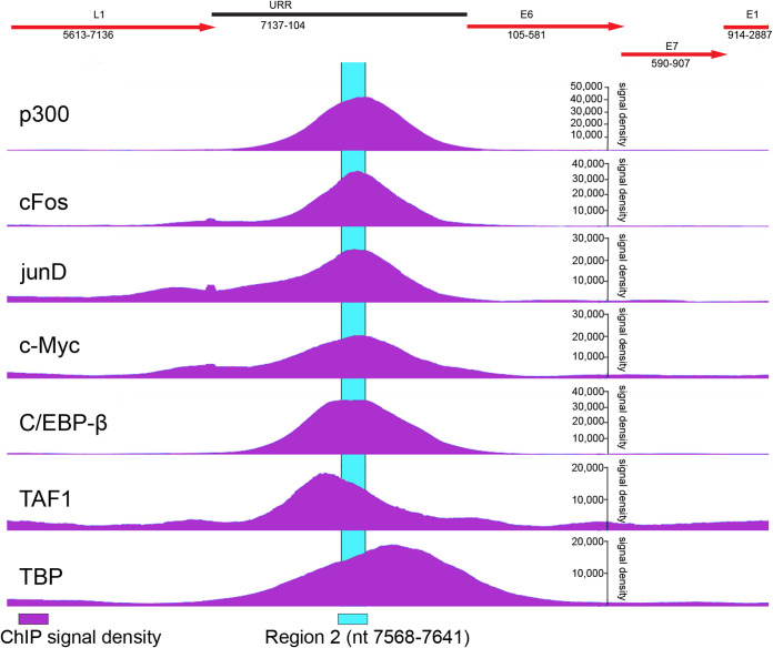
During persistent human papillomavirus infection, the viral genome replicates as an extrachromosomal plasmid that is efficiently partitioned to daughter cells during cell division. We have previously shown that an element which overlaps the human papillomavirus 18 (HPV18) transcriptional enhancer promotes stable DNA replication of replicons containing the viral replication origin. Here, we perform comprehensive analyses to elucidate the function of this maintenance element. We conclude that no unique element or binding site in this region is absolutely required for persistent replication and partitioning and instead propose that the overall chromatin architecture of this region is important to promote efficient use of the replication origin. These results have important implications for the genome partitioning mechanism of papillomaviruses. IMPORTANCE Persistent infection with oncogenic human papillomaviruses (HPVs) is responsible for ∼5% of human cancers. The viral DNA replicates as an extrachromosomal plasmid and is partitioned to daughter cells in dividing keratinocytes. Using a complementation assay that allows us to separate viral transcription and replication, we provide insight into viral sequences that are required for long-term replication and persistence in keratinocytes. Understanding how viral genomes replicate persistently for such long periods of time will guide the development of antiviral therapies.

Keywords: AP1; Alphapapillomavirus; HPV; HPV18; c-MYC; enhancer; maintenance; papillomavirus; partitioning; persistence; persistent infection; replication.


Figures

FIG 1
FIG 1
Diagram of the HPV18 replicon and the HPV18 URR cis element insertion. (A) Diagram of the HPV18 replicon plasmid, pCGneo. Prokaryotic elements are highlighted in orange (R6K ori, I-EC2K, and CER), while eukaryotic elements are highlighted in violet (simian virus 40 [SV40] promoter-enhancer) and green (beta-globin polyA signal). The neomycin resistance gene is in blue. The HPV18 central URR and origin are represented in black, with E1 and E2 binding sites indicated by red and yellow bars, respectively. The BstXI restriction enzyme sites used for Southern blot analysis are indicated. (B) Diagram of the HPV18 central URR and 3′ URR origin sequences inserted into the replicon. The four regions of the central URR are indicated, and nucleotide numbers are from the HPV18 genome. The arrow represents the early viral P105 promoter. (C) A global alignment of the central URR and ori from the nine Alphapapillomavirus 7 species of human papillomaviruses using the Geneious Prime multiple sequence alignment algorithm of the central and 3′-terminal half of the URR. The HPV18 central URR and origin elements are indicated. Nucleotides within the sequences are color-coded as indicated. A mean pairwise identify score (sliding window size of 1) for each nucleotide is shown graphically above the aligned sequences and indicate less than 30% (red), 30% to 99% (yellow), or 100% (green) nucleotide conservation among the sequences.
FIG 2
FIG 2
The HPV18 central URR element, region 4, is unnecessary for replicon maintenance. (A) Diagram of replicons containing the central URR and origin with deletions in region 4 (green). Control replicons include the CpGneo (vector), the CpGneo vectors with the minimum HPV18 origin (ori), region 4 and the origin (Reg4+ori), and the central URR and origin (central URR+ori). (B) Keratinocyte colonies arising from continuous G418 selection were stained with methylene blue approximately 14 days posttransfection. (C) Cellular DNA collected from five passages of cotransfected cells were digested and analyzed by Southern blotting and viral DNA detected by hybridization with a radioactively labeled CpGneo vector probe. The top panel represents DNA samples cut with an enzyme that does not cut the replicon, permitting visualization of extrachromosomal, supercoiled replicon DNA. The bottom panel shows DNA cut with BstXI which separates the replicon into two fragments. Lanes containing a size marker (M) contain two fragments generated from digesting the CpGneo vector with BstXI. The monomeric, supercoiled form (s/c) of the replicon is indicated. (D) HPV18 genome (left) and replicon (right) copy numbers were measured by qPCR. The data shown are representative (panels B and C) or an average of two biological replicates (panel D).
FIG 3
FIG 3
HPV18 central URR element Reg2 alone is necessary but insufficient for replicon maintenance. (A) Diagram of HPV18 replicons. (B) Keratinocyte colonies arising from continuous G418 selection were stained with methylene blue approximately 14 days posttransfection. (C) DNA collected from five passages of transfected cells was analyzed by Southern blotting as described in Fig. 2C. An asterisk marks cellular DNA hybridizing with the beta-globin polyA region present in the radioactive CpGneo vector probe. The monomeric, supercoiled form (s/c) of the replicon is indicated. (D) HPV18 genome (left) and replicon (right) copy numbers were measured by qPCR. Error bars represent standard deviation. The data shown are representative (panels B and C) or an average of two biological replicates (panel D).
FIG 4
FIG 4
Enhancer activity alone cannot complement the HPV18 central URR enhancer element for replicon maintenance. (A) Diagram of enhancer replicons. (B) Keratinocytes were transfected with luciferase reporter plasmids containing the cis elements indicated upstream from a minimal promoter, as well as a positive control (pGL4.13 expressing luciferase from the SV40 enhancer/promoter; shown in green) and a negative control (empty luciferase vector containing a minimal promoter). Cell lysates were collected 48 h after transfection, and luciferase activity was measured and normalized to the total protein concentrations for each cell lysate and the negative control plasmid. Reporter activity for each enhancer is shown relative to the HPV18 central URR enhancer. (C) Keratinocyte colonies arising from continuous G418 selection were stained with methylene blue approximately 14 days posttransfection. (D) DNA collected from five passages of cotransfected cells were analyzed by Southern blotting as described in Fig. 2C. The monomeric, supercoiled form (s/c) of the replicon is indicated. (E) HPV18 genome (left) and replicon (right) copy numbers were measured by qPCR. Error bars represent the standard deviation. The data shown are representative (panels C and D) or an average of two (panel E) biological replicates. The average of three biological replicates is shown in panel B.
FIG 5
FIG 5
The central URR from HPV16 and HPV31 does not fully support HPV18 replicon maintenance. (A) Diagram of enhancer replicons. (B) DNA collected from five passages of cotransfected cells were analyzed by Southern blotting as described in Fig. 2C. The monomeric, supercoiled form (s/c) of the replicon is indicated. (C) HPV18 genome (left) and replicon (right) copy numbers were measured by qPCR. (D) DNA was collected from keratinocytes cotransfected with HPV18 and the indicated test replicon 48 h after transfection. DNA samples were digested with BstXI or BstXI and DpnI and visualized by Southern blotting as described in Fig. 2C. DpnI digestion products are indicated by an asterisk. (E) Keratinocytes were transfected with luciferase reporter plasmids containing the cis elements indicated as well as a positive control (SV40 enhancer) and a negative control (empty luciferase vector containing a minimal promoter). Cell lysates were collected 48 h after transfection, and luciferase activity was measured and normalized to the total protein concentrations for each cell lysate and the negative-control plasmid. Reporter activity for each enhancer is shown relative to the HPV18 full-length enhancer. The luciferase assays shown were conducted at the same time as those reported in Fig. 4C, and the activity of the positive control and HPV18 enhancer luciferase results are replicated here. (F) Keratinocyte colonies arising from continuous G418 selection were stained with methylene blue approximately 14 days posttransfection. Error bars represent the standard deviation. The data shown are representative (panels B, D, and F) or an average of two (panel C) biological replicates. The average of three biological replicates is shown in panel E.
FIG 6
FIG 6
Transcription from the replicon is unnecessary for its extrachromosomal maintenance. (A) Diagram of transcription initiation site mutations in replicons. (B) Keratinocyte colonies arising from continuous G418 selection were stained with methylene blue approximately 14 days posttransfection. (C) DNA collected from five passages of transfected cells was analyzed by Southern blotting as described in Fig. 2C. The monomeric, supercoiled form (s/c) of the replicon is indicated. (D) HPV18 genome (left) and replicon (right) copy numbers were measured by qPCR. Error bars represent the standard deviation. The data shown are representative (panels B and C) or an average (panel D) of two biological replicates.
FIG 7
FIG 7
Linker scanning mutagenesis shows that the 3′ half of region 2 modulates replicon copy number and/or partitioning efficiency. (A) Diagram of the HPV18 central URR and ori. The location of Bgl linker scanning mutations introduced into HPV18 region 2 are indicated by individually numbered white boxes (Bgl 1 to 13). All Bgl linker replicons lack the CER element (Fig. 1B) and are compared to positive controls with and without a CER element. (B) Keratinocyte colonies arising from continuous G418 selection were stained with methylene blue approximately 14 days posttransfection. Neomycin-resistant colonies were counted and averaged from three biological replicates. (C) DNA collected from five passages of transfected cells were analyzed by Southern blotting as described in Fig. 2C. The monomeric, supercoiled form (s/c) of the replicon is indicated. (D) HPV18 genome (left) and replicon (right) copy numbers were measured by qPCR. Error bars represent the standard deviation. The data shown are representative (panels B and C) or an average (panel D) of two or three (panel B) biological replicates. Significance was determined by unpaired t test with unequal variance. n.s. (no significance), P > 0.05.
FIG 8
FIG 8
No single sequence in region 2 is absolutely required for replicon maintenance, but this region modulates replicon copy number and/or partitioning efficiency. (A) Diagram of the HPV18 central URR and ori in addition to the location of important viral (E1, E2) and cellular transcription factor binding sites. The locations of Bgl linker scanning mutations introduced into HPV18 region 2 are indicated by individually numbered white boxes (Bgl 1 to 13). A representative sequence alignment of deletions present in Bgl linker replicons recovered from passaged cells is shown. Deletions are highlighted by gray dashes, while nucleotides are individually colored as indicated. Individual deletions present in select replicons recovered from the Bgl chewback assay are mapped by red bars (labeled Δ1 through Δ5) above the sequence alignment. The frequency with which a region 2 nucleotide was deleted in the recovered replicons is indicated by a heatmap ranging from a maximum 63% (red) to 1% (blue) of the clones showing a deletion at a nucleotide site. (B) Keratinocyte colonies arising from continuous G418 selection were stained with methylene blue approximately 14 days posttransfection. (C) DNA collected from five passages of transfected cells were analyzed by Southern blotting as described in Fig. 2C. The monomeric, supercoiled form (s/c) of the replicon is indicated. (D) HPV18 genome (left) and replicon (right) copy numbers were measured by qPCR. Error bars represent the standard deviation. The data shown is representative (panels B and C) or an average (panel D) of two biological replicates.
FIG 9
FIG 9
Mutation of AP1 binding sites impacts replicon replication. (A) Diagram of AP1 mutations present in replicons. (B) DNA collected from five passages of cotransfected cells were analyzed by Southern blotting as described in Fig. 2C. (C) HPV18 genome (left) and replicon (right) copy numbers were measured by qPCR. (D) DNA was collected from keratinocytes cotransfected with HPV18 and the indicated test replicon 48 h after transfection and analyzed by Southern blotting as described in Fig. 5D. (E) Keratinocytes were cotransfected with the indicated replicons and either empty pMEP vector control plasmids or pMEP E1 and E2 expression plasmids. E1 and E2 expression was induced with CdSO4 24 h posttransfection for 5 h. Low-molecular-weight DNA was extracted 48 h posttransfection and analyzed by Southern blotting as described in Fig. 5D with an additional restriction digest that specifically digests replicating DNA (MboI). MboI-sensitive and DpnI-resistant bands are indicated by green and red asterisks, respectively. DpnI-resistant DNA was quantified by phosphorimaging analysis of three independent experiments. Values shown in the graph are relative to the control central URR+ori replicon. (F) Keratinocyte colonies arising from continuous G418 selection were stained with methylene blue approximately 14 days posttransfection. Error bars represent the standard deviation. The data shown are representative (panels B and E) or an average (panel C) of two biological replicates. The data shown for transient replication are representative of one (panel D) of three (panel E) bioreplicates.
FIG 10
FIG 10
Cellular transcription factors bind to region 2 of the integrated HPV18 URR in HeLa cells. Chromatin immunoprecipitation sequencing (ChIP-seq) signal density profiles (shown in purple) for select cellular proteins binding to integrated copies of HPV18 genome in HeLa cells were analyzed using JBrowse on the PaVE website (https://pave.niaid.nih.gov/). A partial map of the HPV18 viral genome overlapping with the ChIP-seq data is shown above with viral open reading frames in red. Region 2 of the HPV18 URR is highlighted in cyan.

References

    1. Plummer M, de Martel C, Vignat J, Ferlay J, Bray F, Franceschi S. 2016. Global burden of cancers attributable to infections in 2012: a synthetic analysis. Lancet Glob Health 4:e609-16. 10.1016/S2214-109X(16)30143-7. - DOI - PubMed
    1. Doorbar J, Quint W, Banks L, Bravo IG, Stoler M, Broker TR, Stanley MA. 2012. The biology and life-cycle of human papillomaviruses. Vaccine 30(Suppl 5):F55–F70. 10.1016/j.vaccine.2012.06.083. - DOI - PubMed
    1. Coursey TL, McBride AA. 2019. Hitchhiking of viral genomes on cellular chromosomes. Annu Rev Virol 6:275–296. 10.1146/annurev-virology-092818-015716. - DOI - PMC - PubMed
    1. Wu SC, Canarte V, Beeravolu H, Grace M, Sharma S, Munger K. 2020. Finding how human papillomaviruses alter the biochemistry and identity of infected epithelial cells, p 53–65. In Jenkins D, Bosch FX (ed), Human papillomavirus. Academic Press, San Diego, CA. 10.1016/b978-0-12-814457-2.00004-0:53-65. - DOI
    1. Ustav M, Stenlund A. 1991. Transient replication of BPV-1 requires two viral polypeptides encoded by the E1 and E2 open reading frames. EMBO J 10:449–457. 10.1002/j.1460-2075.1991.tb07967.x. - DOI - PMC - PubMed

Publication types

MeSH terms

LinkOut - more resources