Case Reports
. 2021 Jul 7;17(7):e1009639.
doi: 10.1371/journal.pgen.1009639. eCollection 2021 Jul.

A homozygous stop-gain variant in ARHGAP42 is associated with childhood interstitial lung disease, systemic hypertension, and immunological findings

Affiliations
Case Reports

A homozygous stop-gain variant in ARHGAP42 is associated with childhood interstitial lung disease, systemic hypertension, and immunological findings

Qifei Li et al. PLoS Genet. .

Abstract

ARHGAP42 encodes Rho GTPase activating protein 42 that belongs to a member of the GTPase Regulator Associated with Focal Adhesion Kinase (GRAF) family. ARHGAP42 is involved in blood pressure control by regulating vascular tone. Despite these findings, disorders of human variants in the coding part of ARHGAP42 have not been reported. Here, we describe an 8-year-old girl with childhood interstitial lung disease (chILD), systemic hypertension, and immunological findings who carries a homozygous stop-gain variant (c.469G>T, p.(Glu157Ter)) in the ARHGAP42 gene. The family history is notable for both parents with hypertension. Histopathological examination of the proband lung biopsy showed increased mural smooth muscle in small airways and alveolar septa, and concentric medial hypertrophy in pulmonary arteries. ARHGAP42 stop-gain variant in the proband leads to exon 5 skipping, and reduced ARHGAP42 levels, which was associated with enhanced RhoA and Cdc42 expression. This is the first report linking a homozygous stop-gain variant in ARHGAP42 with a chILD disorder, systemic hypertension, and immunological findings in human patient. Evidence of smooth muscle hypertrophy on lung biopsy and an increase in RhoA/ROCK signaling in patient cells suggests the potential mechanistic link between ARHGAP42 deficiency and the development of chILD disorder.


Conflict of interest statement

The authors have declared that no competing interests exist.

Figures
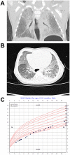
Fig 1
Fig 1. Clinical findings of the proband with homozygous ARHGAP42 stop-gain variant.
(A) Chest X-ray at age 3 months showed bilateral diffuse opacities and low lung volumes; (B) Initial chest CT scan at age 3 months displayed diffuse ground glass opacities with subpleural sparing and patchy subpleural opacities; (C) Patient growth chart for weight through two years of age.
Fig 2
Fig 2. Pathological findings of the proband with homozygous ARHGAP42 stop-gain variant.
Lung histologic findings at age 4 months, by hematoxylin and eosin stain: (A) Small airway obstructive-type changes, with prominent mural smooth muscle and intraluminal macrophages (original magnification, 200x); (B) Intra-alveolar septal extension of smooth muscle (arrows) (original magnification, 400x); (C) Large/maldeveloped airspaces, accentuated subpleurally (original magnification, 100x); (D) Concentric medial hypertrophy in pulmonary arteries (arrowheads) (original magnification, 100x); (E) Pulmonary artery (arrowheads) and airway (asterisk), both with prominent smooth muscle (original magnification, 400x).
Fig 3
Fig 3. Genetic findings in the family with ARHGAP42 stop-gain variant.
(A) Pedigree of the family carrying ARHGAP42 variant. Half-filled symbol: heterozygous; Filled symbol: homozygous; (B) Sanger sequencing chromatogram for the family of the ARHGAP42 variant; (C) Amino acid 157 (arrowhead) is evolutionally conserved in vertebrates; (D) Agarose gel electrophoresis result of ARHGAP42 pre-variant, in-variant, and post-variant amplification by PCR in cDNA samples extracted from blood. The smaller band (arrow) corresponds with exon 5 deletion confirmed by Sanger sequencing. Pt: patient; Mo: mother; Fa: father; C1: control; gDNA: genomic DNA. Pre-variant: Exon 1 to first 7 bp of Exon 5, 279 bp for ARHGAP42 cDNA. A 373 bp band seen with gDNA is from non-specific amplification of another locus; In-variant: Exon 4 to Exon 7, 298 bp for ARHGAP42 cDNA. A 618 bp band was seen with gDNA from non-specific amplification from another locus; Post-variant: Exon 7~8, 204 bp for ARHGAP42 cDNA. (E) Schematic of ARHGAP42 functional domains. The amino acid change is depicted by an arrow, and the skipped exon 5 is depicted as a shaded box in the BAR domain.
Fig 4
Fig 4. ARHGAP42 stop-gain variant causes reduced ARHGAP42 expression.
(A) Immunofluorescence of ARHGAP42 in the patient and her parents lymphoblastoid cells. Lymphoblastoid cells were fixed, permeabilized, and stained with DAPI (blue), phalloidin (green), and ARHGAP42 (red) (scale bar 10 μm); (B) Expression of control, GFP-ARHGAP42 WT and GFP-ARHGAP42 ΔExon5 in transfected HT1080 and U2OS cells by immunoblotting (black arrow indicates a lower level of ARHGAP42 ΔExon5 protein).
Fig 5
Fig 5. ARHGAP42 stop-gain variant contributes to enhanced RhoA activity and Cdc42 expression.
(A) Expression of RhoA and Cdc42 in the patient’s EBV-LCLs compared with her parents and healthy controls by immunoblotting; (B) Measurement of RhoA activity by GST-Rhotekin-RBD (Rho binding domain) pull-down assay in the patient and her parents. The level of RhoA-GTP was determined by western blot with anti-RhoA antibody following a pull-down assay; (C) The RhoA-GTP data are expressed as the mean ± SD of three independent experiments. *p<0.05; **p<0.01.

References

    1. Bai X, Lenhart KC, Bird KE, Suen AA, Rojas M, Kakoki M, et al.. The smooth muscle-selective RhoGAP GRAF3 is a critical regulator of vascular tone and hypertension. Nature communications. 2013;4:2910. doi: 10.1038/ncomms3910 - DOI - PMC - PubMed
    1. Wain LV, Verwoert GC, O’Reilly PF, Shi G, Johnson T, Johnson AD, et al.. Genome-wide association study identifies six new loci influencing pulse pressure and mean arterial pressure. Nature genetics. 2011;43(10):1005. doi: 10.1038/ng.922 - DOI - PMC - PubMed
    1. Ehret GB, Munroe PB, Rice KM, Bochud M, Johnson AD, Chasman DI, et al.. Genetic variants in novel pathways influence blood pressure and cardiovascular disease risk. Nature. 2011;478(7367):103. doi: 10.1038/nature10405 - DOI - PMC - PubMed
    1. Kato N, Loh M, Takeuchi F, Verweij N, Wang X, Zhang W, et al.. Trans-ancestry genome-wide association study identifies 12 genetic loci influencing blood pressure and implicates a role for DNA methylation. Nature genetics. 2015;47(11):1282. doi: 10.1038/ng.3405 - DOI - PMC - PubMed
    1. Bai X, Dee R, Mangum KD, Mack CP, Taylor JM. RhoA signaling and blood pressure: The consequence of failing to “Tone it Down”. World Journal of Hypertension. 2016;6(1):18–35.

Publication types