. 2021 Aug 20;49(14):8177-8188.
doi: 10.1093/nar/gkab591.

Single molecule analysis indicates stimulation of MUTYH by UV-DDB through enzyme turnover

Affiliations

Single molecule analysis indicates stimulation of MUTYH by UV-DDB through enzyme turnover

Sunbok Jang et al. Nucleic Acids Res. .

Abstract

The oxidative base damage, 8-oxo-7,8-dihydroguanine (8-oxoG) is a highly mutagenic lesion because replicative DNA polymerases insert adenine (A) opposite 8-oxoG. In mammalian cells, the removal of A incorporated across from 8-oxoG is mediated by the glycosylase MUTYH during base excision repair (BER). After A excision, MUTYH binds avidly to the abasic site and is thus product inhibited. We have previously reported that UV-DDB plays a non-canonical role in BER during the removal of 8-oxoG by 8-oxoG glycosylase, OGG1 and presented preliminary data that UV-DDB can also increase MUTYH activity. In this present study we examine the mechanism of how UV-DDB stimulates MUTYH. Bulk kinetic assays show that UV-DDB can stimulate the turnover rate of MUTYH excision of A across from 8-oxoG by 4-5-fold. Electrophoretic mobility shift assays and atomic force microscopy suggest transient complex formation between MUTYH and UV-DDB, which displaces MUTYH from abasic sites. Using single molecule fluorescence analysis of MUTYH bound to abasic sites, we show that UV-DDB interacts directly with MUTYH and increases the mobility and dissociation rate of MUTYH. UV-DDB decreases MUTYH half-life on abasic sites in DNA from 8800 to 590 seconds. Together these data suggest that UV-DDB facilitates productive turnover of MUTYH at abasic sites during 8-oxoG:A repair.


Figures

Figure 1.
Figure 1.
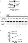
UV-DDB stimulates MUTYH by binding specifically to 8-oxoG:A. (A) Schematic representation of the DNA substrate containing 8oxoG:A and the proposed reaction scheme. Circle represents a 3′-FAM moiety (B) Stimulation of MUTYH excision kinetics by UV-DDB. MUTYH (20 nM) was incubated with dsDNA (50nM) containing 8-oxoG:A in the absence (−) or presence (+) of UV-DDB (50nM) at 37°C. Aliquots were withdrawn at each time point and analyzed on a 10% denaturing polyacrylamide gel. Positions of the un-cleaved full-length substrate and excised product are indicated by arrows. (C) Quantification of the stimulation of MUTYH excision kinetics by UV-DDB in (B). Excision product formation was quantified using ImageJ software. The excision percentage was plotted as mean ± SD from three independent experiments, each run on duplicate gels.
Figure 2.
Figure 2.
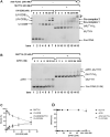
UV-DDB facilitates MUTYH dissociation via co-complex formation. (A) UV-DDB forms co-complexes with MUTYH and displaces MUTYH on abasic sites, shown by EMSA. Binding reactions of THF8oxoG37 and increasing amounts of UV-DDB with or without MUTYH were separated by native PAGE. Protein-DNA complexes were identified based on band migration and labelled accordingly. In lane 15, addition of 250 nM anti-FLAG antibody caused a supershift in UV-DDB containing species, marked with an asterisk (see Supplementary Figure S3). Representative gel shown from three independent experiments. (B) Binding reactions of THF8oxoG37 of APE1 with and without MUTYH were assayed by native PAGE. Even at up to 1 μM APE1, minimal MUTYH dissociation was observed. After running on a sequencing gel (Supplementary Figure S4) MUTYH at 20 nM prevented nearly all APE1 endonuclease activity (C) Quantification of (A) from lane 8 to 14. Percent bound of total DNA by all UV-DDB species (including monomer, dimer, and trimer), MUTYH species (including monomer and dimer), and co-complex (including co-complex 1 and co-complex 2) are plotted as a function of UV-DDB concentration. Data shown as the mean of two measurements from three experiments ± SD. (D) Quantification of (C) from lane 8–15. Data shown as the mean of two measurements from three experiments ± s.d.
Figure 3.
Figure 3.
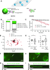
Single molecule analysis reveals that UV-DDB stimulates turnover of MUTYH by facilitated dissociation. (A) Experimental design of DNA tightrope assay to study UV-DDB induced mobility and dissociation of MUTYH (top). Long DNA substrates with defined abasic sites (THF) every 2 kb are suspended between silica beads (bottom, left). His-tagged MUTYH is labelled with primary mouse-anti-His antibody and secondary goat-anti-mouse antibody conjugated to a 605nm Qdot. (bottom, right) un-labeled UV-DDB. (B) Stack bar graph showing the fraction of motile (green) versus stationary (white) and persistent (solid) vs. dissociating (diagonal lines) particles of 605Qdot labelled MUTYH in the absence (−) or presence (+) of un-labeled 1× UV-DDB on abasic (THF) DNA during 300s observation. (****P< 0.0001 by χ2 test). (C) Effects of UV-DDB on the lifetimes of MUTYH-DNA complexes. Data plotted as the mean ± SEM from three independent experiments. For each condition, survival fraction decay is fit to a single exponential decay function to obtain the half-life and errors shown are the errors of the fit. (D) Anomalous diffusion exponent (α) versus diffusion coefficient (log10D) plotted for MUTYH (black filled circles) and MUTYH with 1× UV-DDB (red filled circles). (E) Box and whisker plot (10–90 percentile) of left, the Anomalous diffusion exponent (α) and right, the diffusion coefficient (log10D) calculated for MUTYH only (n = 20 phases) and MUTYH with 1× UV-DDB (n = 61 phases) phases on long DNA substrates with defined abasic sites (THF) every 2 kb. +, sample mean, *P < 0.1, **P < 0.01 by two-tailed Student's t test. (F) Image of 605Qdot-labled MUTYH (green) on abasic (THF) tightrope suspended between beads in the 1× presence of unlabeled UV-DDB. Scale bar represents 2.5 μm. Arrow points to motile MUTYH particle. See corresponding video 1. (G) Kymograph of 605Qdot-labeled MUTYH (green) with motile particle (F). Horizontal and vertical scale bars represent 50 s and 2 kb, respectively. (H) Image of 605Qdot-labled MUTYH (green) on abasic (THF) tightrope suspended between beads in the 1× presence of un-labelled UV-DDB. See corresponding Video 2. Scale bar represents 2.5 μm. Arrow points to motile/ dissociated MUTYH particle. (I) Kymograph of 605Qdot-labeled MUTYH (green) with motile and dissociated particle (H). Horizontal and vertical scale bars represent 50s and 2kb, respectively.
Figure 4.
Figure 4.
Single molecule co-localization of UV-DDB with MUTYH on abasic DNA tightropes. (A) Schematic of the DNA tightrope assay. Long DNA substrates with abasic sites every 2 kb were suspended between 5 μm poly-l-lysine coated silica beads. Anti-His primary antibody was used to link the His-tagged MUTYH to the 605Qdot. Biotin conjugated anti-Flag primary antibody was used to link Flag-tagged UV-DDB to streptavidin-coated 705Qdot. Uniquely labelled MUTYH and UV-DDB were observed interacting on abasic DNA tightropes in real time and their behavior and frequency of co-localization was recorded. (B) Venn diagram showing number of proteins that co-localized (yellow) on abasic (THF) tightropes or were observed separately for 605Qdot-labeled MUTYH (green) with 705Qdot-labeled UV-DDB (red) in the dual-color assay. (C) Image of co-localized (yellow) Qdot-labelled MUTYH (green) and UV-DDB (red) on abasic (THF) tightrope suspended between beads. Scale bar represents 2.5 μm. Arrow points to co-localized particle. See Video 3. (D) Kymograph of co-localized MUTYH and UV-DDB. Top, MUTYH (green); middle, UV-DDB (red); bottom, merged (yellow). Horizontal and vertical scale bars represent 50s and 2 kb, respectively. (E) Stacked bar graph showing the fraction of motile (gray) versus stationary (white) and persistent (solid) versus dissociating (diagonal lines). Results obtained with individual and co-localized particles. (MUTYH; MUTYH behavior in the presence of UV-DDB, co-local; co-localized MUTYH/UV-DDB behavior, UV-DDB; UV-DDB behavior in the presence of MUTYH). Data re-plotted as a sub-set of (B).
Figure 5.
Figure 5.
MUTYH and UV-DDB colocalize on DNA containing 8-oxoG:A damage. (A) Histogram showing distribution of AFM volumes of MUTYH and UV-DDB bound to DNA containing 8-oxoG:A (n = 104). Molecular weights corresponding to MUTYH monomer, MUTYH dimer, UV-DDB monomer, (blue) and UV-DDB-MUTYH complex (red). (B) Representative 3D AFM image of MUTYH bound to 8-oxoG:A DNA. (C) Representative 3D AFM image of UV-DDB bound to 8-oxoG:A DNA. (D) Representative 3D AFM image of MUTYH-UV-DDB bound to 8-oxoG:A DNA.
Figure 6.
Figure 6.
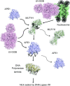
Proposed working model of UV-DDB stimulation of MUTYH in the repair of 8-oxoG. (A) Schematic representation of the proposed BER pathway including UV-DDB is illustrated. UV-DDB is believed to be rapidly recruited to damaged sites in chromatin and help facilitate processing by MUTYH. Biochemical and single molecule data suggest that UV-DDB transiently associates with MUTYH at 8-oxoG:abasic sites to increase its release and turnover. APE1 is expected to be necessary to incise the DNA and has been shown previously to be stimulated by UV-DDB (25). DNA pol λ then undergoes long patch repair (3), and FEN1 (not shown) processes the flap leaving a nick, which is then sealed by DNA ligase I/III.

References

    1. Au K.G., Cabrera M., Miller J.H., Modrich P.. Escherichia coli mutY gene product is required for specific A-G—-C.G mismatch correction. Proc. Natl. Acad. Sci. U.S.A. 1988; 85:9163–9166. - PMC - PubMed
    1. Burak M.J., Guja K.E., Hambardjieva E., Derkunt B., Garcia-Diaz M.. A fidelity mechanism in DNA polymerase lambda promotes error-free bypass of 8-oxo-dG. EMBO J. 2016; 35:2045–2059. - PMC - PubMed
    1. van Loon B., Hubscher U.. An 8-oxo-guanine repair pathway coordinated by MUTYH glycosylase and DNA polymerase lambda. Proc. Natl. Acad. Sci. U.S.A. 2009; 106:18201–18206. - PMC - PubMed
    1. Gu Y., Parker A., Wilson T.M., Bai H., Chang D.Y., Lu A.L.. Human MutY homolog, a DNA glycosylase involved in base excision repair, physically and functionally interacts with mismatch repair proteins human MutS homolog 2/human MutS homolog 6. J. Biol. Chem. 2002; 277:11135–11142. - PubMed
    1. London R.E.The structural basis of XRCC1-mediated DNA repair. DNA Repair. 2015; 30:90–103. - PMC - PubMed

Publication types

MeSH terms