

Clinical value of INSL3 in the diagnosis and development of diabetic nephropathy
- PMID: 34233048
- PMCID: PMC8418484
- DOI: 10.1002/jcla.23898
Clinical value of INSL3 in the diagnosis and development of diabetic nephropathy
Abstract
Background: Insulin-like factor 3 (INSL3) was stated to be an essential regulator in many diseases. This present study aimed to explore the underlying mechanisms of INSL3 in diabetic nephropathy (DN).
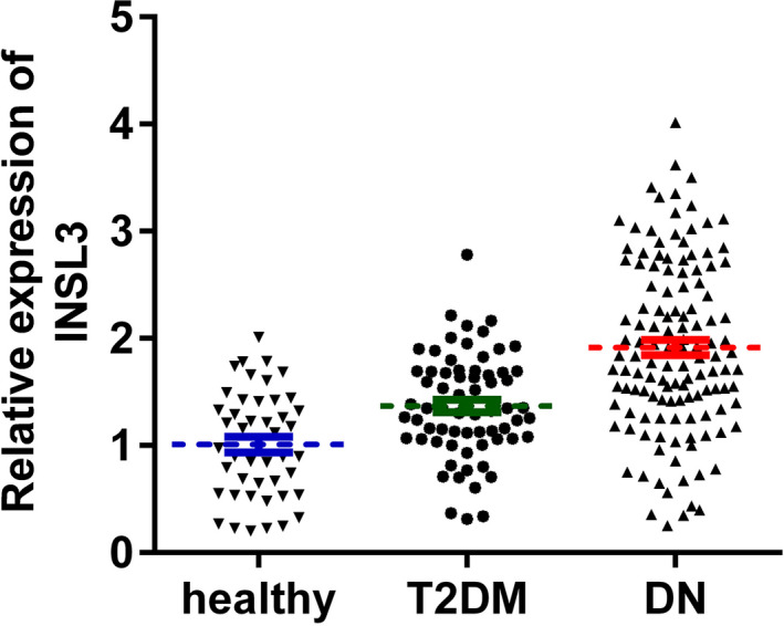
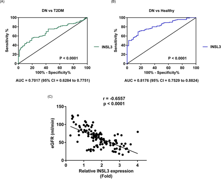
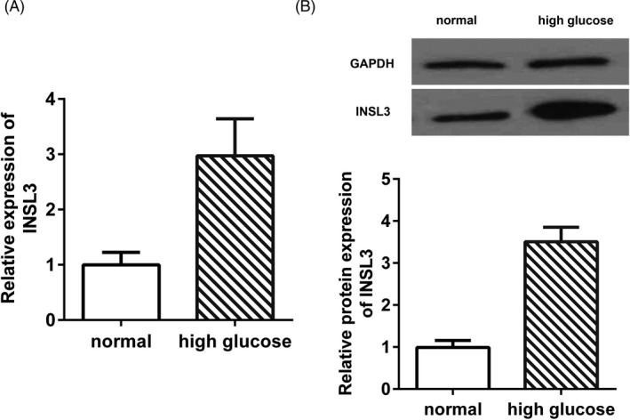
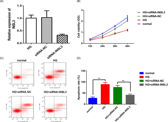
Methods: The serum samples were obtained from 121 DN patients, 67 T2DM patients, and 44 healthy controls. Twenty SD rats were used to establish the DN model in vivo. Quantitative PCR (qPCR) and Western blot were completed to analyze the INSL3 expression in cells, serum samples, and kidney of the rats. The structure of kidney was analyzed by HE staining. The diagnostic values of INSL3 in DN were determined by receiver operating characteristic (ROC) assay. Then, Spearman's correlation analysis was executed to verify the association between INSL3 and glomerular filtration rate (eGFR). Finally, the proliferation and apoptosis status of transfected cells were analyzed by MTT, flow cytometry, and Hoechst33258 staining assay.
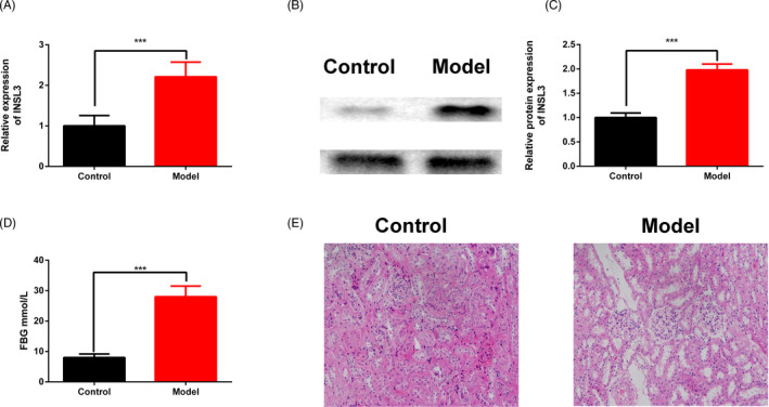
Results: We found that INSL3 expression was up-regulated in DN patients and SV40-MES-13 cells. Furthermore, the correlation analysis elucidated that INSL3 expression was negatively correlated with DN diagnosis golden criterion eGFR. INSL3 knockdown promoted the proliferation rate and inhibited the apoptosis rate of SV40-MES-13 cells after high-glucose treatment. Finally, the INSL3 expression and fast blood glucose were up-regulated in DN rats.
Conclusions: Collectively, this study demonstrated the clinical significance of INSL3 in diagnosing and developing DN.
Keywords: INSL3; diabetic nephropathy; diagnosis; glomerular membrane epithelial cells.
© 2021 The Authors. Journal of Clinical Laboratory Analysis published by Wiley Periodicals LLC.
Conflict of interest statement
None.
Figures
Similar articles
-
Serum LncRNA PANDAR may Act as a Novel Serum Biomarker of Diabetic Nephropathy in Patients with Type 2 Diabetes.Clin Lab. 2020 Jun 1;66(6). doi: 10.7754/Clin.Lab.2019.191032. Clin Lab. 2020. PMID: 32538054
-
miRNA-483-5p Targets HDCA4 to Regulate Renal Tubular Damage in Diabetic Nephropathy.Horm Metab Res. 2021 Aug;53(8):562-569. doi: 10.1055/a-1480-7519. Epub 2021 Jun 14. Horm Metab Res. 2021. PMID: 34126643
-
Significance of serum miR-29a in the occurrence and progression of diabetic nephropathy: A cross-sectional study.J Clin Lab Anal. 2022 Feb;36(2):e24210. doi: 10.1002/jcla.24210. Epub 2021 Dec 28. J Clin Lab Anal. 2022. PMID: 34964177 Free PMC article.
-
Diagnostic Performance of Retinopathy in the Detection of Diabetic Nephropathy in Type 2 Diabetes: A Systematic Review and Meta-Analysis of 45 Studies.Ophthalmic Res. 2019;62(2):68-79. doi: 10.1159/000500833. Epub 2019 Jun 28. Ophthalmic Res. 2019. PMID: 31256153
-
Lessons learned from studies of the natural history of diabetic nephropathy in young type 1 diabetic patients.Pediatr Endocrinol Rev. 2008 Aug;5 Suppl 4:958-63. Pediatr Endocrinol Rev. 2008. PMID: 18806710 Review.
Cited by
-
Association of age, hormonal, and lifestyle factors with the Leydig cell biomarker INSL3 in aging men from the European Male Aging Study cohort.Andrology. 2022 Oct;10(7):1328-1338. doi: 10.1111/andr.13220. Epub 2022 Jul 11. Andrology. 2022. PMID: 35770372 Free PMC article.
-
The Leydig cell biomarker INSL3 as a predictor of age-related morbidity: Findings from the EMAS cohort.Front Endocrinol (Lausanne). 2022 Nov 8;13:1016107. doi: 10.3389/fendo.2022.1016107. eCollection 2022. Front Endocrinol (Lausanne). 2022. PMID: 36425465 Free PMC article.
-
Expression and Role of INSL3 in the Fetal Testis.Front Endocrinol (Lausanne). 2022 Apr 6;13:868313. doi: 10.3389/fendo.2022.868313. eCollection 2022. Front Endocrinol (Lausanne). 2022. PMID: 35464060 Free PMC article. Review.
-
The Mitochondrial-Associated Endoplasmic Reticulum Membrane and Its Role in Diabetic Nephropathy.Oxid Med Cell Longev. 2021 Nov 5;2021:8054817. doi: 10.1155/2021/8054817. eCollection 2021. Oxid Med Cell Longev. 2021. PMID: 34777695 Free PMC article. Review.
References
-
- Parving HH, Smidt UM, Friisberg B, Bonnevie‐Nielsen V, Andersen AR. A prospective study of glomerular filtration rate and arterial blood pressure in insulin‐dependent diabetics with diabetic nephropathy. Diabetologia. 1981;20(4):457‐461. - PubMed
-
- Zhang J, Hu WC, Lin P, Wang R. Decreased serum myonectin concentrations in diabetic nephropathy patients. Clin Exp Med. 2020;27:601‐607. - PubMed
-
- Rossing P. Prediction, progression and prevention of diabetic nephropathy. The Minkowski lecture 2005. Diabetologia. 2006;49(1):11‐19. - PubMed
MeSH terms
Substances
LinkOut - more resources
Full Text Sources
Medical
Research Materials
Miscellaneous
