

Clinical Efficacy of Endocytoscopy for Gastrointestinal Endoscopy
- PMID: 34233111
- PMCID: PMC8357585
- DOI: 10.5946/ce.2021.165
Clinical Efficacy of Endocytoscopy for Gastrointestinal Endoscopy
Abstract
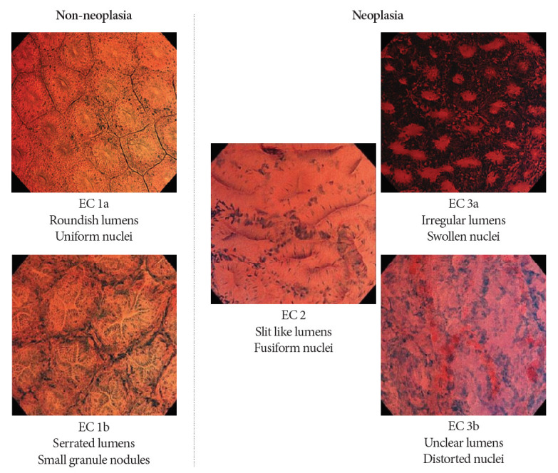
Endocytoscopy (EC) is a contact-type optical endoscope that allows in vivo cellular observation during gastrointestinal endoscopy and is now commercially available not only in Japan but also in Asian, European Union, and Middle Eastern countries. EC helps conduct a highly accurate pathological prediction without biopsy. Initially, EC was reported to be effective for esophageal diseases. Subsequently, its efficacy for stomach and colorectal diseases has been reported. In this narrative review, we searched for clinical studies that investigated the efficacy of EC. EC seems to accurately diagnose gastrointestinal diseases without biopsy. Most of the studies aimed to clarify the relationship between endocytoscopic findings of gastrointestinal neoplasia and pathological diagnosis. Some studies have investigated non-epithelial lesions or diseases, such as inflammatory bowel disease or infectious diseases. However, there are few high-level pieces of evidence, such as randomized trials; thus, further studies are needed.
Keywords: Endocytoscopy; Lower gastrointestinal endoscopy; Magnifying endoscopy; Upper gastrointestinal endoscopy.
Conflict of interest statement
Figures


Similar articles
-
Endocytoscopy: technology and clinical application in upper gastrointestinal tract.Transl Gastroenterol Hepatol. 2020 Apr 5;5:28. doi: 10.21037/tgh.2019.11.12. eCollection 2020. Transl Gastroenterol Hepatol. 2020. PMID: 32258532 Free PMC article. Review.
-
Application and Efficacy of Super-Magnifying Endoscopy for the Lower Intestinal Tract.Clin Endosc. 2016 Jan;49(1):37-40. doi: 10.5946/ce.2016.49.1.37. Epub 2016 Jan 28. Clin Endosc. 2016. PMID: 26855922 Free PMC article. Review.
-
[ENDOCYTOSCOPY--NEW TYPE OF ENDOSCOPIC EXAMINATION OF LOWER GASTROINTESTINAL AND RESPIRATORY TRACT].Eksp Klin Gastroenterol. 2015;(5):58-66. Eksp Klin Gastroenterol. 2015. PMID: 26387172 Review. Russian.
-
Endocytoscopic observation of various esophageal lesions at ×600: can nuclear abnormality be recognized?Dis Esophagus. 2015 Apr;28(3):269-75. doi: 10.1111/dote.12183. Epub 2014 Jan 28. Dis Esophagus. 2015. PMID: 24467464
-
Comprehensive diagnostic ability of endocytoscopy compared with biopsy for colorectal neoplasms: a prospective randomized noninferiority trial.Endoscopy. 2013;45(2):98-105. doi: 10.1055/s-0032-1325932. Epub 2013 Jan 10. Endoscopy. 2013. PMID: 23307149 Clinical Trial.
Cited by
-
Texture and Color Enhancement Imaging-Assisted Endocytoscopy Improves Characterization of Gastric Precancerous Conditions: A Set of Interesting Comparative Images.Diagnostics (Basel). 2025 Jul 31;15(15):1925. doi: 10.3390/diagnostics15151925. Diagnostics (Basel). 2025. PMID: 40804890 Free PMC article.
-
A Review of Colonoscopy in Intestinal Diseases.Diagnostics (Basel). 2023 Mar 27;13(7):1262. doi: 10.3390/diagnostics13071262. Diagnostics (Basel). 2023. PMID: 37046479 Free PMC article. Review.
-
Endoscopic Imaging for the Diagnosis of Neoplastic and Pre-Neoplastic Conditions of the Stomach.Cancers (Basel). 2023 Apr 25;15(9):2445. doi: 10.3390/cancers15092445. Cancers (Basel). 2023. PMID: 37173912 Free PMC article. Review.
References
-
- Inoue H, Kazawa T, Sato Y, et al. In vivo observation of living cancer cells in the esophagus, stomach, and colon using catheter-type contact endoscope, “Endo-Cytoscopy system”. Gastrointest Endosc Clin N Am. 2004;14:589–594. x-xi. - PubMed
-
- Kumagai Y, Monma K, Kawada K. Magnifying chromoendoscopy of the esophagus: in-vivo pathological diagnosis using an endocytoscopy system. Endoscopy. 2004;36:590–4. - PubMed
-
- Sasajima K, Kudo SE, Inoue H, et al. Real-time in vivo virtual histology of colorectal lesions when using the endocytoscopy system. Gastrointest Endosc. 2006;63:1010–1017. - PubMed
Grants and funding
LinkOut - more resources
Full Text Sources
