. 2021 Jul 6;36(1):109312.
doi: 10.1016/j.celrep.2021.109312.

Identification of a class of non-conventional ER-stress-response-derived immunogenic peptides

Affiliations

Identification of a class of non-conventional ER-stress-response-derived immunogenic peptides

Alessia Melacarne et al. Cell Rep. .

Abstract

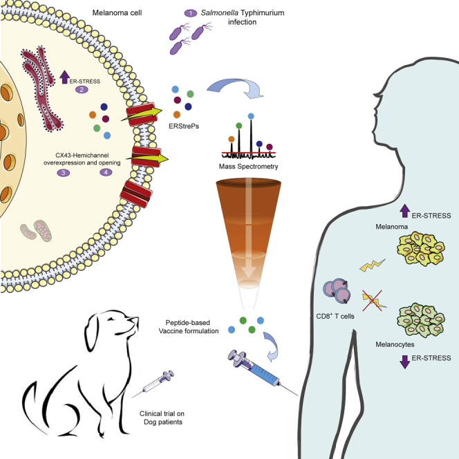
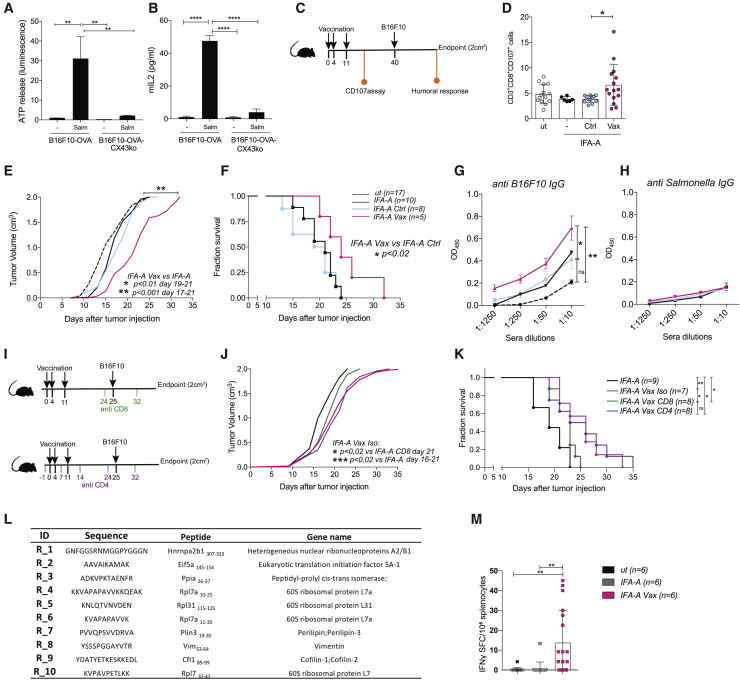
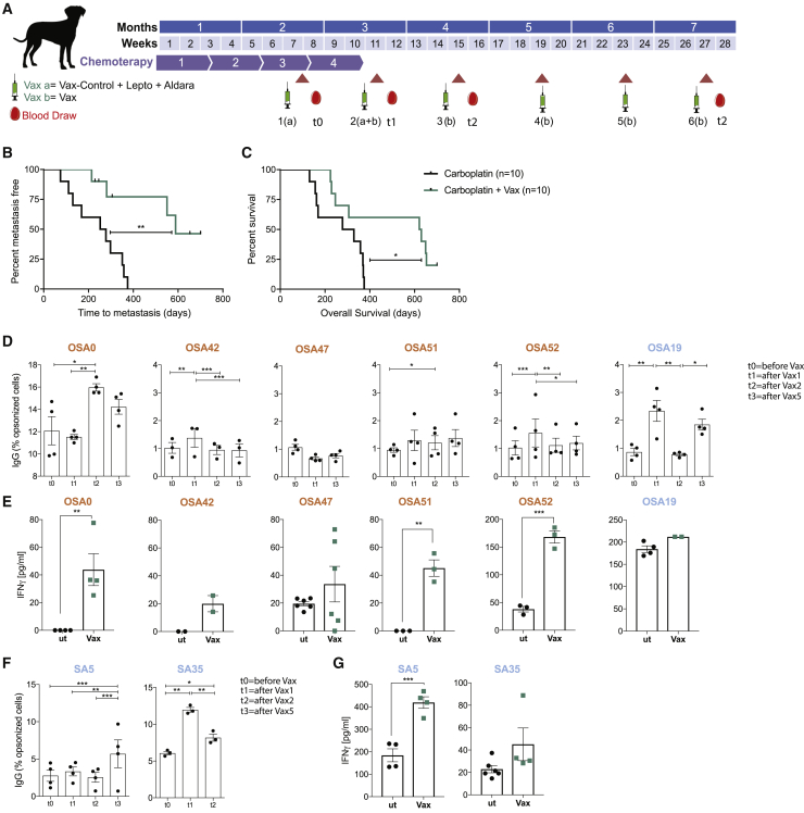
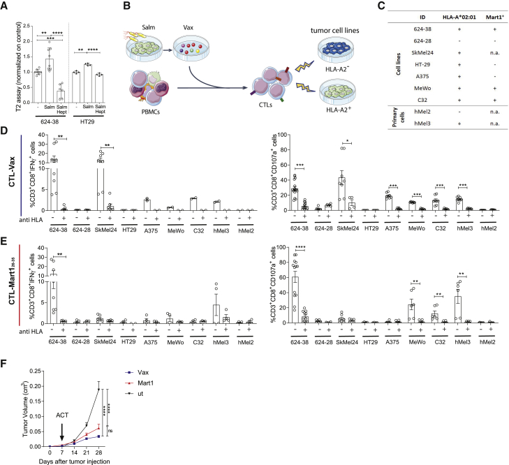
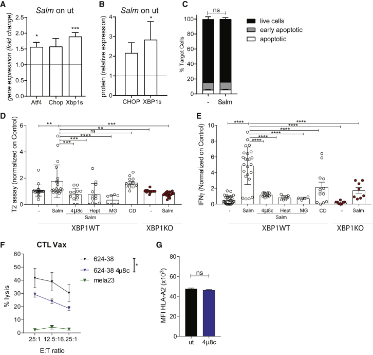
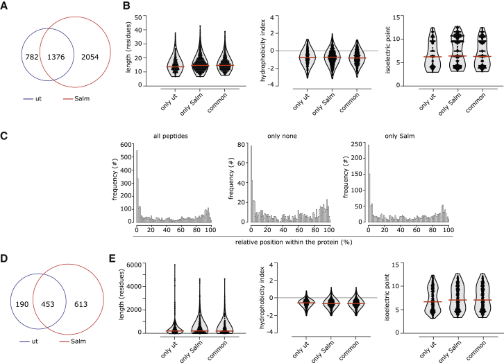
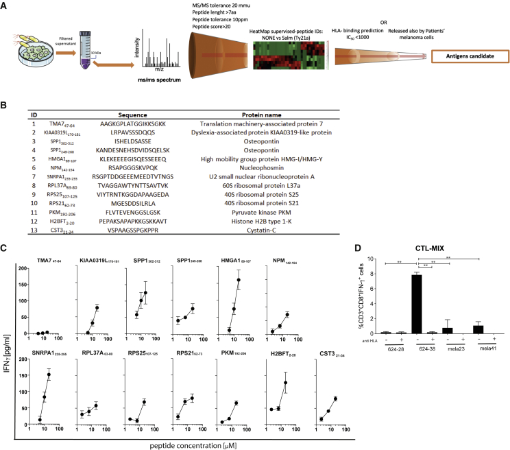
Efforts to overcome resistance to immune checkpoint blockade therapy have focused on vaccination strategies using neoepitopes, although they cannot be applied on a large scale due to the "private" nature of cancer mutations. Here, we show that infection of tumor cells with Salmonella induces the opening of membrane hemichannels and the extracellular release of proteasome-generated peptides by the exacerbation of endoplasmic reticulum (ER) stress. Peptides released by cancer cells foster an antitumor response in vivo, both in mice bearing B16F10 melanomas and in dogs suffering from osteosarcoma. Mass spectrometry analysis on the supernatant of human melanoma cells revealed 12 peptides capable of priming healthy-donor CD8+ T cells that recognize and kill human melanoma cells in vitro and when xenotransplanted in vivo. Hence, we identified a class of shared tumor antigens that are generated in ER-stressed cells, such as tumor cells, that do not induce tolerance and are not presented by healthy cells.

Keywords: ER-stress response; Salmonella; cancer; immunotherapy; tumor antigens; vaccine.


Conflict of interest statement

Declaration of interests The authors declare no competing interests.

Figures

None
Graphical abstract
Figure 1
Figure 1
Peptides released by Salmonella-infected tumor cells by plasma membrane hemichannels induce an anti-tumor response in vivo (A) ATP luminescence test on secretomes of B16F10-OVA and B16F10-OVA CX43KO cells infected with Salmonella (Salm) or left untreated (−). (B) ELISA quantification of mIL2 secretion by OVA257-264-specific B3Z cells. (A and B) Data are represented as mean ± SEM using bar plots (n = 3). (C) Scheme of in vivo immunization experiment; mice were immunized with IFA-Aldara alone (IFA-A), IFA-A combined with secretome derived either from Salmonella-infected B16F10 cells (IFA-A Vax) or untreated cells (IFA-A Ctrl). (D) Frequency of tumor-reactive CD3+CD8+CD107a+ from PBMCs of immunized mice. Data of 2 pooled experiments are represented as mean ± SD using a scatter dot plot. (E and F) Tumor growth (E) and Kaplan-Meier survival curves (F) of vaccinated mice. Data are pooled from two independent experiments (n = 5–9). (G and H) ELISA quantifications of B16F10-specific IgG (G) and Salmonella-specific antibodies (H) in mice sera. Data of 3 pooled experiments are represented as mean ± SEM (n = 6). (I) Scheme of in vivo CD4 and CD8 depletion. Mice were immunized with IFA-A, IFA-A combined with B16F10 secretome and isotype control antibody (IFA-A Vax Iso), or with B16F10 secretome and depleted of either CD8 (IFA-A Vax CD8) or CD4 (IFA-A Vax CD4) T cells. (J and K) Tumor growth (J) and Kaplan-Meier survival curves (K) of vaccinated mice (n = 7–9). (L) Mouse peptides released by B16F10 upon Salmonella infection. (M) ELISpot of IFNγ spot forming cell (SFC)-splenocytes stimulated ex vivo with the mix of identified peptides. Data of 1 experiment are represented as mean ± SE by using a scatter dot plot (n = 6). Statistical analysis was evaluated using two-sided Mann-Whitney test (A, B, and D) or ordinary one-way ANOVA with Bonferroni post-test (D, E, G, H, J, and M); log-rank Mantel-Cox test was performed to assess differences among survival curves (F,K). ns, p > 0.05; p < 0.05, ∗∗p < 0.01, and ∗∗∗p < 0.001. See also Figures S1 and S2 and Table S1.
Figure 2
Figure 2
Secretomes of Salmonella-treated canine primary tumor cells prime an antitumor response in dogs affected by osteosarcoma and high-grade sarcoma (A) Clinical trial scheme for dog patients with OSA and high-grade SA. (B and C) Kaplan-Meier of metastasis free (B) and survival curve of (C) patients with OSA treated with carboplatin alone (black) or in combination with Vax (green). (D and F) Tumor-specific IgG in dog sera shown as frequency of IgG-opsonized tumor cells. (E and G) ELISA quantification of IFN-γ released by dog patient PBMCs upon stimulation with secretome from untreated (ut) or Salmonella-infected (Vax) SA/OSA cells. Immunomonitoring experiments were performed once (D and F) or more (n = 2–3; C and E); each condition was tested at least in triplicate. Data are represented as mean ± SEM using a scatter dot plot. Statistical analysis was evaluated using log-rank Mantel-Cox (B), two-way ANOVA Tukey’s multiple comparison (C and E), and two-sided Mann-Whitney test (D and F). p < 0.05,∗∗p < 0.01,∗∗∗p < 0.001,∗∗∗∗p < 0.0001. See also Figure S3.
Figure 3
Figure 3
Peptides released by 624-38 human melanoma cell line upon Salmonella infection activate tumor-specific CD8+ T cells derived from healthy donor PBMCs (A) MFI (mean fluorescence intensity) of HLA-A02:01 of T2 cells loaded with secretomes of 624-38 and HT-29 cells infected with Salmonella (Salm) with or without Heptanol (Hept) or left untreated (−). (B) Scheme of CTL-Vax and CTL-Mart126-35 expansion from healthy donor PBMCs. (C) Tumor cell targets. HLA-A202:01 and Mart1 antigen expression are indicated. (D and E) Frequency of CD3+CD8+IFN-γ+ (activated) or CD3+CD8+CD107a+ (degranulating) CTL-Vax (D) and CTL-Mart (E) upon co-culture with target cells. Pan HLA-blocker (anti HLA) antibody was added as control, where indicated. (F) Tumor growth curves of NSG mice injected with 624-38 melanoma cells, adoptively transferred with CTL-Vax (blue) or with CTL-Mart1 (red) at day 7 after tumor injection. (A, D, and E) Data are pooled from three or more experiments and are expressed as mean ± SE using scatter dot plot. Statistical analysis was evaluated using one way ANOVA (A and F) or two-sided Mann-Whitney test (D and E). p < 0.05, ∗∗p < 0.01, ∗∗∗p < 0.001, ∗∗∗∗p < 0.0001. See also Figures 4, 5, and 6.
Figure 4
Figure 4
Salmonella-induced secretome peptides are mainly tumor proteasome derived and are released in response to UPR pathway activation along with hemichannel opening (A and B) UPR pathway analysis at gene (A) and protein (B) level in Salmonella-infected 624-38 cells. Data of 3 pooled experiments are normalized to Gapdh (A) or Vinculin (B) and expressed as mean ± SEM of average expression fold change. (C) Frequency of AnnexinPI (live), Annexin+PI (early apoptotic), and Annexin+PI+ (apoptotic) tumor cells Salmonella-infected (Salm) or untreated (−) (n = 2). (D) MFI of HLA-A02:01 on T2 cells loaded with secretomes from untreated (−) or Salmonella-infected (Salm) XBP1WT or XBP1KO cells; from Salmonella-infected XBP1WT in combination with 4μ8c, Heptanol (Hept), MG132 (MG); supernatant of dead cells (C and D). (E) ELISA of IFN-γ released by CTL-Vax stimulated with secretomes differently derived. (D and E) Data of 3 pooled experiments normalized on (−) are represented as mean ± SEM using a scatter dot plot. (F) Delfia assay; lysis of target cells by CTL-Vax (effector) at different effector-to-target (E:T) ratios (n = 3). (G) MFI of HLA-A02:01 on 624-38 cells. Data of 3 pooled experiments are represented. Statistical analysis was evaluated using two-sided Mann-Whitney test (A, B, C, and G), one-way ANOVA followed by multiple comparisons (Dunnett) against XBP1WT-Salm (D and E), or two-way ANOVA followed by multiple comparisons (Tukey) (F). p < 0.05,∗∗p < 0.01,∗∗∗p < 0.001,∗∗∗∗p < 0.0001. See also Figures S4, S5, and S6.
Figure 5
Figure 5
Characteristics of melanoma primary cell secretome (A) Frequency of peptides identified only in the secretome of Salmonella-infected (Salm) or untreated (ut) melanoma primary cultures or present in both (common). (B) Distribution of length, hydrophobicity, and isoelectric point of peptides. (C) Relative distribution of the secretome’s peptides within the cognate protein sequence. (D) Frequency of proteins having at least one peptide identified in the secretome. (E) Distribution of length, hydrophobicity, and isoelectric point of proteins. In (B)–(E), unique peptides or proteins identified in at least one melanoma patient secretome are reported. In (B) and (E), the violin plots indicate the frequency of the peptides/proteins, with all single peptides/proteins indicated as dots. Red lines indicate the median. See also Figure S7 and Table S2.
Figure 6
Figure 6
Identification of peptides released by human melanoma cells upon Salmonella infection (A) Strategy to identify candidate antigens in secretome derived from Salmonella-infected melanoma cells. (B) List of the selected antigen candidates. (C) ELISA of IFN-γ released by CTL-Vax upon stimulation with single peptides. Results are representative of 5 independent experiments on CTL-Vax derived from 2 different donors. (D) Frequency of CD3+CD8+IFN-γ+ CTL-MIX upon co-culture with target cells. Pan HLA-blocker (anti HLA) antibody was added, where indicated. Data of 2 pooled experiments are represented as mean ± SEM using bar plot. Statistical analysis was evaluated using one-way ANOVA followed by multiple comparisons (Dunnett). p < 0.05,∗∗p < 0.01. See also Table S2.

References

    1. Antoniou A.N., Lenart I., Kriston-Vizi J., Iwawaki T., Turmaine M., McHugh K., Ali S., Blake N., Bowness P., Bajaj-Elliott M. Salmonella exploits HLA-B27 and host unfolded protein responses to promote intracellular replication. Ann. Rheum. Dis. 2019;78:74–82. - PMC - PubMed
    1. Bartoszewski R., Brewer J.W., Rab A., Crossman D.K., Bartoszewska S., Kapoor N., Fuller C., Collawn J.F., Bebok Z. The unfolded protein response (UPR)-activated transcription factor X-box-binding protein 1 (XBP1) induces microRNA-346 expression that targets the human antigen peptide transporter 1 (TAP1) mRNA and governs immune regulatory genes. J. Biol. Chem. 2011;286:41862–41870. - PMC - PubMed
    1. Boegel S., Löwer M., Bukur T., Sahin U., Castle J.C. A catalog of HLA type, HLA expression, and neo-epitope candidates in human cancer cell lines. OncoImmunology. 2014;3:e954893. - PMC - PubMed
    1. Cerezo M., Lehraiki A., Millet A., Rouaud F., Plaisant M., Jaune E., Botton T., Ronco C., Abbe P., Amdouni H. Compounds triggering ER stress exert anti- melanoma effects and overcome BRAF inhibitor resistance. Cancer Cell. 2016;29:805–819. - PubMed
    1. Chapiro J., Claverol S., Piette F., Ma W., Stroobant V., Guillaume B., Gairin J.E., Morel S., Burlet-Schiltz O., Monsarrat B. Destructive cleavage of antigenic peptides either by the immunoproteasome or by the standard proteasome results in differential antigen presentation. J. Immunol. 2006;176:1053–1061. - PubMed

Publication types

MeSH terms