. 2021 Jul 6;36(1):109317.
doi: 10.1016/j.celrep.2021.109317.

Structure of the TELO2-TTI1-TTI2 complex and its function in TOR recruitment to the R2TP chaperone

Affiliations

Structure of the TELO2-TTI1-TTI2 complex and its function in TOR recruitment to the R2TP chaperone

Mohinder Pal et al. Cell Rep. .

Abstract

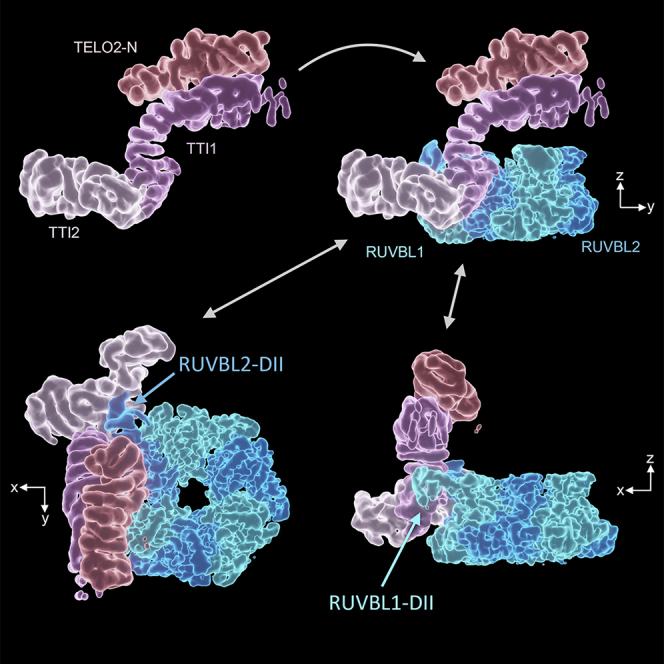
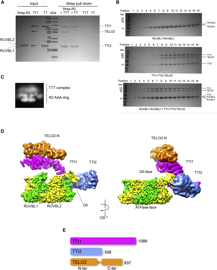
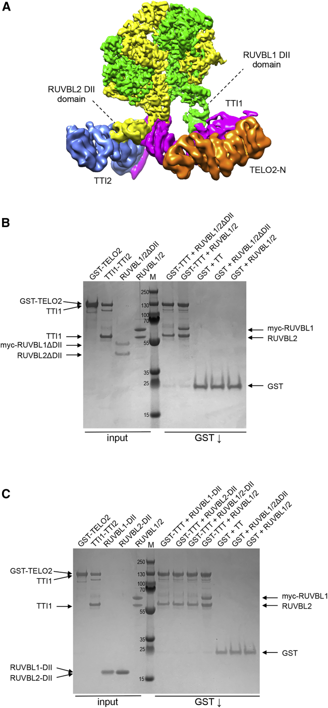
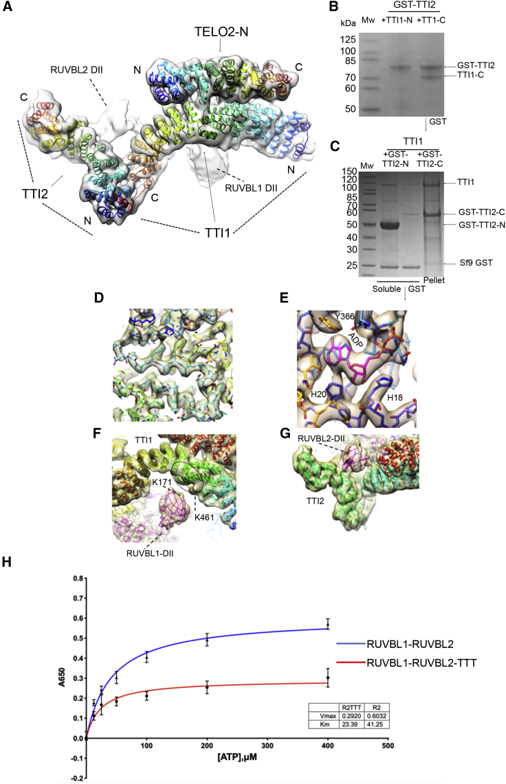
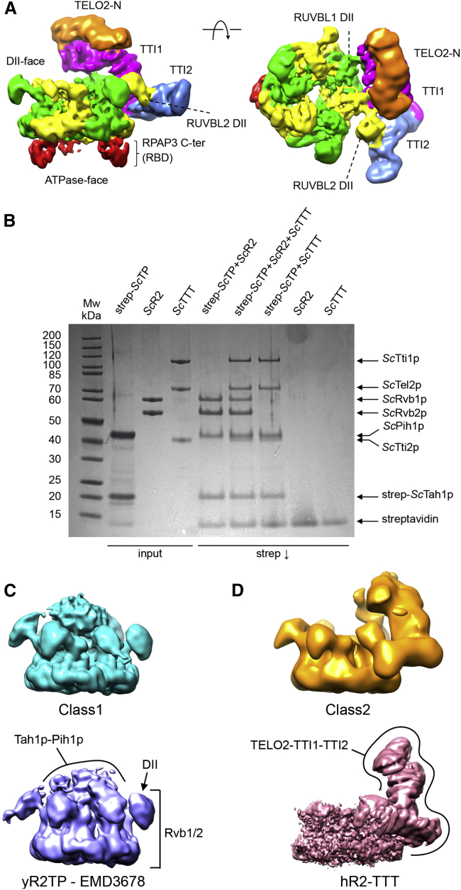
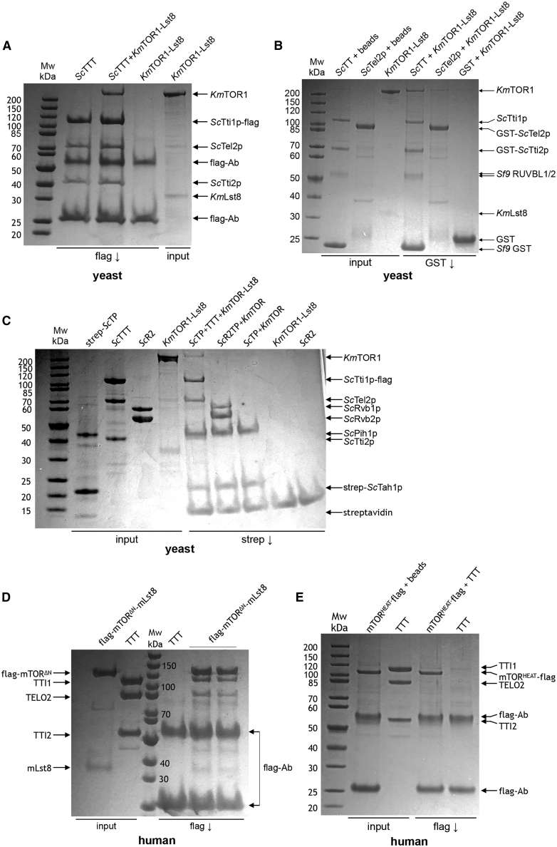
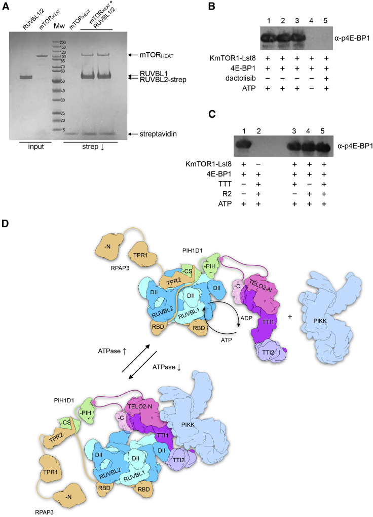
The R2TP (RUVBL1-RUVBL2-RPAP3-PIH1D1) complex, in collaboration with heat shock protein 90 (HSP90), functions as a chaperone for the assembly and stability of protein complexes, including RNA polymerases, small nuclear ribonucleoprotein particles (snRNPs), and phosphatidylinositol 3-kinase (PI3K)-like kinases (PIKKs) such as TOR and SMG1. PIKK stabilization depends on an additional complex of TELO2, TTI1, and TTI2 (TTT), whose structure and function are poorly understood. The cryoelectron microscopy (cryo-EM) structure of the human R2TP-TTT complex, together with biochemical experiments, reveals the mechanism of TOR recruitment to the R2TP-TTT chaperone. The HEAT-repeat TTT complex binds the kinase domain of TOR, without blocking its activity, and delivers TOR to the R2TP chaperone. In addition, TTT regulates the R2TP chaperone by inhibiting RUVBL1-RUVBL2 ATPase activity and by modulating the conformation and interactions of the PIH1D1 and RPAP3 components of R2TP. Taken together, our results show how TTT couples the recruitment of TOR to R2TP with the regulation of this chaperone system.

Keywords: HSP90 chaperone; PIKK; R2TP; RUVBL1; RUVBL2; TELO2; TTI1; TTI2; TTT; mTOR.


Conflict of interest statement

Declaration of interests The authors declare no competing interests.

Figures

None
Graphical abstract
Figure 1
Figure 1
Cryo-EM of the human RUVBL1-RUVBL2-TTT complex (A) Coomassie-stained SDS-PAGE gel showing analysis of interaction of RUVBL1-RUVBL2 (R2), TTI1-TTI2-TELO2 (TTT), and TTI1-TTI2 (TT) subcomplexes. Pull-down on the strep-tag attached to the C terminus of RUVBL2 co-precipitates TTT and TT subcomplexes, showing that neither RPAP3, PIH1D1, nor TELO2 is required to couple TTI1-TTI2 to R2. TTT and TT are not pulled down in the absence of R2. (B) Coomassie-stained SDS-PAGE of fractions from a size-exclusion chromatography column for the human RUVBL1-RUVBL2 complex, R2, (top panel), the TTT complex (middle panel), and the mixture of R2 and TTT (bottom panel). In the mixture, both R2 and TTT elute in earlier fractions than when loaded separately. (C) Representative 2D average of R2-TTT cryo-EM data. (D) Views of human R2-TTT volume. RUVBL1-RUVBL2 hexamer, green and yellow respectively. Three distinct α-helical segments (TTI1, magenta; TTI2, blue; and TELO2-N, orange) are identifiable in the cryo-EM reconstruction after multi-body refinement of the R2-TTT complex. (E) Schematic showing the relative lengths of the TTT components TTI1, TTI2, and TELO2. All three proteins are known or thought to be entirely composed of HEAT repeats, which are interrupted by an ~70-residue flexible linker in TELO2 that connects the N- and C-terminal domains.
Figure 2
Figure 2
RUVBL1-RUVBL2 DII domains mediate TTT interaction (A) View of human R2-TTT volume colored as in Figure 1D, highlighting the contacts between the DII domains in two consecutive RUVBL subunits and the elongated TTT subcomplex. (B) Coomassie-stained SDS-PAGE gel showing analysis of dependence of the interaction of TTI1-TTI2-TELO2 (TTT) with RUVBL1-RUVBL2 on the RUVBL1/2 DII domains. Pull-down on the GST-tag attached to the N terminus of TELO2 co-precipitates TTI1-TTI2 and wild-type RUVBL1/2, but not the RUVBL1/2ΔDII complex where the DII domains have been excised. (C) As in (B), but showing that the isolated DII domains of RUVBL1 and RUVBL2, individually or in combination, are not co-precipitated by GST-TELO2-TTI1-TTI2 (GST-TTT) when not presented as part of the RUVBL1/2 heterohexamer.
Figure 3
Figure 3
Molecular modeling of TTT and its interaction with RUVBL1-RUVBL2 (A) Molecular model of TTT fitted within the cryo-EM density of R2-TTT (transparent density). Individual helices of TTI1 and TTI2 were fitted manually into density using COOT and connected into a continuous structure by taking advantage of the repetitious linear right-handed α-solenoid architecture of HEAT repeats. The polarity of the two polypeptides was determined by the results of the interaction mapping experiments shown in (C) and (D). The fit of the poly-alanine structures was optimized by real-space refinement in Phenix (Adams et al., 2010). A homology model of the N-terminal domain of human TELO2 based on the crystal structure of S. cerevisiae Tel2p (Takai et al., 2010) was manually adjusted to fit the experimental density using COOT (Emsley and Cowtan, 2004). The TTT model was optimized by real-space refinement in Phenix (Adams et al., 2010). Each protein is colored as a rainbow from N- to C-terminal ends. (B) Co-expression of GST-tagged full-length TTI2 with TTI1 constructs spanning residues 1–459 (TTI1-N) and residues 468–1089 in Sf9 insect cells. Only the TTI1-C construct was co-precipitated as a soluble species with GST-TTI2, suggesting that TTI2 primarily interacts with the C-terminal half of TTI1. (C) Co-expression of full-length TTI1 with GST-tagged TTI2 constructs spanning residues 1–193 (TTI2-N) and residues 194–508 (TTI2-C) in Sf9 insect cells. The GST-tagged TTI2-N was well expressed as a soluble species and co-precipitated TTI1 in GST pull-downs, whereas co-expressed TTI1 and GST-TTI2-C were primarily found in the insoluble fraction. This suggests that TTI1 primarily interacts with the N-terminal half of TTI2. (D) Close-up of electron density in the core of the RUVBL2 subunit that contacts TTI1 and TTI2, showing clear electron density for side chains. The fit of the all-atom model of the RUVBL1-RUVBL2 heterohexamer was optimized by real-space refinement in Phenix. (E) Close-up of electron density in the nucleotide-binding site of the RUVBL1 subunit that contacts TTI1, showing bound ADP. All six nucleotide-binding sites in the R2 ring are fully occupied by ADP, and the His-Ser-His motif that closes off the nucleotide-binding site as part of the N-terminal “gatekeeper” segment (Munoz-Hernandez et al., 2019) is also fully ordered in all six ATPase subunits. (F) Interaction of TTI1 (rainbow-colored helices) with the DII domain of a RUVBL1 subunit (magenta). The fit of the all-atom model of the RUVBL1-RUVBL2 heterohexamer and the TELO2-N domain as well as the poly-Ala models of TTI1 and TTI2 were simultaneously optimized by real-space refinement in Phenix against a “consensus” electron density map comprising maps from separate multibody and focused refinements of the R2 rings and TTT with the interacting DII domains. In an XL-MS analysis of R2TP-TTT, a single cross-link was observed between RUVBL1-Lys171 (black circle) and Lys461 of TTI1. Although the resolution of the TTI1 density does not permit fitting of the sequence, the crosslink helps localize the approximate location of TTI1-Lys461 (red ellipse). (G) Interaction of TTI1 (cyan helices) and TTI2 (green helices) with the DII domain of a RUVBL2 subunit. The DII domain packs into the cleft formed at the junction of the α-helical solenoids of the two HEAT repeat proteins. (H) Human RUVBL1-RUVBL2 displays a weak inherent ATPase activity with a maximal rate of ~0.6 mol/min/mol. This is significantly diminished in the presence of TTT at a 3:1 molar ratio. Error bars indicate the standard error of the mean of three experiments.
Figure 4
Figure 4
Cryo-EM of human and yeast R2TP-TTT complexes (A) Two views of human R2TP-TTT volume: RUVBL1-RUVBL2 hexamer, green and yellow respectively; TTI1, magenta; TTI2, blue; TELO2-N, orange. The RPAP3 C-terminal RUVBL2-binding domain (RBD) is identifiable in the ATPase face of each of the RUVBL2 subunits (red). (B) Coomassie-stained SDS-PAGE gel showing analysis of interaction of yeast Tah1p-Pih1p (yTP), Rvb1p-Rvb2p (yR2), and Tel2p-Tti1p-Tti2p (yTTT) subcomplexes. Pull-down on the tandem strep-tag attached to the N terminus of Tah1p within the yTP complex co-precipitates yR2 and yTTT simultaneously and as separate co-complexes. (C) Cryo-EM analysis of co-precipitated Tah1p-Pih1p-Rvb1p-Rvb2p-Tel2p-Tti1p-Tti2p (yeast R2TP-TTT). 3D classification yields two main classes: one class contained particles with strong central density between the DII insertion domains of the R2 ring comprising two-thirds of the particles (top), and the other class resembled the previously described yeast R2TP complex (EMD-3678) (Rivera-Calzada et al., 2017) (bottom). (D) As in (C), but showing the second main class comprising two-thirds of the particles, which lack the central density attributed to Tah1p-Pih1p, but display strong peripheral density at one edge of the R2 ring (top) that strongly resembles the density for the TTT complex in the higher resolution human R2TTT structure (bottom). No significant class of particles was identified in which the central and peripheral density features were simultaneously present.
Figure 5
Figure 5
Mapping the interactions between TTT and TOR (A) Coomassie-stained SDS-PAGE gel showing interaction of yeast (Kluyveromyces marxianus) TOR1-Lst8 with yeast (S. cerevisiae) TTT. FLAG-tagged Tti1p co-immunoprecipitates TOR1, whereas anti-FLAG beads alone do not. Lst8, which is evident as a weakly staining band in the input, was not evident in the immunoprecipitated TTT-TOR1. (B) As in (A), but for interaction of KmTOR-Lst8 with GST-tagged Tti2p-Tti1p or GST-tagged Tel2p. KmTOR was co-precipitated in GST pull-down with Tti1p-GST-TTI2p, but not with Tel2p. (C) As in (A), but for interaction of KmTOR-Lst8 with TP and R2TP complexes containing strep-tagged Tah1p. Neither TP nor R2TP complexes were able to co-precipitate KmTOR directly. However, tagged TP could co-precipitate KmTOR when TTT was present, confirming that TP and KmTOR do not compete for binding to TTT. (D) As in (A), but for interaction of a FLAG-tagged N-terminally truncated human mTOR construct (mTORΔN) co-expressed with mLST8, with human TTT. TTT is co-immunoprecipitated by FLAG-tagged mTORΔN, but not by FLAG beads alone. (E) As in (A), but for interaction of a FLAG-tagged soluble N-terminal fragment of human mTOR encompassing the major segment of HEAT repeats (mTORHEAT) with human TTT. Flag-tagged mTORHEAT was precipitated by FLAG beads, but it did not co-immunoprecipitate TTT.
Figure 6
Figure 6
TTT couples mTOR recruitment to R2TP to regulation of the chaperone (A) Coomassie-stained SDS-PAGE gel showing the interaction of an mTOR fragment comprising the N-terminal HEAT region (residues 1–929) with the strep-tagged RUVBL1-RUVBL2 complex. (B) Western blot showing phosphorylation of 4E-BP1 by the yeast (K. marxianus) TOR1-Lst8 complex. A strong signal was detected by a phosphospecific antibody to pThr37/46 in the presence of ATP, but not when ATP was omitted from the reaction, or when the ATP-competitive mTOR inhibitor dactolisib (NVP-BEZ235) was present. (C) As in (B), but with the addition of TTT, R2, or both. Neither of these chaperone components, alone or in combination, affects its ability to phosphorylate a substrate. (D) Model of the functions of TTT. Flexible tethering to the R2 ring allows for facile exchange of TP and TTT components at the main interaction site on the DII domain face of the ring. Interaction of the PIH1D1 part of TP with a RUVBL1 DII domain facilitates partial ring opening and nucleotide exchange (Munoz-Hernandez et al., 2019), accelerating the basal ATPase activity of the ring (Rivera-Calzada et al., 2017). The CK2 phosphorylation site within the inherently disordered linker connecting the N- and C-terminal domains of TELO2 (Horejsí et al., 2010) tethers the TTT subcomplex to the R2 ring through interaction with the PIH domain of PIH1D1 (Hořejší et al., 2014; Pal et al., 2014) and thereby facilitates recruitment of a PIKK client to the core complex (top). Rearrangement of the complex allows TTT to bind to the DII domains of consecutive RUVBL1-RUVBL2 domains, fixing them in an ADP-bound closed state that downregulates the basal ATPase, potentially bringing the PIKK client into closer association. Steric hindrance of the DII domains, as well as allosteric incompatibility, prevents simultaneous binding of PIH1D1 to the DII face of the ring. Although not engaged, TP remains tethered to the complex through the interaction of the PIH domain with the TELO2 linker segment (see above) and the interaction of the C-terminal domain of RPAP3 with the ATPase face of the R2 ring (bottom).

Similar articles

Cited by

References

    1. Adams P.D., Afonine P.V., Bunkóczi G., Chen V.B., Davis I.W., Echols N., Headd J.J., Hung L.W., Kapral G.J., Grosse-Kunstleve R.W. PHENIX: A comprehensive Python-based system for macromolecular structure solution. Acta Crystallogr. D Biol. Crystallogr. 2010;66:213–221. - PMC - PubMed
    1. Afonine P.V., Poon B.K., Read R.J., Sobolev O.V., Terwilliger T.C., Urzhumtsev A., Adams P.D. Real-space refinement in PHENIX for cryo-EM and crystallography. Acta Crystallogr. D Struct. Biol. 2018;74:531–544. - PMC - PubMed
    1. Baretić D., Berndt A., Ohashi Y., Johnson C.M., Williams R.L. Tor forms a dimer through an N-terminal helical solenoid with a complex topology. Nat. Commun. 2016;7:11016. - PMC - PubMed
    1. Casanal A., Lohkamp B., Emsley P. Current developments in Coot for macromolecular model building of electron cryo-microscopy and crystallographic data. Protein Sci. 2020;29:1069–1078. - PMC - PubMed
    1. Emsley P., Cowtan K. Coot: model-building tools for molecular graphics. Acta Crystallogr. D Biol. Crystallogr. 2004;60:2126–2132. - PubMed

Publication types

MeSH terms

Substances