. 2021 Aug;19(8):1990-2001.
doi: 10.1111/jth.15407. Epub 2021 Jul 7.

Pathologically stiff erythrocytes impede contraction of blood clots

Affiliations

Pathologically stiff erythrocytes impede contraction of blood clots

Valerie Tutwiler et al. J Thromb Haemost. 2021 Aug.

Abstract

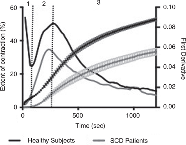
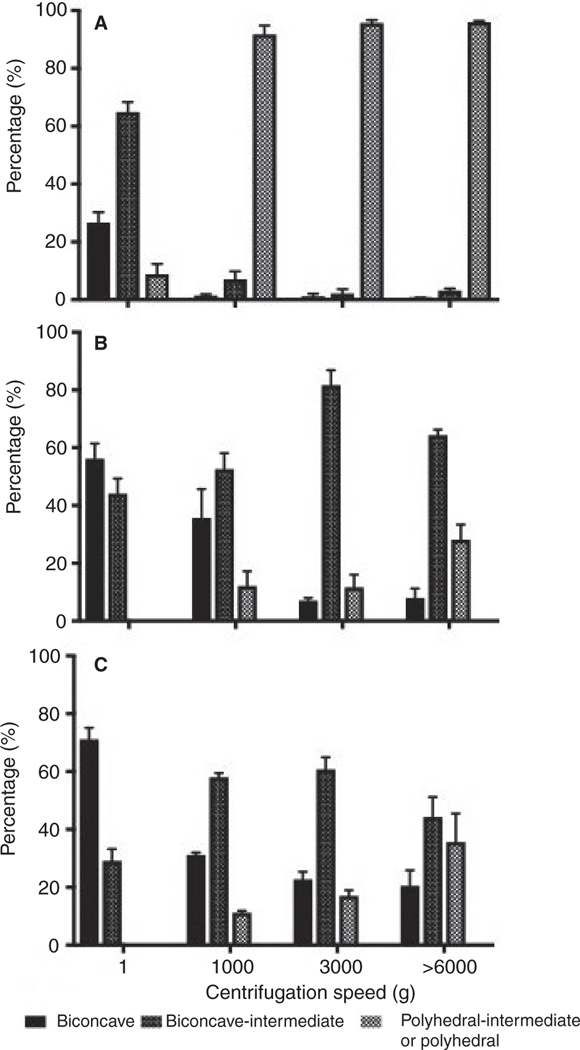
Background: Blood clot contraction, volume shrinkage of the clot, is driven by platelet contraction and accompanied by compaction of the erythrocytes and their gradual shape change from biconcave to polyhedral, with the resulting cells named polyhedrocytes.

Objectives: Here, we examined the role of erythrocyte rigidity on clot contraction and erythrocyte shape transformation.

Methods: We used an optical tracking methodology that allowed us to quantify changes in contracting clot size over time.

Results and conclusions: Erythrocyte rigidity has been shown to be increased in sickle cell disease (SCD), and in our experiments erythrocytes from SCD patients were 4-fold stiffer than those from healthy subjects. On average, the final extent of clot contraction was reduced by 53% in the clots from the blood of patients with SCD compared to healthy individuals, and there was significantly less polyhedrocyte formation. To test if this reduction in clot contraction was due to the increase in erythrocyte rigidity, we used stiffening of erythrocytes via chemical cross-linking (glutaraldehyde), rigidifying Wrightb antibodies (Wrb ), and naturally more rigid llama ovalocytes. Results revealed that stiffening erythrocytes result in impaired clot contraction and fewer polyhedrocytes. These results demonstrate the role of erythrocyte rigidity in the contraction of blood clots and suggest that the impaired clot contraction/shrinkage in SCD is due to the reduced erythrocyte deformability, which may be an underappreciated mechanism that aggravates obstructiveness of erythrocyte-rich (micro)thrombi in SCD.

Keywords: blood clotting; clot retractions; coagulation; sickle cell disease; thrombosis.


Conflict of interest statement

CONFLICTS OF INTEREST

The authors have no competing interests to disclose.

Figures

FIGURE 1
FIGURE 1
Parameters of blood clot contraction in sickle cell disease (SCD) patients and healthy individuals. Optical tracking of contracting blood clots was used to compare (A) extent of clot contraction at 20 min, (B) lag time, (C) average velocity, and (D) area under the kinetic curve. Parameters for healthy individuals (n = 52) and patients with SCD (n = 16) were compared using an unpaired, 2- tailed student t- test. ****P < .0001
FIGURE 2
FIGURE 2
Averaged kinetic curves of clot contraction for sickle cell disease (SCD) patients (n = 16) and healthy subjects (n = 52). Optical tracking was used to measure the extent of clot contraction every 15 s over the course of 20 min. The instantaneous first derivative was used to define transitions between phases and calculate rate constants of each phase of contraction. Data points in the curves represent mean ± standard error of the mean from individual kinetic curves. Dashed vertical lines denote the transitions between phases
FIGURE 3
FIGURE 3
Representative scanning electron microscopy images of the core of contracted clots formed from the blood of (A,C) healthy subjects and (B,D) sickle cell disease (SCD) patients, showing the different degree of compaction of red blood cells and their transformation to polyhedrocytes, as well as absence (A, C) and presence (B, D) of fibrin in the interior of the blood clots. Scale bar for (A,B) is 30 μm and for (C,D) is 10 μm
FIGURE 4
FIGURE 4
Representative scanning electron micrographs showing reduced polyhedral deformation in chemically rigidified erythrocytes. Centrifugation of normal untreated erythrocytes (control) revealed a transition from biconcave to polyhedral similar to what is observed in contracted blood clots. Polyhedral- intermediate forms are largely observed due to the anisotropic application of forces during centrifugation. This transition was mitigated in erythrocytes pretreated with 0.03% and 0.06% glutaraldehyde, which increased erythrocyte rigidity and reduced deformability (see Figure 5 for quantification)
FIGURE 5
FIGURE 5
Quantification of polyhedrocyte formation in chemically rigidified erythrocytes. Erythrocytes were quantified for their shape transformation from biconcave to biconcave-intermediate, and polyhedral- intermediate or polyhedral following centrifugation. Deformations induced by centrifugation of untreated erythrocytes (A) and erythrocytes rigidified by incubation with (B) 0.03% or (C) 0.06% glutaraldehyde
FIGURE 6
FIGURE 6
Clot contraction with naturally rigid llama ovalocytes. A, Representative scanning electron micrographs showing llama ovalocytes inside blood clots that were uncontracted, contracted with normal platelet count (~250,000/μl), or contracted using a higher platelet count (~400,000/μl). B, Kinetic curves of clot contraction in reconstituted blood to assess clot contraction in samples containing human erythrocytes or llama ovalocytes. C, Average extent of clot contraction at 20 mins, (D) average velocity, and (E) the rates of phases 2 and 3 were quantified in contracting reconstituted blood samples containing human or llama ovalocytes at various platelet counts
FIGURE 7
FIGURE 7
Contraction of blood clots formed in the presence of erythrocyte- rigidifying Wrightb antibodies. Representative single donor experiment revealed that (A) Wrb antibodies induced an increase in erythrocyte rigidity, which corresponded to (B) a decrease in the extent of contraction. Representative scanning electron microscopy showing erythrocyte deformation in the core of clots made (C) without or (D) with addition of Wrb antibodies and following centrifugation of washed erythrocytes in the (E) absence or (F) presence of Wrb antibodies. Distributions of red blood cell (RBC) shapes quantified from scanning electron microscopy images for (G) contracted clots and (H) centrifuged RBCs

Comment in

References

    1. Cines DB, Lebedeva T, Nagaswami C, et al. Clot contraction: compression of erythrocytes into tightly packed polyhedra and redistribution of platelets and fibrin. Blood. 2014;123(10):1596–1603. - PMC - PubMed
    1. Muthard R, Diamond S. Blood clots are rapidly assembled hemodynamic sensors: flow arrest triggers intraluminal thrombus contraction. Arterioscler Thromb Vasc Biol. 2012;32(12):2938–2945. - PMC - PubMed
    1. Tutwiler V, Peshkova AD, Le Minh G, et al. Blood clot contraction differentially modulates internal and external fibrinolysis. J Thromb Haemost. 2019;17(2):361–370. - PubMed
    1. Peshkova AD, Malyasyov DV, Bredikhin RA, et al. Reduced contraction of blood clots in venous thromboembolism is a potential thrombogenicand embologenic mechanism. TH Open. 2018;02(01):e104–e115. - PMC - PubMed
    1. Tutwiler V, Peshkova AD, Andrianova IA, Khasanova DR, Weisel JW, Litvinov RI. Contraction of blood clots is impaired in ischemic stroke. Arterioscler Thromb Vasc Biol. 2017;37(2):271–279. - PMC - PubMed

Publication types