. 2021 Oct 1;94(1126):20210236.
doi: 10.1259/bjr.20210236. Epub 2021 Jul 8.

Soft tissue masses of the epitrochlear region

Affiliations

Soft tissue masses of the epitrochlear region

William Tilden et al. Br J Radiol. .

Abstract

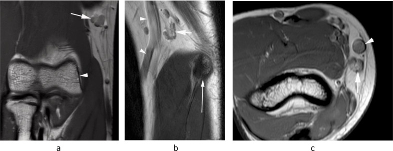
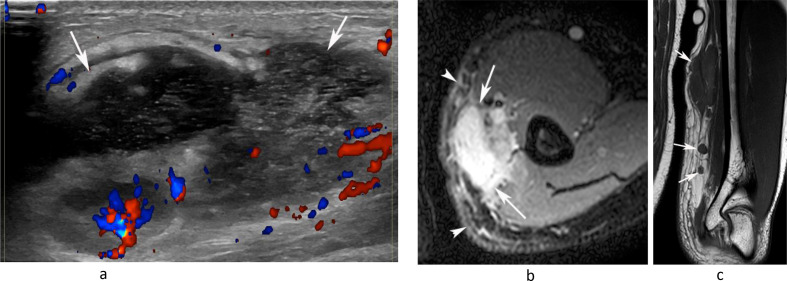
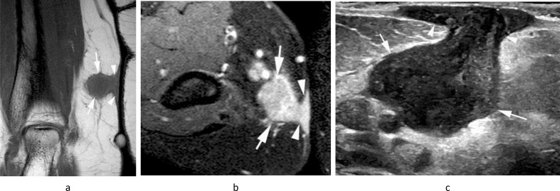
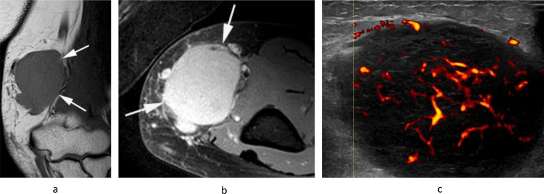
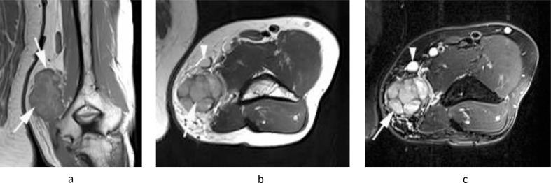
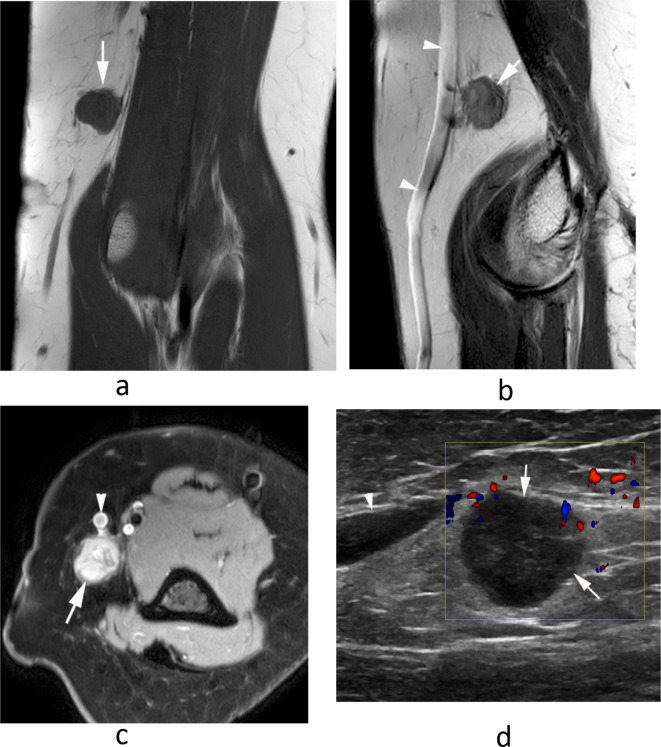
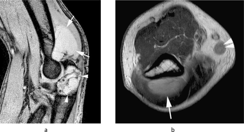
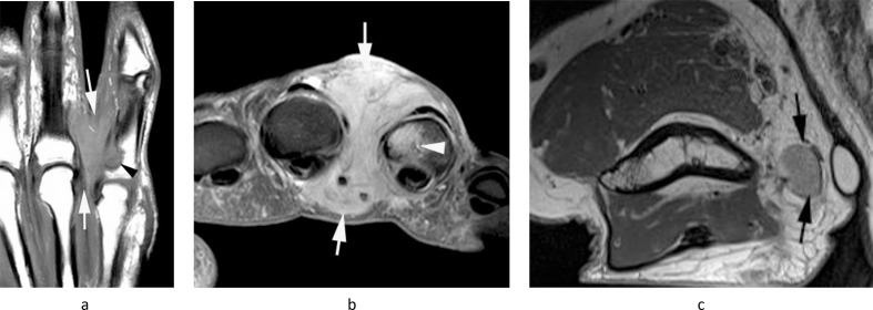
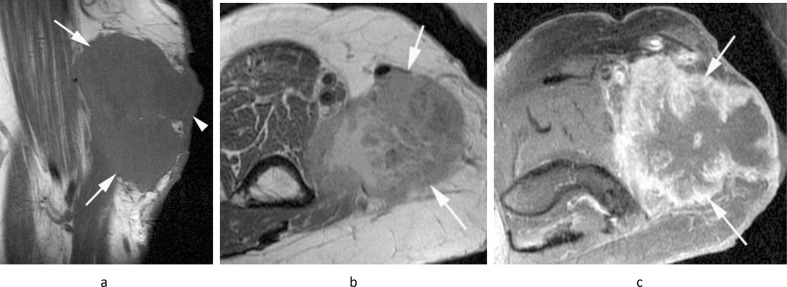
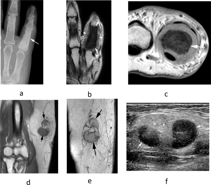
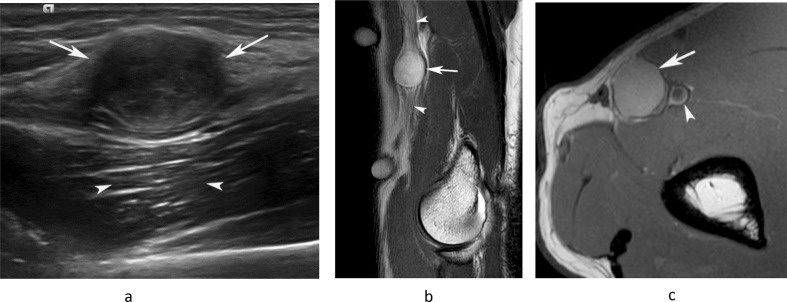
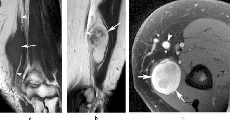
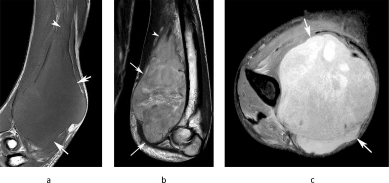
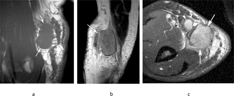
The epitrochlear lymph nodes (ELN) are rarely examined clinically and are difficult to identify radiologically in healthy patients. They are, therefore, generally under appreciated as a source of significant pathology. Despite this, enlargement of an ELN is almost always secondary to a pathological process, the differential for which is relatively narrow. The following pictorial review illustrates the spectrum of infectious, inflammatory and malignant conditions affecting the ELN, some of which are quite specific to this location. We also emphasise the importance of distinguishing enlarged ELNs from benign and malignant non-nodal soft tissue masses, which can have very similar clinical presentation and imaging appearances.


Figures

Figure 1.
Figure 1.
A 16-year-old boy being imaged for right elbow pain. (a) Coronal PDW FSE MR image shows the normal epitrochlear lymph node (arrow) located ~2 cm proximal to the medial epicondyle (arrowhead). (b) Sagittal T2W FSE and (c) axial PDW FSE MR images show the epitrochlear lymph node (arrows) located directly posterior to the basilic vein (arrowheads) and ~2 cm proximal to the medial epicondyle (thin arrow-b).
Figure 2.
Figure 2.
A 20-year-old male presenting with a painful swelling of the medial distal arm. (a) Longitudinal Doppler US demonstrates a poorly defined mixed echogenicity hypervascular mass with internal fluid collection (arrows). (b) Axial PD-SPAIR and (c) coronal T1W TSE MR images show a large heterogeneously T1 isointense/SPAIR hypeintense mass with a fluid collection and multiple smaller enlarged medial epitrochlear lymph nodes (arrows-c) with inflammatory oedema-like signal and stranding in the surrounding soft tissues (arrowheads-b). Histology confirmed necrotising granulomatous lymphadenitis.
Figure 3.
Figure 3.
A 49-year-old female presenting with an ulcerating mass on the inner margin of the right arm just above the elbow. (a) Coronal T1W TSE, (b) axial PD-SPAIR MR images and (c) longitudinal US show a lobular hypoechoic mass (arrows), isointense on T1 and heterogeneously hyperintense on SPAIR images, which is forming a sinus to the skin (arrowheads-a,b), the features being consistent with a “collar stud” abscess. Biopsy revealed chronic necrotising granulomatous infection consistent with tuberculosis.
Figure 4.
Figure 4.
A 66-year-old female presenting with a large mass in the medial epitrochlear space. (a) Coronal T1W TSE and (b) axial PD-SPAIR MR images show a somewhat poorly defined mass (arrows) displacing the basilic vein which is homogeneously isointense on T1 and hyperintense on SPAIR. (c) Longitudinal Doppler US shows a homogeneous hypoechoic hypervascular mass. Histology confirmed a diagnosis of lymphoma.
Figure 5.
Figure 5.
A 61-year-old male with a previous history of melanoma presenting with a mass in the medial epitrochlear space. (a) Coronal T1W TSE, (b) axial T2W FSE and (c) axial PD-SPAIR MR images show a lobular and heterogenous mildly T1 and T2 hyperintense mass (arrows) contacting the basilic vein (arrowheads-b,c). Histology confirmed melanoma metastasis.
Figure 6.
Figure 6.
A 39-year-old female with a history of clear cell sarcoma of the hand treated 3 years previously, now presenting with a small mass in the medial epitrochlear space. (a) Coronal T1W TSE, (b) sagittal T2W FSE and (c) axial PD-SPAIR MR images show a small lobular mass (arrows) located directly posterior to the basilic vein (arrowheads-b,c), isointense on T1 and T2 and heterogeneously hyperintense on SPAIR, consistent with a nodal mass. (d) Longitudinal US shows a homogeneous hypoechoic mass (arrows) located adjacent to the basilic vein (arrowhead). Histologically confirmed clear cell sarcoma metastasis.
Figure 7.
Figure 7.
A 77-year-old male with a painful swollen right elbow. (a) Sagittal T2W gradient echo and (b) axial PDW FSE MR images show a homogeneously T2 hyperintense soft tissue mass in the posterior recess of the elbow (arrows) with erosion of the olecranon (arrowheads-a) and enlargement of the epitrochlear lymph node (double arrowhead-b). The patient had a known intra-articular synovial sarcoma of the elbow joint.
Figure 8.
Figure 8.
A 74-year-old male presenting with a painful swelling on the ulnar-side of the right hand and a small mass in the medial epitrochlear space. (a) Coronal T1W TSE and (b) axial PD-SPAIR MR images show a poorly defined T1 isointense, SPAIR hyperintense soft tissue mass in the fourth webspace (arrows) which is eroding the little finger proximal phalanx (arrowhead-b). (c) Axial PDW FSE MR image through the distal arm shows a small oval mildly hyperintense mass (arrows) in the location of the epitrochlear lymph node. Histologically confirmed undifferentiated pleomorphic sarcoma at both sites.
Figure 9.
Figure 9.
An 86-year-old female with a previous history of squamous cell carcinoma of the lung who developed a large fungating mass in the medial epitrochlear space. (a) Sagittal T1W TSE, (b) axial T2W FSE and (c) axial fat suppressed post-gadolinium T1W TSE MR images show a poorly defined mass with a solid enhancing outer component and fluid signal intensity necrotic centre (arrows). Histologically confirmed squamous cell carcinoma metastasis.
Figure 10.
Figure 10.
A 69-year-old male presenting with painful swelling of the little finger. (a) Dorso-palmar radiograph shows diffuse sclerosis of the little finger proximal phalanx (arrow). (b) Coronal T2W FSE and (c) axial PDW FSE MR images show diffuse reduction of marrow signal in the little finger proximal phalanx (arrows) with circumferential soft tissue extension (arrowheads). (d) Coronal T1W TSE and (e) sagittal T2W FSE MR images as well as (f) longitudinal US show lobular enlargement of the epitrochlear lymph node (arrows), isointense on T1 and slightly hyperintense on T2, with a maintained fatty hilum on US (arrowhead-f). Histologically confirmed metastatic prostate carcinoma at both sites.
Figure 11.
Figure 11.
A 46-year-old male presenting with a small mass in the medial distal arm. (a) Longitudinal US study demonstrates a homogeneous hypoechoic mass (arrows) with posterior acoustic enhancement (arrowheads). (b) Sagittal T2W FSE and (c) axial PDW FSE MR images show a well-defined, homogeneously hyperintense oval mass (arrows) arising in relation to the median nerve (arrowheads-b) and brachial artery (arrowhead-c). (c). Histologically confirmed schwannoma.
Figure 12.
Figure 12.
A 62-year-old female presenting with a swelling in the medial distal arm. (a) Coronal T1W TSE, (b) sagittal T2W FSE and (c) axial fat-suppressed PDW FSE MR images show a lobular homogeneous T1 isointense and heterogeneous T2 hyperintense oval mass (arrows) arising in relation to the ulnar nerve (arrowheads-a). The lesion demonstrates a “target” sign’, with low central T2 signal (arrowhead-b) and is separated from the basilic vein (arrowhead-c) consistent with its extra nodal origin. Histologically confirmed schwannoma.
Figure 13.
Figure 13.
A 35-year-old male presenting with a large painful swelling in the medial distal arm which had rapidly grown over the previous month, along with new paresthesia in the hand. (a) Coronal T1W TSE, (b) sagittal T2W FSE and (c) axial PD-SPAIR MR images show a poorly defined heterogeneous T1 isointense and T2/SPAIR iso-hyperintense mass (arrows) extending proximally along the median nerve (arrowheads-a,b). Histologically confirmed high-grade MPNST.
Figure 14.
Figure 14.
A 63-year-old male presenting with a painful mass in the medial distal arm. (a) Coronal T1W TSE, (b) sagittal T2W FSE and (c) axial PD-SPAIR MR images show a poorly defined heterogeneous intermediate T1 and T2 signal intensity mass (arrows) with prominent reactive peri-lesional oedema and a metastasis to the adjacent epitrochlear lymph node (arrowhead-c). Histologically confirmed undifferentiated pleomorphic sarcoma.
Figure 15.
Figure 15.
A 48-year-old male presenting with a large mass in the medial distal arm. (a) Coronal T1W TSE, (b) sagittal T2W FSE and (c) axial PDW FSE MR images show a well-defined lobular mass (arrows) located posterior to the basilic vein (arrowhead-c), which demonstrates extensive haemorrhagic necrosis, as demonstrated by regions of T1 hyperintensity with a fluid–fluid level and a peripheral T1/T2 isointense solid component. Histologically confirmed high-grade leiomyosarcoma.

Similar articles

References

    1. Bernard SA, Walker EA, Carroll JF, Klassen-Fischer M, Murphey MD. Epitrochlear cat scratch disease: unique imaging features allowing differentiation from other soft tissue masses of the medial arm. Skeletal Radiol 2016; 45: 1227–34. doi: 10.1007/s00256-016-2407-6 - DOI - PubMed
    1. Crum NF. Tuberculosis presenting as epitrochlear lymphadenitis. Scand J Infect Dis 2003; 35(11-12): 888–90. doi: 10.1080/00365540310016899 - DOI - PubMed
    1. Selby CD, Marcus HS, Toghill PJ. Enlarged epitrochlear lymph nodes: an old physical sign revisited. J R Coll Physicians Lond 1992; 26: 159–61. - PMC - PubMed
    1. Choi J-A, Lee G-K, Kong KY, Hong SH, Suh JS, Ahn JM, et al. . Imaging findings of kimura’s disease in the soft tissue of the upper extremity. Am J Roentgenol 2005; 184: 193–9. doi: 10.2214/ajr.184.1.01840193 - DOI - PubMed
    1. Chang BK, Backstrand KH, Ng AK, Silver B, Hitchcock SL, Mauch PM. Significance of epitrochlear lymph node involvement in Hodgkin disease. Cancer 2001; 91: 1213–8. doi: 10.1002/1097-0142(20010401)91:7<1213::AID-CNCR1121>3.0.CO;2-M - DOI - PubMed