. 2021 Jul 7;12(1):383.
doi: 10.1186/s13287-021-02425-3.

Aldo-keto reductase family 1 member C1 regulates the osteogenic differentiation of human ASCs by targeting the progesterone receptor

Affiliations

Aldo-keto reductase family 1 member C1 regulates the osteogenic differentiation of human ASCs by targeting the progesterone receptor

Xuenan Liu et al. Stem Cell Res Ther. .

Abstract

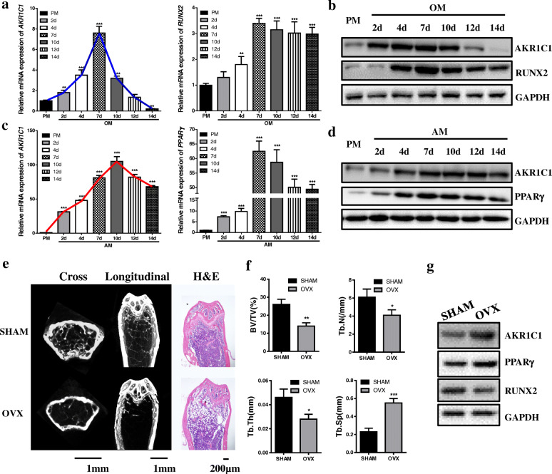
Background: As a promising way to repair bone defect, bone tissue engineering has attracted a lot of attentions from researchers in recent years. Searching for new molecular target to modify the seed cells and enhance their osteogenesis capacity is one of the hot topics in this field. As a member of aldo-keto reductase family, aldo-keto reductase family 1 member C1 (AKR1C1) is reported to associate with various tumors. However, whether AKR1C1 takes part in regulating differentiation of adipose-derived mesenchymal stromal/stem cells (ASCs) and its relationship with progesterone receptor (PGR) remain unclear.

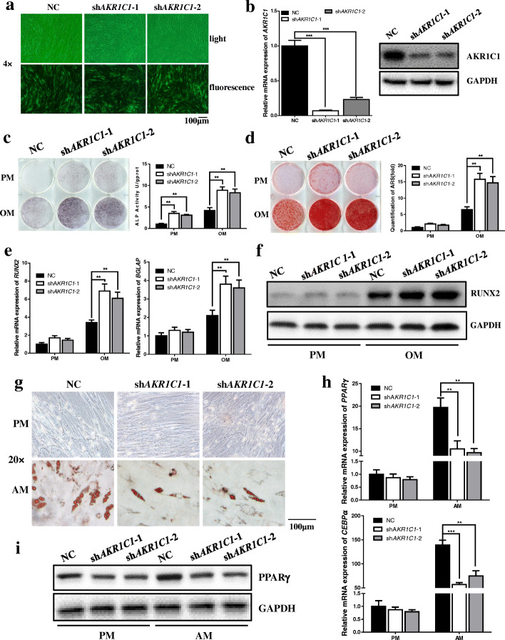
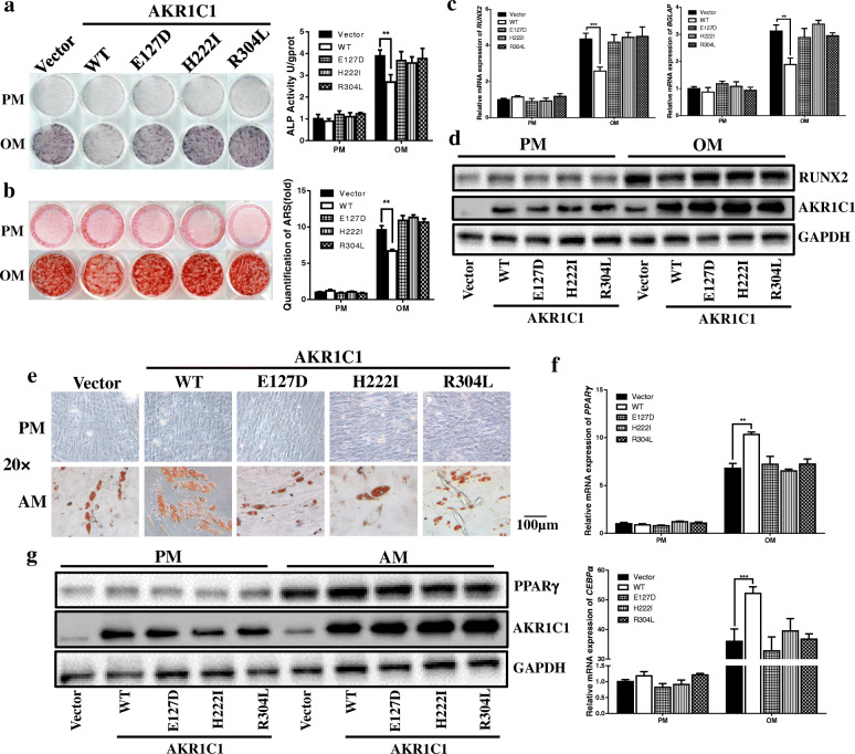
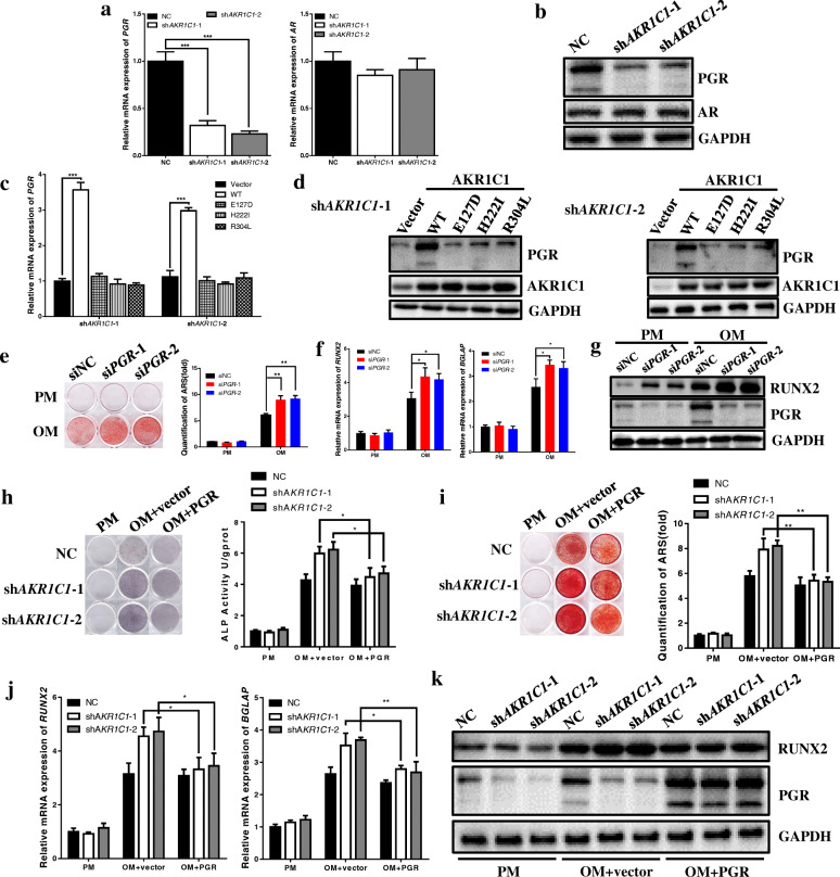
Methods: Lost-and-gain-of-function experiments were performed using knockdown and overexpression of AKR1C1 to identify its role in regulating osteogenic and adipogenic differentiation of hASCs in vitro. Heterotypic bone and adipose tissue formation assay in nude mice were used to conduct the in vivo experiment. Plasmid and siRNA of PGR, as well as western blot, were used to clarify the mechanism AKR1C1 regulating osteogenesis.

Results: Our results demonstrated that AKR1C1 acted as a negative regulator of osteogenesis and a positive regulator of adipogenesis of hASCs via its enzyme activity both in vitro and in vivo. Mechanistically, PGR mediated the regulation of AKR1C1 on osteogenesis.

Conclusions: Collectively, our study suggested that AKR1C1 could serve as a regulator of osteogenic differentiation via targeting PGR and be used as a new molecular target for ASCs modification in bone tissue engineering.

Keywords: AKR1C1; Adipose-derived mesenchymal stromal/stem cells; Osteogenesis; Progesterone receptor.


Conflict of interest statement

The authors declare that they have no competing interests.

Figures

Fig. 1
Fig. 1
AKR1C1 was a potential target for lineage commitment regulation of MSCs. a, b The expression of AKR1C1 on different days during the osteogenic induction process tested by qRT-PCR (a) and western blot (b). c, d The expression of AKR1C1 on different days during the adipogenic induction process tested by qRT-PCR (c) and western blot (b). e Micro-CT and H&E staining images of femurs in SHAM and OVX mice. f Bone morphology analysis of femurs in SHAM and OVX mice based on micro-CT. g Western blot of protein expression of AKR1C1, PPARγ, and RUNX2 in BMMSCs obtained from SHAM and OVX mice. *p < 0.05, **p < 0.01, ***p < 0.001
Fig. 2
Fig. 2
Knockdown of AKR1C1 enhanced the osteogenic capacity and impaired the adipogenic capacity of hASCs in vitro. a Transfection efficiency showed by fluorescence microscopy. b Knockdown efficiency of AKR1C1 determined by qRT-PCR and western blot. c Knockdown of AKR1C1 enhanced the ALP activity after 7 days of osteogenic induction as shown by ALP staining and quantification. d Knockdown of AKR1C1 accelerated mineralization after 21 days of osteogenic induction as shown by ARS staining and quantification. e Knockdown of AKR1C1 promoted the mRNA expression of RUNX2 on the 7th day of osteogenic induction and BGLAP on the 14th day of osteogenic induction as determined by qRT-PCR. f The protein expression of RUNX2 tested by western blot was consistent with the result of qRT-PCR. g Knockdown of AKR1C1 inhibited lipid droplet formation after 21 days of adipogenic induction as shown by oil red O staining. h Knockdown of AKR1C1 inhibited the mRNA expression of PPARγ and CEBPα on the 7th day of adipogenic induction as determined by qRT-PCR. i The protein expression of PPARγ tested by western blot was consistent with the result of qRT-PCR. **p < 0.01, ***p < 0.001
Fig. 3
Fig. 3
The regulation of AKR1C1 on hASCs differentiation in vitro depended on its enzyme activity. a ALP staining indicated that overexpression of AKR1C1 by wild type plasmid (WT) in the AKR1C1 knockdown cells (shAKR1C1-1) downregulated the ALP activity whereas mutant plasmids (E127D, H222I and R304L) had no significant influence. The result of ALP quantification was consistent with the result of ALP staining. b The results of ARS staining and quantification were consistent with the results of ALP staining and quantification. c Overexpression of AKR1C1 by wild type plasmid (WT) in the AKR1C1 knockdown cells (shAKR1C1-1) downregulated the mRNA expression of RUNX2 and BGLAP whereas mutant plasmids (E127D, H222I and R304L) made no significant difference. d Western blot showed that transfection of wild type plasmid and mutant plasmids upregulated the protein expression of AKR1C1. The protein expression of RUNX2 was inhibited by transfection of wildtype plasmid but not mutant plasmids. e Oil red O staining revealed that in the AKR1C1 knockdown cells (shAKR1C1-1), more lipid droplet formed in WT group, whereas mutant plasmids (E127D, H222I and R304L) made no significant difference. f Overexpression of AKR1C1 by wild type plasmid (WT) in the AKR1C1 knockdown cells (shAKR1C1-1) upregulated the mRNA expression of PPARγ and CEBPα whereas mutant plasmids (E127D, H222I, and R304L) had no significant influence. g Western blot showed that transfection of wild type plasmid and mutant plasmids upregulated the protein expression of AKR1C1. The protein expression of PPARγ was promoted by transfection of wild type plasmid but not mutant plasmids. **p < 0.01, ***p < 0.001
Fig. 4
Fig. 4
AKR1C1 regulated the osteogenic and adipogenic capacity of hASCs in vivo and the effect relied on the enzyme activity. a H&E staining of NC, shAKR1C1-1, and shAKR1C1-2 groups in heterotopic bone formation assay. b Masson staining of NC, shAKR1C1-1, and shAKR1C1-2 groups in heterotopic bone formation assay. c H&E staining of NC, shAKR1C1-1, and shAKR1C1-2 groups in heterotopic adipose tissue formation assay. Cells were separately loaded on Collagen Sponge scaffolds after 7 days of adipogenic induction and then implanted in the nude mice. d Oil red O staining of NC, shAKR1C1-1, and shAKR1C1-2 groups in heterotopic adipose tissue formation assay. e H&E staining of vector, wild type (WT) and mutant groups of AKR1C1 knockdown cells (shAKR1C1-1) in heterotopic bone formation assay. f Masson staining of vector, wild type (WT) and mutant groups of AKR1C1 knockdown cells (shAKR1C1-1) in heterotopic bone formation assay. g H&E staining of vector, wild type (WT) and mutant groups of AKR1C1 knockdown cells (shAKR1C1-1) in heterotopic adipose tissue formation assay. Cells were separately loaded on Collagen Sponge scaffolds after 7 days of adipogenic induction and then implanted in the nude mice. h Oil red O staining of vector, wild type (WT), and mutant groups of AKR1C1 knockdown cells (shAKR1C1-1) in heterotopic adipose tissue formation assay
Fig. 5
Fig. 5
Knockdown of AKR1C1 promoted osteogenesis of hASCs in vitro through targeting PGR. a, b AKR1C1 knockdown inhibited expression of PGR but not AR validated by qRT-PCR and western blot. c, d QRT-PCR and western blot indicated that overexpression of AKR1C1 by wild type plasmid (WT) in the AKR1C1 knockdown cells upregulated expression of PGR whereas mutant plasmids (E127D, H222I, and R304L) had no significant influence. e ARS staining and quantification showed PGR knockdown in ASCs accelerated mineral accumulation after 21 days of osteogenic induction. f, g QRT-PCR and western blot showed that PGR knockdown in ASCs promoted expression of RUNX2 and BGLAP after 14 days of osteogenic induction. h Expression of PGR in AKR1C1 knockdown cells slightly downregulated the ALP activity. i Expression of PGR in AKR1C1 knockdown cells slowed down mineral accumulation. j, k QRT-PCR and western blot showed that expression of PGR in AKR1C1 knockdown cells downregulated the expression of osteogenic markers (RUNX2, 7 day and BGLAP, 14 day). *p < 0.05, **p < 0.01, ***p < 0.001

References

    1. Wang W, Yeung KWK. Bone grafts and biomaterials substitutes for bone defect repair: A review. Bioact Mater. 2017;2(4):224–247. doi: 10.1016/j.bioactmat.2017.05.007. - DOI - PMC - PubMed
    1. Tae SK, Lee SH, Park JS, Im GI. Mesenchymal stem cells for tissue engineering and regenerative medicine. Biomed Mater. 2006;1(2):63–71. doi: 10.1088/1748-6041/1/2/003. - DOI - PubMed
    1. Marolt D, Knezevic M, Novakovic GV. Bone tissue engineering with human stem cells. Stem Cell Res Ther. 2010;1(2):10. doi: 10.1186/scrt10. - DOI - PMC - PubMed
    1. Torres-Torrillas M, Rubio M, Damia E, Cuervo B, Del Romero A, Pelaez P, et al. Adipose-derived mesenchymal stem cells: a promising tool in the treatment of musculoskeletal diseases. Int J Mol Sci. 2019;20(12):3105. - PMC - PubMed
    1. Pittenger MF, Mackay AM, Beck SC, Jaiswal RK, Douglas R, Mosca JD, Moorman MA, Simonetti DW, Craig S, Marshak DR. Multilineage potential of adult human mesenchymal stem cells. Science. 1999;284(5411):143–147. doi: 10.1126/science.284.5411.143. - DOI - PubMed

Publication types

Substances