

An Optimized Integrin α6-Targeted Magnetic Resonance Probe for Molecular Imaging of Hepatocellular Carcinoma in Mice
- PMID: 34235103
- PMCID: PMC8244641
- DOI: 10.2147/JHC.S312921
An Optimized Integrin α6-Targeted Magnetic Resonance Probe for Molecular Imaging of Hepatocellular Carcinoma in Mice
Abstract
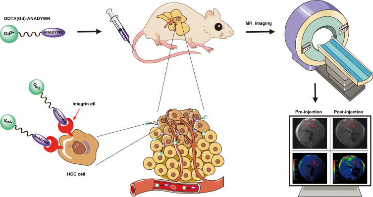
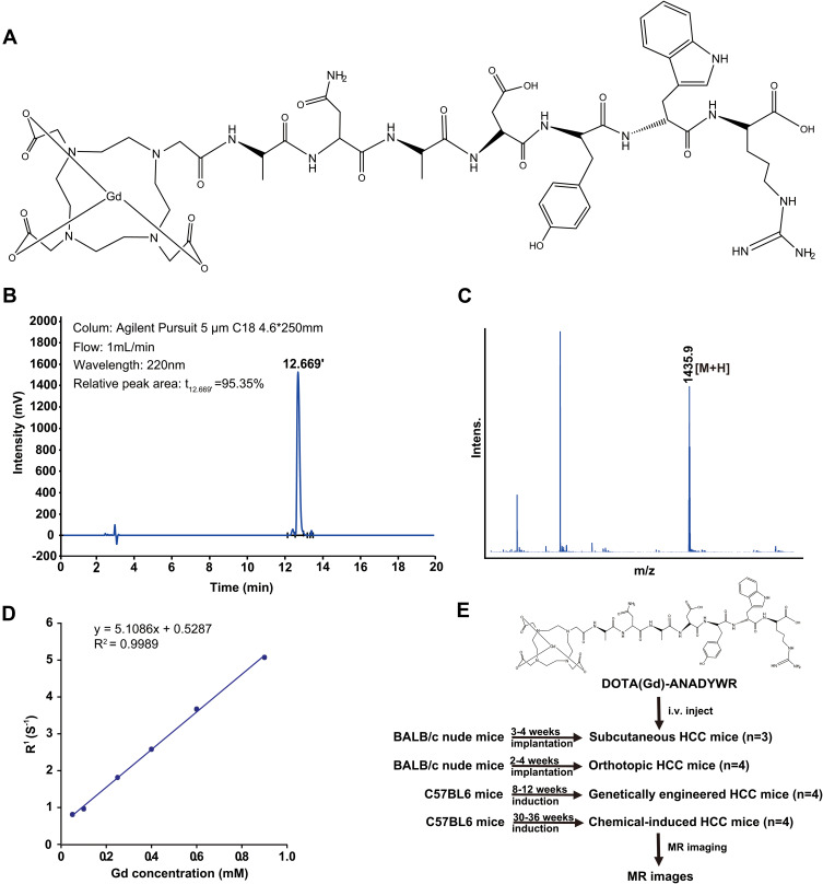
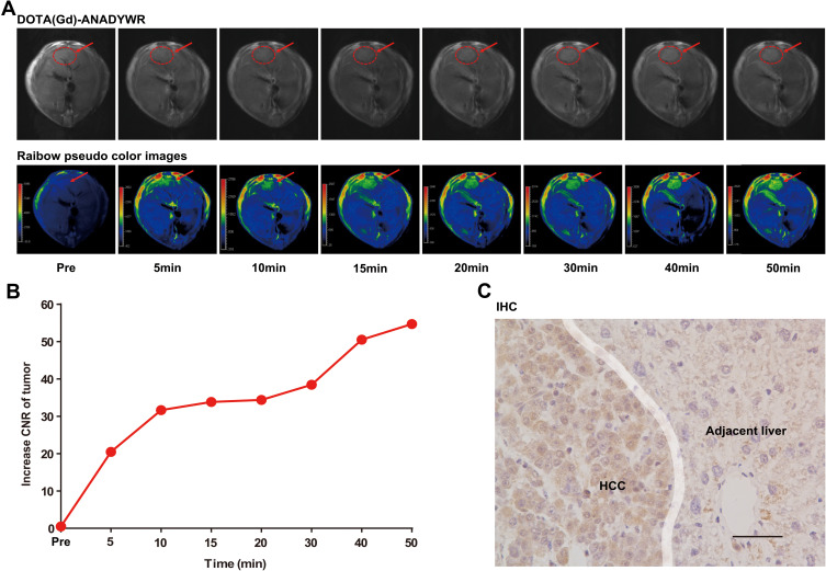
Introduction: Integrin α6 is an attractive diagnostic biomarker for molecular imaging of hepatocellular carcinoma (HCC) as it has an extremely high positive rate (approximately 94%) in clinical early-stage HCC. In this study, based on our previously identified integrin α6-targeted peptide, we developed an optimized integrin α6-targeted magnetic resonance (MR) probe dubbed DOTA(Gd)-ANADYWR for MR imaging of HCC in mice.
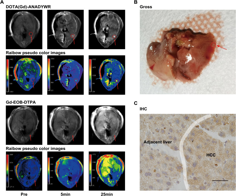
Materials and methods: The longitudinal (R1) relaxivity of DOTA(Gd)-ANADYWR was measured on a 3.0 T MR system . The specific tumor enhancement of the agent was investigated in four distinct mouse models, including subcutaneous, orthotopic, genetically engineered and chemically induced HCC mice.
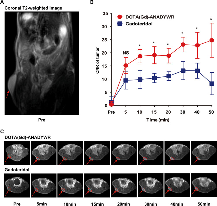
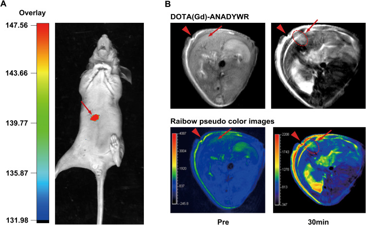
Results: The R1 relaxivity value of DOTA(Gd)-ANADYWR is 5.11 mM-1s-1 at 3.0 T, which is similar to that of the nonspecific clinical agent Gadoteridol. DOTA(Gd)-ANADYWR generated superior enhanced MR signal in HCC lesions and provided complementary enhancement MR signals to the clinically available hepatobiliary MR contrast agent gadoxetate disodium (Gd-EOB-DTPA). Importantly, DOTA(Gd)-ANADYWR could efficiently visualize small HCC lesion (approximately 1 mm) which was hardly detected by the clinical Gd-EOB-DTPA.
Conclusion: These findings suggest the potential application of this integrin α6-targeted MR probe for the detection of HCC, particularly for small HCC.
Keywords: hepatocellular carcinoma; integrin α6; magnetic resonance; peptide; probe.
© 2021 Lin et al.
Conflict of interest statement
The authors declare that they have no known competing financial interests or personal relationships that could have appeared to influence the work reported in this paper.
Figures
Similar articles
-
Integrin α6-Targeted Molecular Imaging of Central Nervous System Leukemia in Mice.Front Bioeng Biotechnol. 2022 Feb 23;10:812277. doi: 10.3389/fbioe.2022.812277. eCollection 2022. Front Bioeng Biotechnol. 2022. PMID: 35284414 Free PMC article.
-
Integrin α6-Targeted Magnetic Resonance Imaging of Hepatocellular Carcinoma in Mice.Mol Imaging Biol. 2020 Aug;22(4):864-872. doi: 10.1007/s11307-019-01437-z. Mol Imaging Biol. 2020. PMID: 31792839
-
Integrin α6 targeted positron emission tomography imaging of hepatocellular carcinoma in mouse models.J Control Release. 2019 Sep 28;310:11-21. doi: 10.1016/j.jconrel.2019.08.003. Epub 2019 Aug 7. J Control Release. 2019. PMID: 31400382
-
Application of Gd-EOB-DTPA-enhanced magnetic resonance imaging (MRI) in hepatocellular carcinoma.World J Surg Oncol. 2020 Aug 22;18(1):219. doi: 10.1186/s12957-020-01996-4. World J Surg Oncol. 2020. PMID: 32828123 Free PMC article. Review.
-
Diagnostic performance of contrast-enhanced multidetector computed tomography and gadoxetic acid disodium-enhanced magnetic resonance imaging in detecting hepatocellular carcinoma: direct comparison and a meta-analysis.Abdom Radiol (NY). 2016 Oct;41(10):1960-72. doi: 10.1007/s00261-016-0807-7. Abdom Radiol (NY). 2016. PMID: 27318936 Free PMC article.
Cited by
-
Identification of an IGF2BP2-Targeted Peptide for Near-Infrared Imaging of Esophageal Squamous Cell Carcinoma.Molecules. 2022 Nov 6;27(21):7609. doi: 10.3390/molecules27217609. Molecules. 2022. PMID: 36364436 Free PMC article.
-
Hovenia dulcis Suppresses the Growth of Huh7-Derived Liver Cancer Stem Cells by Inducing Necroptosis and Apoptosis and Blocking c-MET Signaling.Cells. 2023 Dec 21;13(1):22. doi: 10.3390/cells13010022. Cells. 2023. PMID: 38201226 Free PMC article.
-
Integrin α6-Targeted Molecular Imaging of Central Nervous System Leukemia in Mice.Front Bioeng Biotechnol. 2022 Feb 23;10:812277. doi: 10.3389/fbioe.2022.812277. eCollection 2022. Front Bioeng Biotechnol. 2022. PMID: 35284414 Free PMC article.
-
Promising therapeutic efficacy and safety of a novel integrin α6-targeting peptide-drug conjugate in lung adenocarcinoma.Mol Cancer. 2025 Jul 5;24(1):190. doi: 10.1186/s12943-025-02395-7. Mol Cancer. 2025. PMID: 40616126 Free PMC article.
-
Integrin α6 Targeted Near Infrared Fluorescent Imaging and Photoacoustic Imaging of Hepatocellular Carcinoma in Mice.J Clin Transl Hepatol. 2023 Feb 28;11(1):110-117. doi: 10.14218/JCTH.2021.00414. Epub 2022 Apr 12. J Clin Transl Hepatol. 2023. PMID: 36406330 Free PMC article.
References
LinkOut - more resources
Full Text Sources
