. 2021 Jun 13:2021:5545331.
doi: 10.1155/2021/5545331. eCollection 2021.

Antischistosomal Activity of Origanum majorana, Ziziphus spina-christi, and Salvia fruticosa Plant Extracts on Hamster Infected with Schistosoma haematobium

Affiliations

Antischistosomal Activity of Origanum majorana, Ziziphus spina-christi, and Salvia fruticosa Plant Extracts on Hamster Infected with Schistosoma haematobium

Yousef Abdal Jalil Fadladdin. Biomed Res Int. .

Abstract

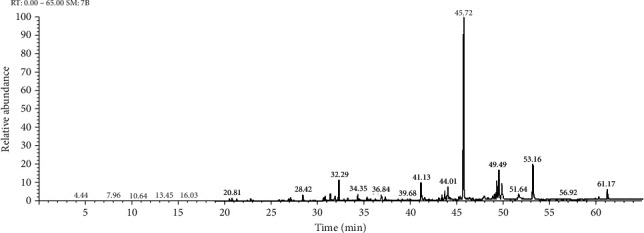
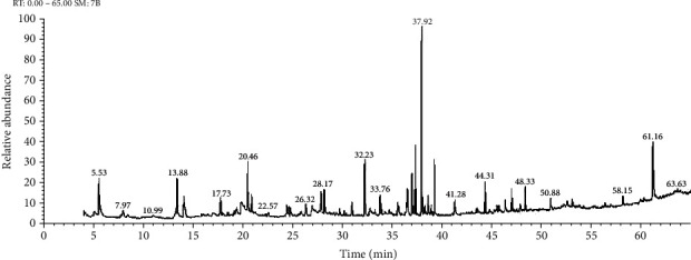
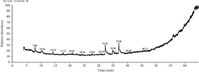
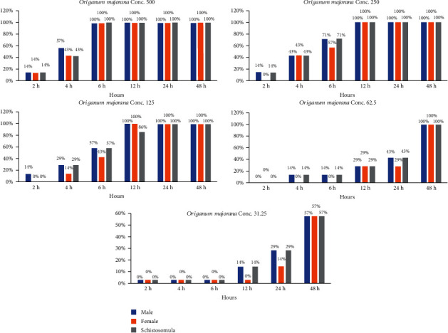
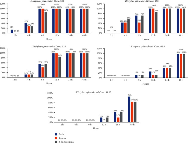
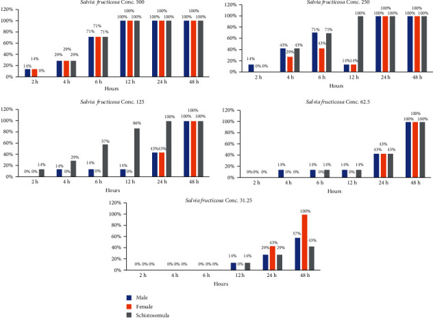
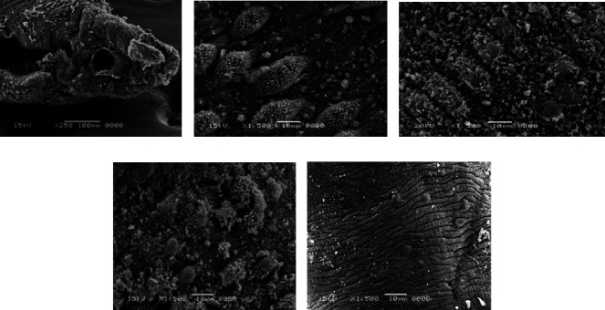
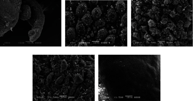
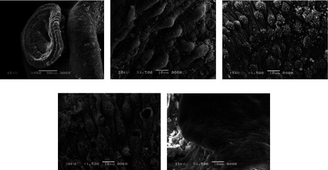
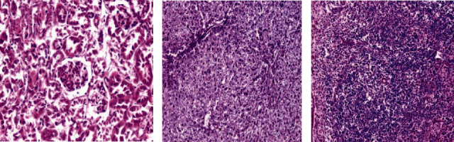
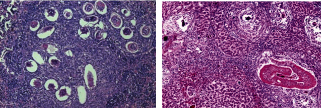
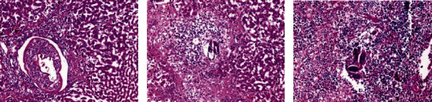
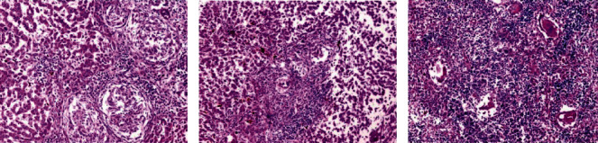
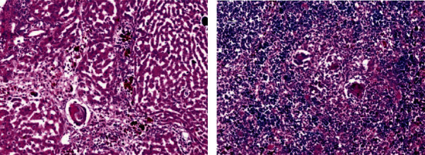
World Health Organization (WHO) has approved only one treatment for schistosomiasis, praziquantel (PZQ), but some poor efficacy was noticed in patients during the early stage of infection. Therefore, researchers have intensified their efforts to research new alternative medicines to treat schistosomiasis. In the present study, in vitro as well as in vivo studies have been accomplished to evaluate the effect of Origanum majorana, Ziziphus spina-christi, and Salvia fruticosa extracts in a different concentration 500, 250, 125, 62.5, and 31.25 μg/ml on golden hamster infected by Egyptian strains of schistosome (Schistosoma haematobium). In vitro, the adult worms and schistosomula of S. haematobium were investigated in RPMI-1640 medium for 48 hrs. The results showed that the concentration 500, 250, and 125 μg/ml of Origanum majorana, and Ziziphus spina-christi caused dead of 100% of Egyptian Schistosoma strains of adult worm and schistosomula of S. haematobium within 6 to 12 hrs of incubation. On the other hand, the extract of Salvia fruticosa at concentrations 500, 250, and 125 μg/ml showed death 100% parasites after 12 to 24 hrs of incubation. Inclusion, Origanum majorana, and Ziziphus spina-christi showed effectiveness against Egyptian Schistosoma strains (S. haematobium), a slight decrease in Salvia fruticosa was observed. Therefore, these medical plant extracts may be used as a safe and effective treatment for schistosomiasis.


Conflict of interest statement

The authors declare that they have no conflicts of interest.

Figures

Figure 1
Figure 1
GC-MS chromatogram of aqueous extract of Origanum majorana.
Figure 2
Figure 2
GC-MS chromatogram of aqueous plant extract of Ziziphus spina-christi.
Figure 3
Figure 3
GC-MS chromatogram of aqueous plant extract of Salvia fruticosa.
Figure 4
Figure 4
Statistical outcome of the effectiveness of Origanum majorana with different times and different concentrations on S. haematobium.
Figure 5
Figure 5
Statistical outcome of the effectiveness of Ziziphus spina-christi with different times and different concentrations on S. haematobium.
Figure 6
Figure 6
Statistical outcome of the effectiveness of Salvia fruticosa with different times and different concentrations on S. haematobium.
Figure 7
Figure 7
Normal tegument of S. haematobium from the hamster tissue.
Figure 8
Figure 8
Effect PZQ on adult worms of Schistosoma.
Figure 9
Figure 9
Scanning electron microscopy of S. haematobium (adult worms and schistosomula) after exposure to different concentrations of Origanum majorana. (a) Destroyed sucker. (b, c, d) Dorsal surface of male showing tegumental exfoliation with damage and exfoliation of spines and tubercles. (e) Schistosomula.
Figure 10
Figure 10
Scanning electron microscopy of S. haematobium (adult worms and schistosomula) after exposure to different concentrations of Ziziphus spina-christi. (a) Destroyed sucker. (b, c, d) Dorsal surface of male showing tegumental exfoliation with damage and exfoliation of spines and tubercles. (e) Schistosomula.
Figure 11
Figure 11
Scanning electron microscopy of S. haematobium (adult worms and schistosomula) after exposure to different concentrations of Salvia fruticosa. (a) Destroyed sucker. (b, c, d) Dorsal surface of male showing tegumental exfoliation with damage and exfoliation of spines and tubercles. (e) Schistosomula.
Figure 12
Figure 12
Histological section staining with hematoxylin and eosin for a healthy control (negative control). (a) Normal cortical structure in kidney control (×200). (b) Normal hepatic lobular architecture in liver control (×100). (c) Normal architecture in spleen control (×100).
Figure 13
Figure 13
Histological section staining with hematoxylin and eosin for infected untreated control. (a) Aggregate of deposited ova of Schistosoma in spleen, enclosed by lympho-epithelioid tissue reaction (×200). (b) A worm affect inside a portal vein and multiple egg granulomas in liver (×100).
Figure 14
Figure 14
Histological section staining with H&E of infected treated with 600 mg/kg Origanum majorana group. (a) Liver showing worm inside portal vein radical with moderate inflammatory changes within the hepatic lobule. (b) Liver showing intralobular ova enclosed by dense inflammatory cellular reaction and focal necrosis. (c) Some ova surrounded by inflammatory cellular reaction in the spleen (×200).
Figure 15
Figure 15
Histological section staining with H&E of infected treated with 600 mg/kg Ziziphus spina-christi group. (a) Many lympho-epithelioid granulomas in the liver. (b) Amalgamated ova granulomas in the liver. (c) Many fresh deposited and degenerated egg in the spleen (×200).
Figure 16
Figure 16
Histological section staining with H&E of infected treated with 800 mg/kg Salvia fruticosa group. (a) Degenerated ova with mild infiltration of the hepatic lobule by mononuclear inflammatory cells in the liver. (b) Some degenerated ova, enclosed by lympho-epithelioid cellular inflammatory cellular infiltration (×200).

References

    1. Steinmann P., Keiser J., Bos R., Tanner M., Utzinger J. Schistosomiasis and water resources development: systematic review, meta-analysis, and estimates of people at risk. The Lancet Infectious Diseases. 2006;6(7):411–425. doi: 10.1016/S1473-3099(06)70521-7. - DOI - PubMed
    1. Gryseels B., Polman K., Clerinx J., Kestens L. Human schistosomiasis. The Lancet. 2006;368(9541):1106–1118. doi: 10.1016/S0140-6736(06)69440-3. - DOI - PubMed
    1. World Health Organization (WHO) Schistosomiasis fact sheet. 2014. https://www.who.int/mediacentre/factsheets/fs115/en.
    1. World Health Organization (WHO) Schistosomiasis fact sheet. 2019. https://www.who.int/news-room/fact-sheets/detail/schistosomiasis.
    1. Ruffer M. Note on the presence of “Bilharzia haematobia” in Egyptian mummies of the twentieth dynasty [1250-1000 B.C.] British Medical Journal. 1910;1(2557):p. 16. doi: 10.1136/bmj.1.2557.16-a. - DOI - PMC - PubMed

LinkOut - more resources