. 2021 Jun 23;7(6):1009-1018.
doi: 10.1021/acscentsci.1c00010. Epub 2021 Jun 2.

Heparan Sulfate Proteoglycans as Attachment Factor for SARS-CoV-2

Affiliations

Heparan Sulfate Proteoglycans as Attachment Factor for SARS-CoV-2

Lin Liu et al. ACS Cent Sci. .

Abstract

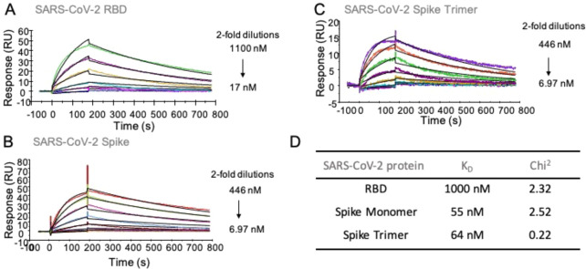
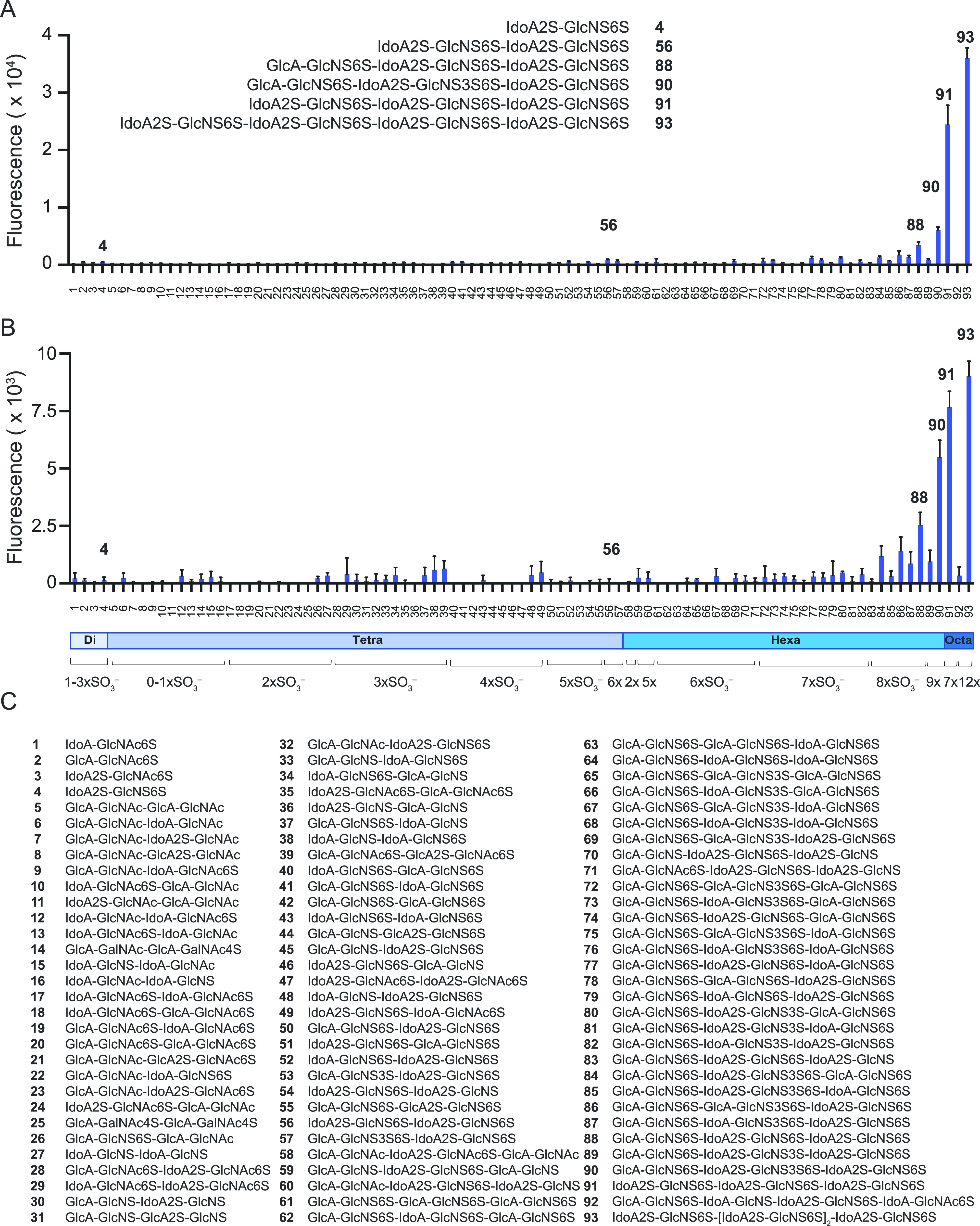
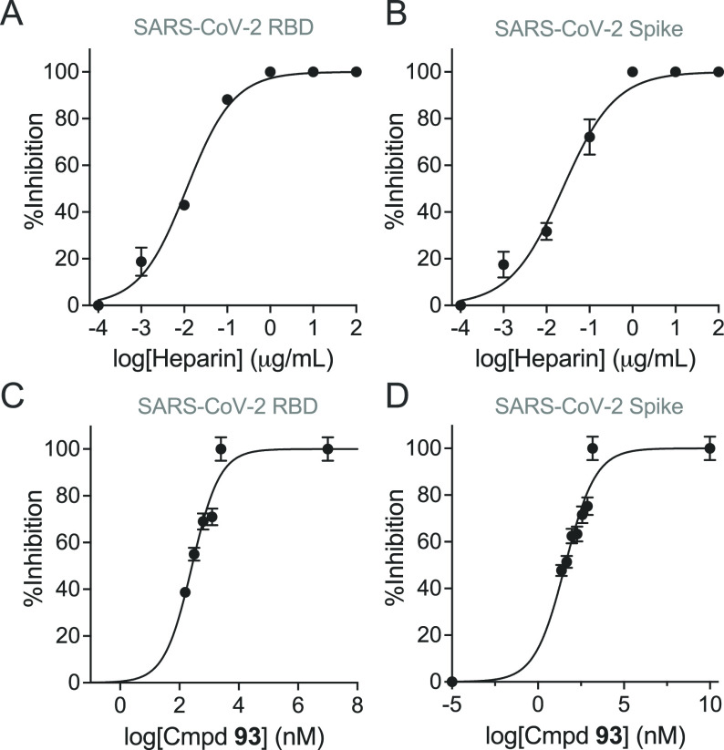
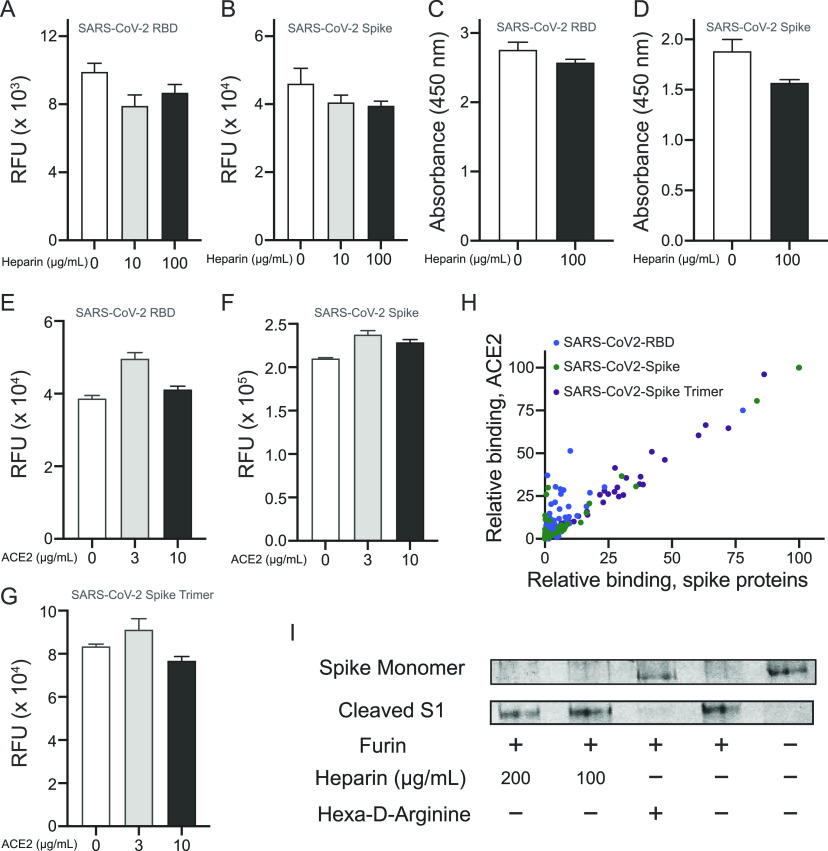
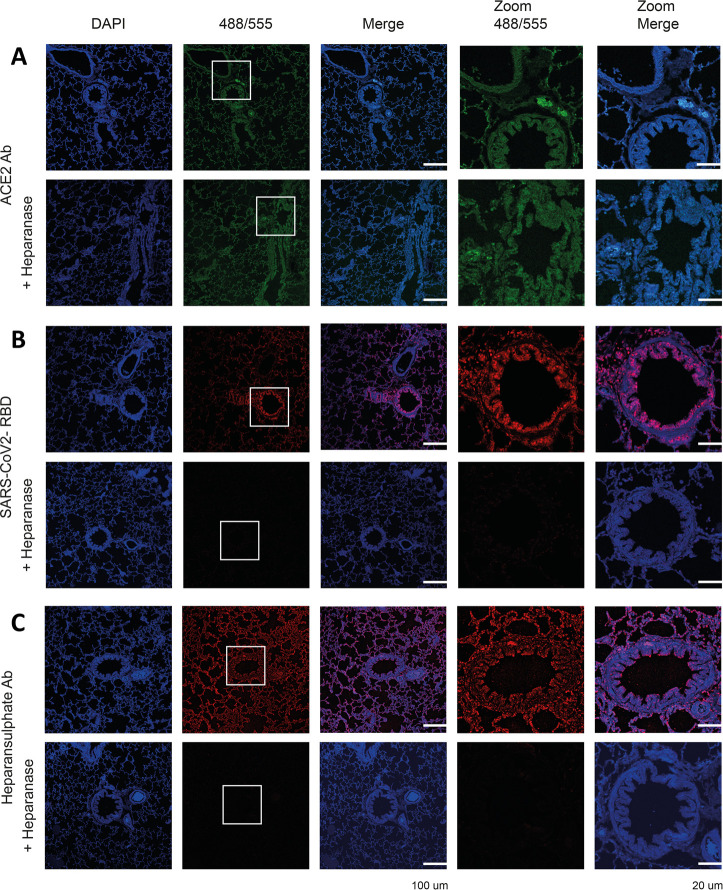
Severe acute respiratory syndrome-related coronavirus 2 (SARS-CoV-2) is causing an unprecedented global pandemic demanding the urgent development of therapeutic strategies. Microarray binding experiments, using an extensive heparan sulfate (HS) oligosaccharide library, showed that the receptor binding domain (RBD) of the spike of SARS-CoV-2 can bind HS in a length- and sequence-dependent manner. A hexasaccharide composed of IdoA2S-GlcNS6S repeating units was identified as the minimal binding epitope. Surface plasmon resonance showed the SARS-CoV-2 spike protein binds with a much higher affinity to heparin (K D = 55 nM) compared to the RBD (K D = 1 μM) alone. It was also found that heparin does not interfere in angiotensin-converting enzyme 2 (ACE2) binding or proteolytic processing of the spike. However, exogenous administered heparin or a highly sulfated HS oligosaccharide inhibited RBD binding to cells. Furthermore, an enzymatic removal of HS proteoglycan from physiological relevant tissue resulted in a loss of RBD binding. The data support a model in which HS functions as the point of initial attachment allowing the virus to travel through the glycocalyx by low-affinity high-avidity interactions to reach the cell membrane, where it can engage with ACE2 for cell entry. Microarray binding experiments showed that ACE2 and HS can simultaneously engage with the RBD, and it is likely no dissociation between HS and RBD is required for binding to ACE2. The results highlight the potential of using HS oligosaccharides as a starting material for therapeutic agent development.


Conflict of interest statement

The authors declare no competing financial interest.

Figures

Figure 1
Figure 1
SPR sensorgrams representing the concentration-dependent kinetic analysis of the binding of immobilized heparin with SARS-CoV-2 related proteins. (A) RBD, (B) spike monomer, (C) spike trimer, (D) summary table of equilibrium dissociation constant (KD) and chi-square (Chi2) goodness-of-fit values. Data were analyzed using Biacore T100 evaluation software, and representative data are shown, which was repeated at least three times.
Figure 2
Figure 2
Binding analysis of synthetic HS oligosaccharides to SARS-CoV-2 related proteins by microarray. (A) Spike protein (10 μg/mL); structures of strongest binders are shown as insets. (B) RBD protein (30 μg/mL). (C) Compound numbering and structures. All compounds have a linker at reducing end, R = O(CH2)5NH2. Data are presented as mean ± SD (n = 4). Representative data are shown, which was repeated at least three times.
Figure 3
Figure 3
SPR-based competition assays on heparin-immobilized surface. (A) RBD protein in the presence of UFH. (B) Spike protein in the presence of UFH. (C) RBD protein in the presence of HS-octasaccharide (93). (D) Spike protein in the presence of 93. Concentrations of RBD and spike proteins were 5 μM and 150 nM, respectively. The IC50 values were calculated using dose–response equations [nonlinear regression, log(inhibitor) vs response-variable slope (four parameters)] built in Prism software 9 (GraphPad Software, Inc.). Experiments were performed (in duplicate) three times at the minimum.
Figure 4
Figure 4
Interplay of interactions of HS and ACE2 with spike/RBD. (A) Influence of heparin on the binding of His-tagged RBD or (B) His-tagged spike monomer to biotinylated human ACE2 immobilized on streptavidin-coated microarray slides. Detection of RBD and spike was accomplished using an anti-His antibody labeled with AlexaFluor 647. (C) Influence of heparin on the binding of biotinylated human ACE2 to RBD and (D) to immobilized spike monomer immobilized to high surface microtiter plates. Binding was detected by treatment with streptavidin-horseradish peroxidase (HRP) followed by an addition of a colorimetric HRP substrate. (E). Effect of ACE2 on binding of RBD, (F) spike monomer, and (G) spike trimer to the HS microarray. Binding intensity corresponding to compound 93 was used. (H) Binding of ACE2-spike protein complex to HS oligosaccharides on the microarray, by detecting spike proteins (x-axis) and ACE2 (y-axis). Each spot represents an individual compound on the HS array. (I) SDS-PAGE analysis of furin-mediated cleavage of spike monomer in the presence and absence of heparin or a known furin inhibitor (hexa-d-arginine).
Figure 5
Figure 5
Binding of SARS-CoV-2 RBD pretreated with GAGs to Vero-E6 cells. (A) (top to bottom) Untreated RBD, HA (250 μg/mL), UFH (10 μg/mL), NACH (10 μg/mL), and octasaccharide 93 (100 μg/mL), complete dilution series are shown in Figure S7 and Figure S8. (B) ACE2 antibody pretreated with GAGs.
Figure 6
Figure 6
Binding of ACE2 antibody, SARS-CoV-2 RBD, and heparan sulfate antibody to ferret lung serial tissue slides. (A) ACE2 antibody staining without and after HPSE treatment. (B) SARS-CoV-2 RBD staining without and after HPSE treatment. (C) Heparan sulfate antibody (10E4) staining without and after HPSE treatment. HPSE treatment was achieved by an overnight incubation of the tissues with HPSE (0.2 μg/mL) at 37 °C.

Update of

References

    1. Dimitrov D. S. Virus entry: molecular mechanisms and biomedical applications. Nat. Rev. Microbiol. 2004, 2 (2), 109–122. 10.1038/nrmicro817. - DOI - PMC - PubMed
    1. Walls A. C.; Park Y. J.; Tortorici M. A.; Wall A.; McGuire A. T.; Veesler D. Structure, function, and antigenicity of the SARS-CoV-2 spike glycoprotein. Cell 2020, 181 (2), 281–292. 10.1016/j.cell.2020.02.058. - DOI - PMC - PubMed
    1. Li F.; Li W.; Farzan M.; Harrison S. C. Structure of SARS coronavirus spike receptor-binding domain complexed with receptor. Science 2005, 309 (5742), 1864–1868. 10.1126/science.1116480. - DOI - PubMed
    1. Monteil V.; Kwon H.; Prado P.; Hagelkruys A.; Wimmer R. A.; Stahl M.; Leopoldi A.; Garreta E.; Hurtado Del Pozo C.; Prosper F.; Romero J. P.; Wirnsberger G.; Zhang H.; Slutsky A. S.; Conder R.; Montserrat N.; Mirazimi A.; Penninger J. M. Inhibition of SARS-CoV-2 infections in engineered human tissues using clinical-grade soluble human ACE2. Cell 2020, 181 (4), 905–913. 10.1016/j.cell.2020.04.004. - DOI - PMC - PubMed
    1. Li W.; Hulswit R. J. G.; Widjaja I.; Raj V. S.; McBride R.; Peng W.; Widagdo W.; Tortorici M. A.; van Dieren B.; Lang Y.; van Lent J. W. M.; Paulson J. C.; de Haan C. A. M.; de Groot R. J.; van Kuppeveld F. J. M.; Haagmans B. L.; Bosch B. J. Identification of sialic acid-binding function for the Middle East respiratory syndrome coronavirus spike glycoprotein. Proc. Natl. Acad. Sci. U. S. A. 2017, 114 (40), E8508–E8517. 10.1073/pnas.1712592114. - DOI - PMC - PubMed