. 2020 Apr 5:2:35-44.
doi: 10.1016/j.crstbi.2020.03.001. eCollection 2020.

Isoelectric point-amyloid formation of α-synuclein extends the generality of the solubility and supersaturation-limited mechanism

Affiliations

Isoelectric point-amyloid formation of α-synuclein extends the generality of the solubility and supersaturation-limited mechanism

Koki Furukawa et al. Curr Res Struct Biol. .

Abstract

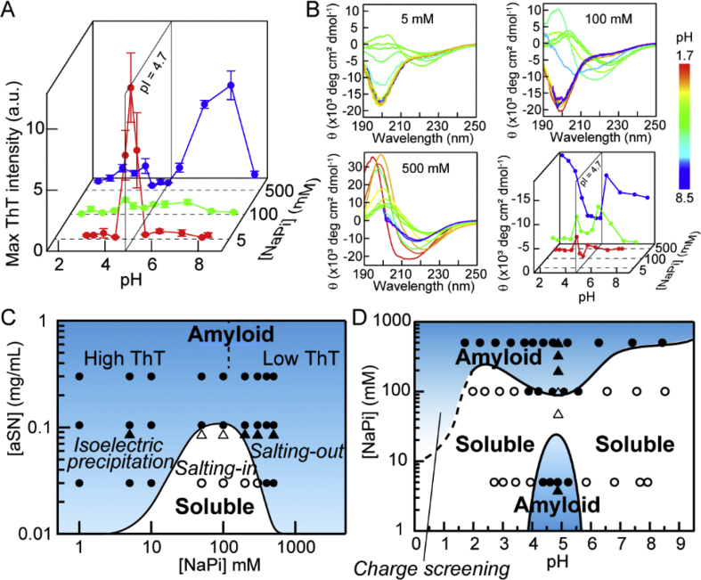
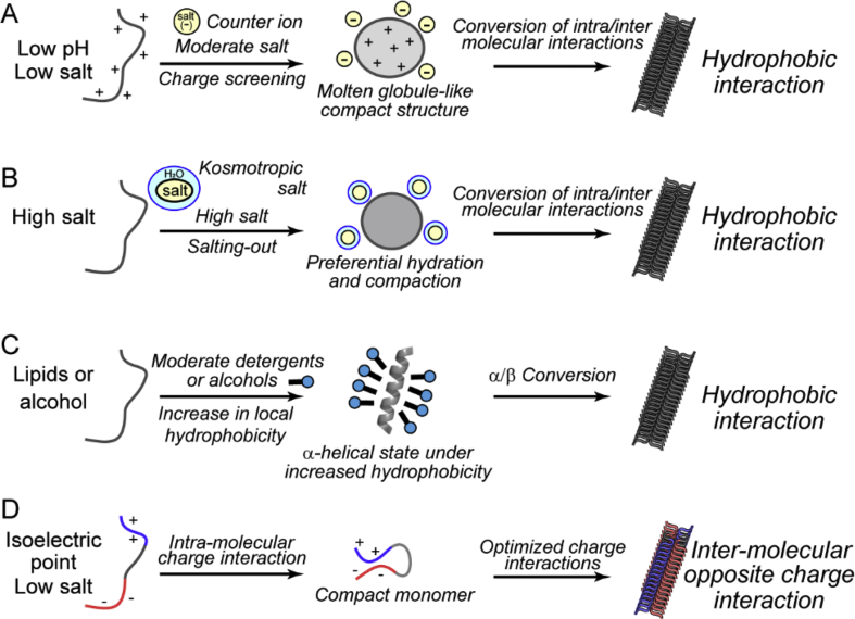
Proteins in either a native or denatured conformation often aggregate at an isoelectric point (pI), a phenomenon known as pI precipitation. However, only a few studies have addressed the role of pI precipitation in amyloid formation, the crystal-like aggregation of denatured proteins. We found that α-synuclein, an intrinsically disordered protein of 140 amino acid residues associated with Parkinson's disease, formed amyloid fibrils at pI (= 4.7) under the low-sodium phosphate conditions. Although α-synuclein also formed amyloid fibrils at a wide pH range under high concentrations of sodium phosphate, the pI-amyloid formation was characterized by marked amyloid-specific thioflavin T fluorescence and clear fibrillar morphology, indicating highly ordered structures. Analysis by heteronuclear NMR in combination with principal component analysis suggested that amyloid formation under low and high phosphate conditions occurred by distinct mechanisms. The former was likely to be caused by the intermolecular attractive charge-charge interactions, where α-synuclein has +17 and -17 charges even with the zero net charge. On the other hand, the latter was caused by the phosphate-dependent salting-out effects. pI-amyloid formation may play a role in the membrane-dependent amyloid formation of α-synuclein, where the negatively charged membrane surface reduces the local pH to pI and the membrane hydrophobic environment enhances electrostatic interactions. The results extend the supersaturation-limited mechanism of amyloid formation: Amyloid fibrils are formed under a variety of conditions of decreased solubility of denatured proteins triggered by the breakdown of supersaturation.

Keywords: Amyloid fibrils; Isoelectric point precipitation; Nuclear magnetic resonance (NMR); Principal component analysis; Salting-out effects; α-synuclein.


Conflict of interest statement

The authors declare that they have no conflicts of interest with the contents of this article.

Figures

Image 1
Graphical abstract
Fig. 1
Fig. 1
Amyloid formation of αSN at pI. (A) Amino acid sequence of αSN with N-terminus (1–60 residues), NAC (61–95 residues), and C-terminus (96–140 residues) domains indicated. Acidic and basic residues are highlighted in red and blue, respectively. (B–D) Kinetics of αSN amyloid formation in the presence of different concentrations of NaPi at 0.3 (B), 0.1 (C), or 0.03 mg/mL of (D) αSN. (E–J) Dependence of the maximum ThT value (E–G) and lag time (H–J) on NaPi concentration. Insets show log plots of the same data to highlight data at low ThT values and NaPi concentrations. Data is shown as the average ± s.d. (n = 5). (K–M) CD spectra after amyloid formation. (N–Q) TEM images of fibrils of 0.1 mg/mL of αSN. Scale bars indicate 200 nm.
Fig. 2
Fig. 2
pH-dependent amyloid formation of αSN at different NaPi concentrations and conformational phase diagrams. (A) Maximum ThT values at different pH and NaPi concentrations. Data is shown as the average ± s.d. (n = 5). Kinetic data are shown in Fig. S3. (B) CD spectra and the intensities at 220 nm. Different colors in CD spectra indicate different pH as defined by a color scale bar. (C) Conformational phase diagram at pI (=4.7) dependent on the concentrations of NaPi and αSN. (D) Conformational phase diagram at 0.1 mg/mL αSN dependent on pH and NaPi concentration. Data points obtained from CD (circle) and soluble and insoluble fraction measurements (triangle) were also plotted.
Fig. 3
Fig. 3
Dependence of NMR spectra at different concentrations of NaPi at pI and principal component analysis of the NMR data against NaPi concentration. (A–C) 1H–15N HSQC spectra at different NaPi concentrations were overlaid in different colors. Arrows indicate the direction of peak movement. (D) The change in principal components 1 and 2 depending on the NaPi concentration. The numbers indicate 3 principal conformational states of αSN. (E–J) Profiles of the peak intensity and chemical shift changes in three concentration regions. (E, G, I) The peak intensity changes between 5 and 50 mM (E), 50 and 200 mM (G), and 200 and 500 mM NaPi (I). (F, H, J) The chemical shift changes between 5 and 50 mM (F), 50 and 200 mM (H), and 200 and 500 mM (J). Domains of αSN are shown on top.
Fig. 4
Fig. 4
Distinct mechanisms of amyloid formation based on solubility and supersaturation. (A) Counter ion-binding mechanism observed under acidic conditions in the presence of moderate concentrations of salts. (B) Salting-out mechanism observed under high salt conditions independent of pH. (C) Hydrophobic additive-binding mechanism observed in the presence of moderate concentrations of alcohols or detergents like SDS. (D) pI-precipitation mechanism. The hydrophobic additive-binding mechanism is similar to the salting-out-dependent mechanism in that additives without charges strengthen the hydrophobic interactions, leading to the decrease in solubility and thus to amyloid formation.

References

    1. Adachi M., Noji M., So M., Sasahara K., Kardos J., Naiki H., Goto Y. Aggregation-phase diagrams of β2-microglobulin reveal temperature and salt effects on competitive formation of amyloids versus amorphous aggregates. J. Biol. Chem. 2018;293:14775–14785. - PMC - PubMed
    1. Araki K., Yagi N., Aoyama K., Choong C.-J., Hayakawa H., Fujimura H., Nagai Y., Goto Y., Mochizuki H. Parkinson's disease is a type of amyloidosis featuring accumulation of amyloid fibrils of α-synuclein. Proc. Natl. Acad. Sci. U.S.A. 2019:201906124. doi: 10.1073/pnas.1906124116. - DOI - PMC - PubMed
    1. Bjellqvist B., Basse B., Olsen E., Celis J.E. Reference points for comparisons of two-dimensional maps of proteins from different human cell types defined in a pH scale where isoelectric points correlate with polypeptide compositions. Electrophoresis. 1994;15:529–539. - PubMed
    1. Bousset L., Pieri L., Ruiz-Arlandis G., Gath J., Jensen P.H., Habenstein B., Madiona K., Olieric V., Böckmann A., Meier B.H. Structural and functional characterization of two alpha-synuclein strains. Nat. Commun. 2013;4:2575. - PMC - PubMed
    1. Brender J.R., Krishnamoorthy J., Sciacca M.F.M., Vivekanandan S., D'Urso L., Chen J., La Rosa C., Ramamoorthy A. Probing the sources of the apparent irreproducibility of amyloid formation: drastic changes in kinetics and a switch in mechanism due to micellelike oligomer formation at critical concentrations of IAPP. J. Phys. Chem. B. 2015;119:2886–2896. - PMC - PubMed