. 2021 Dec;13(1_suppl):318S-325S.
doi: 10.1177/19476035211029704. Epub 2021 Jul 8.

Acute Cartilage Injury Induced by Trans-Articular Sutures

Affiliations

Acute Cartilage Injury Induced by Trans-Articular Sutures

Matic Ciglič et al. Cartilage. 2021 Dec.

Abstract

Objective: To determine the extent of acute cartilage injury by using trans-articular sutures.

Methods: Five different absorbable sutures, monofilament polydioxanone (PDS) and braided polyglactin (Vicryl), were compared on viable human osteochondral explants. An atraumatic needle with 30 cm of thread was advanced through the cartilage with the final thread left in the tissue. A representative 300 μm transversal slice from the cartilage midportion was stained with Live/Dead probes, scanned under the confocal laser microscope, and analyzed for the diameters of (a) central "Black zone" without any cells, representing in situ thread thickness and (b) "Green zone," including the closest Live cells, representing the maximum injury to the tissue. The exact diameters of suture needles and threads were separately measured under an optical microscope.

Results: The diameters of the Black (from 144 to 219 µm) and the Green zones (from 282 to 487 µm) varied between the different sutures (P < 0.001). The Green/Black zone ratio remained relatively constant (from 1.9 to 2.2; P = 0.767). A positive correlation between thread diameters and PDS suturing material, toward the Black and Green zone, was established, but needle diameters did not reveal any influence on the zones.

Conclusions: The width of acute cartilage injury induced by the trans-articular sutures is about twice the thread thickness inside of the tissue. Less compressible monofilament PDS induced wider tissue injury in comparison to a softer braided Vicryl. Needle diameter did not correlate to the extent of acute cartilage injury.

Keywords: articular cartilage; live/dead staining; polydioxanone; polyglactin; suture.


Conflict of interest statement

Declaration of Conflicting Interests: The author(s) declared no potential conflicts of interest with respect to the research, authorship, and/or publication of this article.

Figures

Figure 1.
Figure 1.
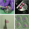
Demonstration of the trans-articular suture passage protocol: in-side out free-hand suture passing (A), transversal cartilage slicing with microtome (B), a representative 300 µm cartilage tissue slice with the thread in situ (C), Live/Dead molecular probes staining (D).
Figure 2.
Figure 2.
Image analysis of a transversal articular cartilage slice with an absorbable suture (PDS 4-0) after it had been stained with Live/Dead molecular probes and scanned under a confocal laser microscope. (A) The retained suture was identified in the image and a central longitudinal line was drawn to mark the suture passage canal (red line). A perpendicular line was drawn from the center to the maximal noncellular area on each side of the red line, as highlighted by white line. This represents the diameter of the missing articular tissue occupied by the suture material—the Black zone. (B) The region-of-interest for viable cells measurements was limited to one suture diameter around the suture mid-portion—the area between the 2 red perpendicular lines. (C) The Green zone diameter (the closest aggregated viable cells within the region-of-interest) was measured on each side of the central longitudinal line, as depicted by the 2 while lines. It represents the diameter of maximal chondrocyte injury by the suture passage and its in-tissue compressive forces.

References

    1. Årøen A, Løken S, Heir S, Alvik E, Ekeland A, Granlund OG, et al.. Articular cartilage lesions in 993 consecutive knee arthroscopies. Am J Sports Med. 2004;32(1_suppl):211-5. doi:10.1177/0363546503259345 - DOI - PubMed
    1. Curl WW, Krome J, Gordon ES, Rushing J, Smith BP, Poehling GG. Cartilage injuries: a review of 31,516 knee arthroscopies. Arthroscopy. 1997;13(4):456-60. doi:10.1016/S0749-8063(97)90124-9 - DOI - PubMed
    1. Bedi A, Feeley BT, Williams RJ, 3rd. Management of articular cartilage defects of the knee. J Bone Joint Surg Am. 2010;92(4):994-1009. doi:10.2106/JBJS.I.00895 - DOI - PubMed
    1. Brittberg M, Lindahl A, Nilsson A, Ohlsson C, Isaksson O, Peterson L. Treatment of deep cartilage defects in the knee with autologous chondrocyte transplantation. N Engl J Med. 1994;331(14):889-95. doi:10.1056/NEJM199410063311401 - DOI - PubMed
    1. Drobnic M, Kregar-Velikonja N, Radosavljevic D, Gorensek M, Koritnik B, Malicev E, et al.. The outcome of autologous chondrocyte transplantation treatment of cartilage lesions in the knee. Cell Mol Biol Lett. 2002;7(2):361-3. - PubMed

Publication types