. 2022 Jan;37(1):152-160.
doi: 10.1007/s00380-021-01900-4. Epub 2021 Jul 8.

SAHA could inhibit TGF-β1/p38 pathway in MI-induced cardiac fibrosis through DUSP4 overexpression

Affiliations

SAHA could inhibit TGF-β1/p38 pathway in MI-induced cardiac fibrosis through DUSP4 overexpression

Kaihao Wang et al. Heart Vessels. 2022 Jan.

Abstract

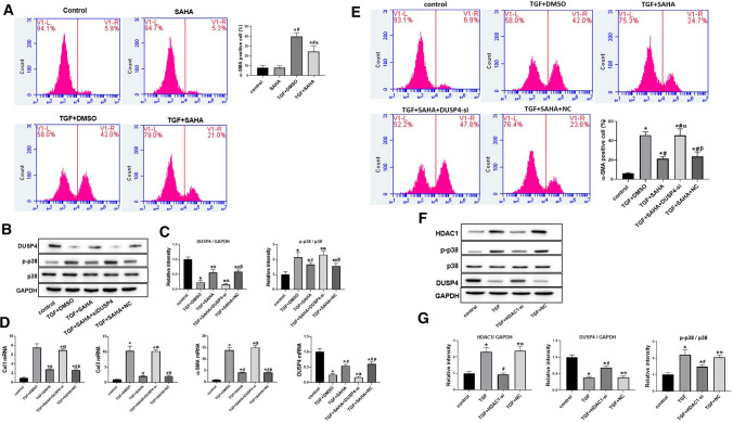
Growing evidences have revealed that a histone deacetylase inhibitor (HDACi), suberoylanilide hydroxamic acid (SAHA) has anti-fibrotic effect in different diseases. In this study, we first evaluated whether SAHA could suppress cardiac fibrosis. Mice with MI-induced cardiac fibrosis were treated with SAHA by intraperitoneal injection and their cardiac function was improved after SAHA treatment. Results of western blotting and qRT-PCR in heart tissues suggested that TGFβ1/P38 pathway was activated in MI mice, and this effect was reversed by SAHA. Cell proliferation assay suggested that SAHA could suppress TGF-β1-induced cardiac fibroblasts proliferation. Furthermore, results of western blotting and qRT-PCR in cardiac fibroblasts depicted that SAHA may exert its anti-fibrotic effect through inhibiting TGF-β1-induced P38 phosphorylation by promoting DUSP4 expression. Our findings may substantiate SAHA as a promising treatment for MI-induced cardiac fibrosis.

Keywords: Cardiac fibrosis; Dual-specificity phosphatase 4 (DUSP4); Suberoylanilide hydroxamic acid (SAHA); TGF-β1.


Conflict of interest statement

The authors declare that they have no conflict of interest.

Figures

Fig. 1
Fig. 1
Treatment of SAHA improved cardiac function in mice after MI a Echocardiographic analysis of sham mice, MI + DMSO mice and MI + SAHA mice. LVEF (left ventricular eject fraction), LVFS(left ventricular fractional shortening), AWTd (left ventricular anterior wall thickness in diastole), AWTs(left ventricular anterior wall thickness in systole), LVDd(left ventricular diameter in diastole) and LVDs(left ventricular diameter in systole) were calculated according to the guidelines. N = 12 for all groups. One-way ANOVA with Tukey’s post-hoc multiple-group comparisons was used in statistical analysis *p < 0.05 vs. Sham, #p < 0.05 vs. MI + DMSO. b Myocardial infarction size assessed by Masson staining and Sirius Red. c IL(infarction length)/TL(total cross-sectional left heart length) and NICR(collage ratio in non-infarct area) were analyzed. Unpaired two tailed student’s t test comparison was used for statistical analysis. Bar graphs show group means ± SD. N = 6 for each group. One-way ANOVA with Tukey’s post-hoc multiple-group comparisons was used in statistical analysis *p < 0.05 vs. Sham, #p < 0.05 vs. MI + DMSO.
Fig.2
Fig.2
Treatment of SAHA reversed effect of P38 activation and ECM generation induced by MI in mice a Protein level of Ac-H3, DUSP4, TGF-β1 and p-p38 analyzed by Western blotting in each group of heart tissue. bThe intensity of the blots for DUSP4, Ac-H3/H3, TGF-β1 and p-p38/p38, standardized by GAPDH. c Expression of Col1a, Col3a, α-SMA and DUSP4mRNA in heart tissue analyzed by qRT-PCR. N = 6 for each group. One-way ANOVA with Tukey’s post-hoc multiple-group comparisons was used for statistical analysis. *p < 0.05 vs. Sham, #p < 0.05 vs. MI + DMSO. Bar graphs show group mean ± SD.
Fig.3
Fig.3
SAHA treatment could inhibit TGF-β1-induced cell proliferation expression, P38 phosphorylation and ECM generation in cardiac fibroblasts a Protein level of Acetyl-Histone H3, DUSP4, TGF-β1 and p-p38 analyzed by Western blotting in fibroblasts. b Intensity of the blots for Ac-H3/H3, DUSP4, and p-p38/p38standardized by GAPDH; c Expression of Col1a, Col3a, α-SMA and DUSP4 mRNA in fibroblasts analyzed by qRT-PCR; N = 4 for each group. One-way ANOVA with Tukey’s post-hoc multiple-group comparisons was used for statistical analysis. *p < 0.05 vs. control, #p < 0.05 vs. only SAHA, αp < 0.05 vs. TGF + DMSO group. d Cell viability changes induced by SAHA for cardiac fibroblasts analyzed by CCK-8 assay. e HDAC1 activity changes induced by SAHA for cardiac fibroblasts analyzed by Results was expressed as percentage of activity compared to control group (no TGF- β1 and no SAHA). N = 4 for each group. One-way ANOVA with Tukey’s post-hoc multiple-group comparisons was used for statistical analysis. *p < 0.05 vs. Sham, #p < 0.05 vs. only SAHA group.
Fig. 4
Fig. 4
SAHA inhibited TGF-β1/p38 pathway and α-SMA expression by increasing DUSP4 expression in fibroblasts a α-SMA expression and calculated α-SMA-positive rate of myocardial fibroblasts analyzed by flow cytometry in each group. Bar graphs show group means ± SD. N = 4 for each group. b Protein level of DUSP4 and p-p38 analyzed by Western blotting in each group in fibroblasts. c Intensity of the blots for DUSP4 and p-p38/p38standardized by GAPDH; d Expression of Col1a, Col3a, α-SMA and DUSP4 mRNA in fibroblasts analyzed by qRT-PCR. One-way ANOVA with Tukey’s post-hoc multiple-group comparisons was used for statistical analysis. *p < 0.05 vs. control, #p < 0.05 vs. only SAHA, αp < 0.05 vs. TGF + DMSO group. βp < 0.05 vs. TGF + SAHA + DUSP4-si group; NC: siRNA negative control. e α-SMA expression and α-SMA-positive rate in fibroblasts analyzed by flow cytometry in each group. Bar graphs show group means ± SD. f Protein level of HDAC1, DUSP4 and p-p38 analyzed by Western blotting in each group in fibroblasts. g Intensity of the blots for HDAC1, DUSP4 and p-p38/p38standardized by GAPDH. N = 4 for each group. One-way ANOVA with Tukey’s post-hoc multiple-group comparisons was used for statistical analysis. *p < 0.05 vs. control, #p < 0.05 vs. TGF, αp < 0.05 vs. TGF + HDAC1-si; NC: siRNA negative control.

Similar articles

Cited by

References

    1. Leask A. Getting to the heart of the matter: new insights into cardiac fibrosis. Circ Res. 2015;116(7):1269–1276. doi: 10.1161/CIRCRESAHA.116.305381. - DOI - PubMed
    1. Lin CY, Hsu YJ, Hsu SC, Chen Y, Lee HS, Lin SH. CB1 cannibalised receptor antagonist attenuates left ventricular hypertrophy and Akt-mediated cardiac fibrosis in experimental uremia. J Mol Cell Cardiol. 2015;85:249–261. doi: 10.1016/j.yjmcc.2015.06.010. - DOI - PubMed
    1. Tao X, Fan J, Kao G, Zhang X, Su L, Yin Y. Angiotensin-(1–7) attenuates angiotensin II-induced signalling associated with activation of a tyrosine phosphatase in Sprague-Dawley rats cardiac fibroblasts. Biol Cell. 2014;106(6):182–192. doi: 10.1111/boc.201400015. - DOI - PubMed
    1. Krishna M, Narang H. The complexity of mitogen-activated protein kinases (MAPKs) made simple. Cell Mol Life Sci. 2008;65(22):3525–3544. doi: 10.1007/s00018-008-8170-7. - DOI - PMC - PubMed
    1. Cook SA, Sugden PH, Clerk A. Activation of c-Jun N-terminal kinases and p38-mitogen-activated protein kinases in human heart failure secondary to ischaemic heart disease. J Mol Cell Cardiol. 1999;31(8):1429–1434. doi: 10.1006/jmcc.1999.0979. - DOI - PubMed