

Diagnosis and follow-up evaluation of central nervous system vasculitis: an evaluation of vessel-wall MRI findings
- PMID: 34236502
- PMCID: PMC8264821
- DOI: 10.1007/s00415-021-10683-7
Diagnosis and follow-up evaluation of central nervous system vasculitis: an evaluation of vessel-wall MRI findings
Abstract
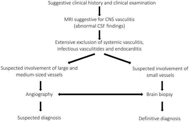
Objective: To approach the clinical value of MRI with vessel wall imaging (VWI) in patients with central nervous system vasculitis (CNSV), we analyzed patterns of VWI findings both at the time of initial presentation and during follow-up.
Methods: Stenoocclusive lesions, vessel-wall contrast enhancement (VW-CE) and diffusion-restricted lesions were analyzed in patients with a diagnosis of CNSV. On available VWI follow-up, progression, regression or stability of VW-CE were evaluated and correlated with the clinical status.
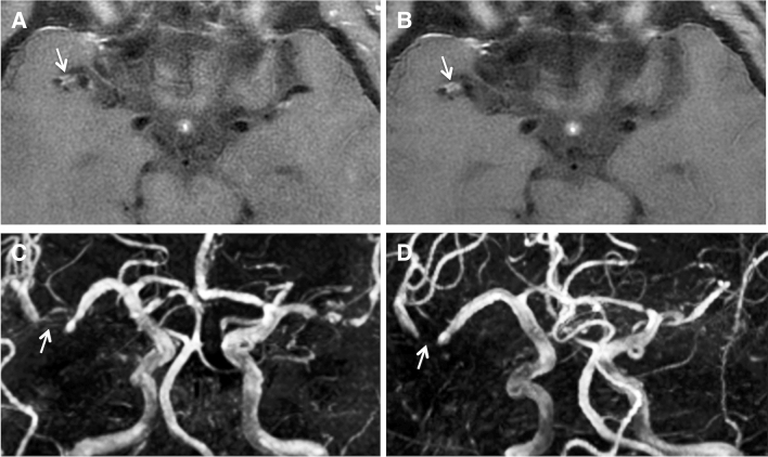
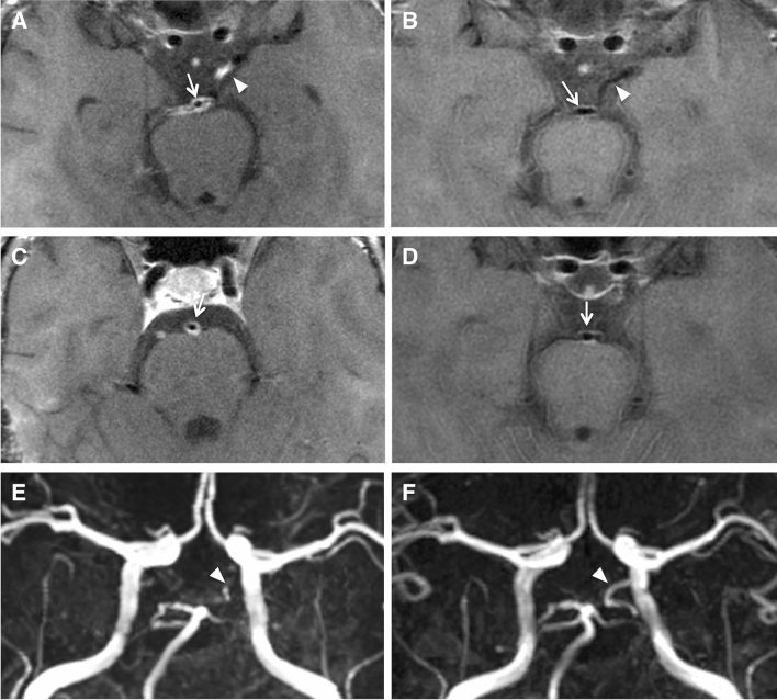
Results: Of the 45 patients included, 28 exhibited stenoses visible on MR angiography (MRA-positive) while 17 had no stenosis (MRA-negative). VW-CE was found in 2/17 MRA-negative and all MRA-positive patients (p < 0.05). 79.1% (53/67) of stenoses showed VW-CE. VW-CE was concentric in 88.3% and eccentric in 11.7% of cases. Diffusion-restricted lesions were found more frequently in relation to stenoses with VW-CE than without VW-CE (p < 0.05). 48 VW-CE lesions in 23 patients were followed over a median time of 239.5 days. 13 VW-CE lesions (27.1%) resolved completely, 14 (29.2%) showed partial regression, 17 (35.4%) remained stable and 4 (8.3%) progressed. 22/23 patients received immunosuppressive therapy for the duration of follow-up. Patients with stable or progressive VW-CE were more likely (p < 0.05) to have a relapse (14/30 cases) than patients with partial or complete regression of VW-CE (5/25 cases).
Conclusion: Concentric VW-CE is a common finding in medium/large-sized vessel CNSV. VW-CE might represent active inflammation in certain situations. However, follow-up VWI findings proved ambiguous as persisting VW-CE despite immunosuppressive therapy and clinical remission was a frequent finding.
Keywords: Cerebral vasculitis; Follow-up; MRI; Stroke; Vessel wall imaging.
© 2021. The Author(s).
Conflict of interest statement
The authors declare that they have no conflict of interest.
Figures


Similar articles
-
Evaluation of vessel-wall contrast-enhancement on high-resolution MRI in European patients with Moyamoya disease.J Stroke Cerebrovasc Dis. 2023 Jun;32(6):107135. doi: 10.1016/j.jstrokecerebrovasdis.2023.107135. Epub 2023 Apr 18. J Stroke Cerebrovasc Dis. 2023. PMID: 37079960
-
Predictors of improvement for patients with CNS vasculitis stenoses: A high-resolution vessel wall MRI follow-up study.Eur J Radiol. 2023 Jan;158:110619. doi: 10.1016/j.ejrad.2022.110619. Epub 2022 Nov 25. Eur J Radiol. 2023. PMID: 36463705
-
Assessment of central nervous system vasculitis in children based on high-resolution vascular wall imaging.Rheumatol Adv Pract. 2024 Mar 7;8(2):rkae038. doi: 10.1093/rap/rkae038. eCollection 2024. Rheumatol Adv Pract. 2024. PMID: 38605731 Free PMC article.
-
Vessel wall MR imaging of central nervous system vasculitis: a systematic review.Neuroradiology. 2022 Jan;64(1):43-58. doi: 10.1007/s00234-021-02724-9. Epub 2021 May 3. Neuroradiology. 2022. PMID: 33938989
-
Intracranial vessel wall magnetic resonance imaging features of infectious vasculitis.Clin Imaging. 2023 Jun;98:26-35. doi: 10.1016/j.clinimag.2023.03.014. Epub 2023 Mar 29. Clin Imaging. 2023. PMID: 36996597 Review.
Cited by
-
Percutaneous Cerebral Angioplasty for Refractory Middle Cerebral Artery Stenosis Due to Varicella-Zoster Virus-Related Vasculopathy: A Case Report.Cureus. 2024 Sep 20;16(9):e69773. doi: 10.7759/cureus.69773. eCollection 2024 Sep. Cureus. 2024. PMID: 39429375 Free PMC article.
-
Vessel Wall Magnetic Resonance Imaging in Cerebrovascular Diseases.Diagnostics (Basel). 2022 Jan 20;12(2):258. doi: 10.3390/diagnostics12020258. Diagnostics (Basel). 2022. PMID: 35204348 Free PMC article. Review.
-
[Inflammatory causes of stroke-Diagnostics and treatment].Nervenarzt. 2024 Oct;95(10):909-919. doi: 10.1007/s00115-024-01711-8. Epub 2024 Jul 30. Nervenarzt. 2024. PMID: 39080056 Free PMC article. German.
-
Clinical and Radiological Outcomes of Angiographically Proven Central Nervous System Arteriopathy.Cureus. 2025 Jan 23;17(1):e77897. doi: 10.7759/cureus.77897. eCollection 2025 Jan. Cureus. 2025. PMID: 39996226 Free PMC article.
-
Vasculitic Moyamoya syndrome in a patient with paroxysmal nocturnal hemoglobinuria: A case report and literature review.CNS Neurosci Ther. 2022 Sep;28(9):1464-1468. doi: 10.1111/cns.13891. Epub 2022 Jul 11. CNS Neurosci Ther. 2022. PMID: 35815532 Free PMC article. Review. No abstract available.
References
MeSH terms
LinkOut - more resources
Full Text Sources
