

Accuracy of Using Generative Adversarial Networks for Glaucoma Detection: Systematic Review and Bibliometric Analysis
- PMID: 34236992
- PMCID: PMC8493455
- DOI: 10.2196/27414
Accuracy of Using Generative Adversarial Networks for Glaucoma Detection: Systematic Review and Bibliometric Analysis
Abstract
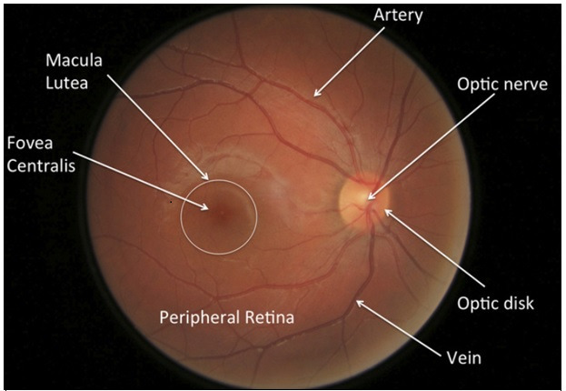
Background: Glaucoma leads to irreversible blindness. Globally, it is the second most common retinal disease that leads to blindness, slightly less common than cataracts. Therefore, there is a great need to avoid the silent growth of this disease using recently developed generative adversarial networks (GANs).
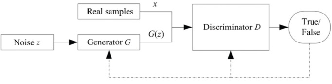
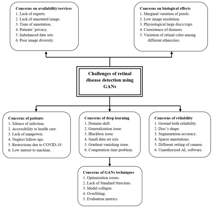
Objective: This paper aims to introduce a GAN technology for the diagnosis of eye disorders, particularly glaucoma. This paper illustrates deep adversarial learning as a potential diagnostic tool and the challenges involved in its implementation. This study describes and analyzes many of the pitfalls and problems that researchers will need to overcome to implement this kind of technology.
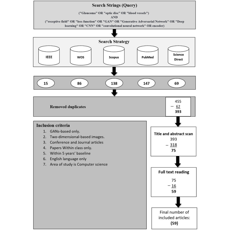
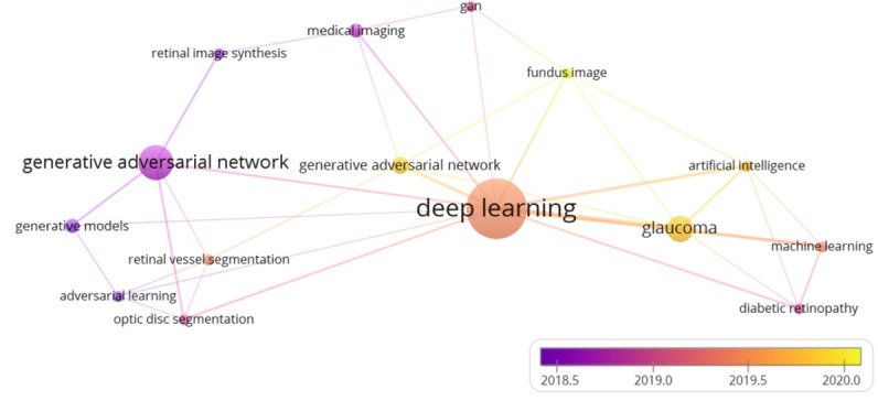
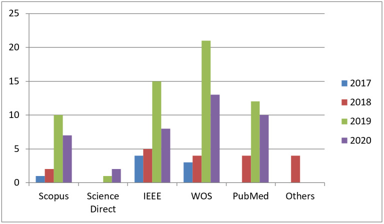
Methods: To organize this review comprehensively, articles and reviews were collected using the following keywords: ("Glaucoma," "optic disc," "blood vessels") and ("receptive field," "loss function," "GAN," "Generative Adversarial Network," "Deep learning," "CNN," "convolutional neural network" OR encoder). The records were identified from 5 highly reputed databases: IEEE Xplore, Web of Science, Scopus, ScienceDirect, and PubMed. These libraries broadly cover the technical and medical literature. Publications within the last 5 years, specifically 2015-2020, were included because the target GAN technique was invented only in 2014 and the publishing date of the collected papers was not earlier than 2016. Duplicate records were removed, and irrelevant titles and abstracts were excluded. In addition, we excluded papers that used optical coherence tomography and visual field images, except for those with 2D images. A large-scale systematic analysis was performed, and then a summarized taxonomy was generated. Furthermore, the results of the collected articles were summarized and a visual representation of the results was presented on a T-shaped matrix diagram. This study was conducted between March 2020 and November 2020.
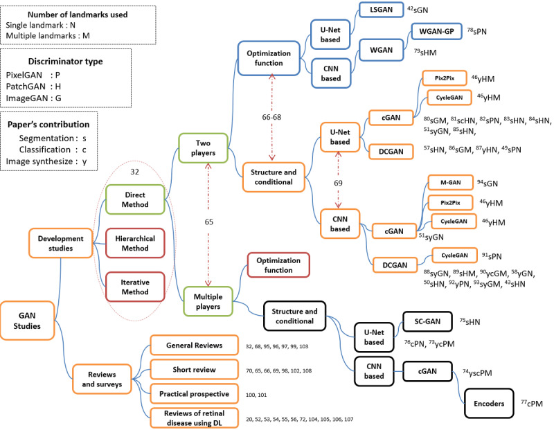
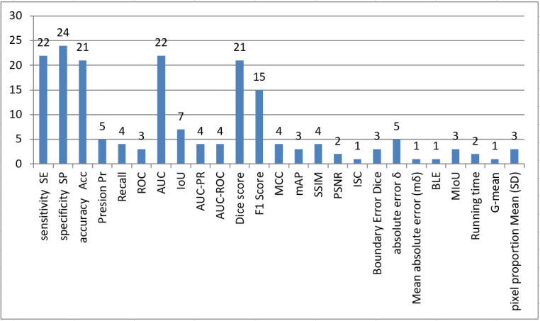
Results: We found 59 articles after conducting a comprehensive survey of the literature. Among the 59 articles, 30 present actual attempts to synthesize images and provide accurate segmentation/classification using single/multiple landmarks or share certain experiences. The other 29 articles discuss the recent advances in GANs, do practical experiments, and contain analytical studies of retinal disease.
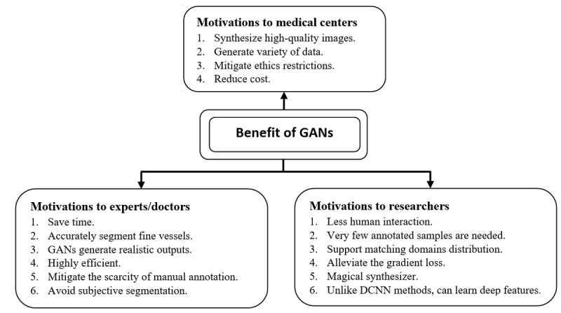
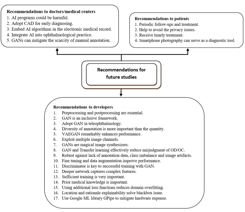
Conclusions: Recent deep learning techniques, namely GANs, have shown encouraging performance in retinal disease detection. Although this methodology involves an extensive computing budget and optimization process, it saturates the greedy nature of deep learning techniques by synthesizing images and solves major medical issues. This paper contributes to this research field by offering a thorough analysis of existing works, highlighting current limitations, and suggesting alternatives to support other researchers and participants in further improving and strengthening future work. Finally, new directions for this research have been identified.
Keywords: blood vessels; deep learning; generative adversarial network; glaucoma; optic disc; retinal disease; systematic literature review.
©Ali Q Saeed, Siti Norul Huda Sheikh Abdullah, Jemaima Che-Hamzah, Ahmad Tarmizi Abdul Ghani. Originally published in the Journal of Medical Internet Research (https://www.jmir.org), 21.09.2021.
Conflict of interest statement
Conflicts of Interest: None declared.
Figures
References
-
- Flaxman SR, Bourne RRA, Resnikoff S, Ackland P, Braithwaite T, Cicinelli MV, Das A, Jonas JB, Keeffe J, Kempen JH, Leasher J, Limburg H, Naidoo K, Pesudovs K, Silvester A, Stevens GA, Tahhan N, Wong TY, Taylor HR, Vision Loss Expert Group of the Global Burden of Disease Study Global causes of blindness and distance vision impairment 1990-2020: a systematic review and meta-analysis. Lancet Glob Health. 2017 Dec;5(12):e1221–e1234. doi: 10.1016/S2214-109X(17)30393-5. https://linkinghub.elsevier.com/retrieve/pii/S2214-109X(17)30393-5 S2214-109X(17)30393-5 - DOI - PubMed
-
- Pascolini D, Mariotti SP. Global estimates of visual impairment: 2010. Br J Ophthalmol. 2012 May;96(5):614–8. doi: 10.1136/bjophthalmol-2011-300539. https://pubmed.ncbi.nlm.nih.gov/22133988/ bjophthalmol-2011-300539 - DOI - PubMed
-
- de Carvalho Junior ASV, Carvalho ED, de Carvalho Filho AO, de Sousa AD, Corrêa Silva A, Gattass M. Automatic methods for diagnosis of glaucoma using texture descriptors based on phylogenetic diversity. Computers & Electrical Engineering. 2018 Oct;71:102–114. doi: 10.1016/j.compeleceng.2018.07.028. https://www.sciencedirect.com/science/article/abs/pii/S0045790617338570 - DOI
-
- Wood K. Glaucoma: the silent thief of sight. The Lamp. 1995 Sep;52(8):15. https://www.semanticscholar.org/paper/Glaucoma%3A-the-silent-thief-of-si... - PubMed
-
- Villain MA. [The epidemiology of glaucoma] J Fr Ophtalmol. 2005 Jun;28 Spec No 2:2S9–2S12. https://pubmed.ncbi.nlm.nih.gov/16208234/ MDOI-JFO-06-2005-28-HS2-0181-5512-101019-200506342 - PubMed
Publication types
MeSH terms
LinkOut - more resources
Full Text Sources
Medical
