. 2021 Nov 5;22(6):bbab141.
doi: 10.1093/bib/bbab141.

Identification of viral-mediated pathogenic mechanisms in neurodegenerative diseases using network-based approaches

Affiliations

Identification of viral-mediated pathogenic mechanisms in neurodegenerative diseases using network-based approaches

Anna Onisiforou et al. Brief Bioinform. .

Abstract

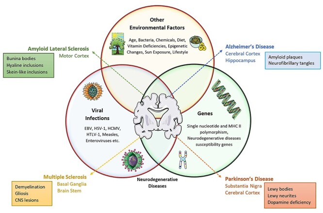
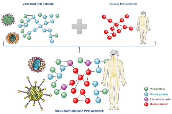
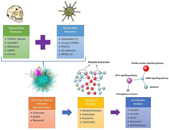
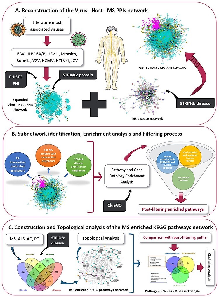
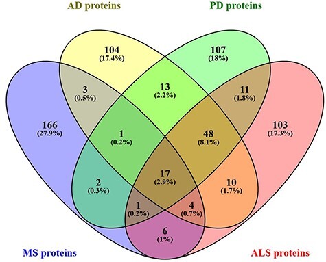
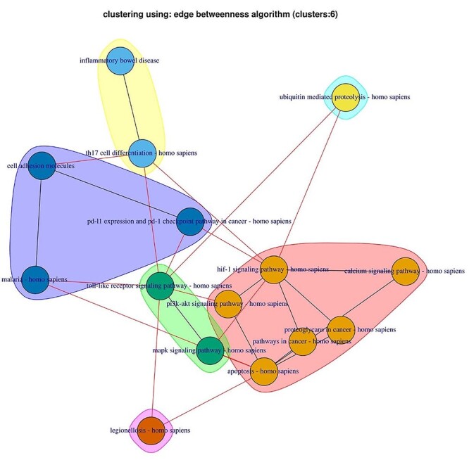
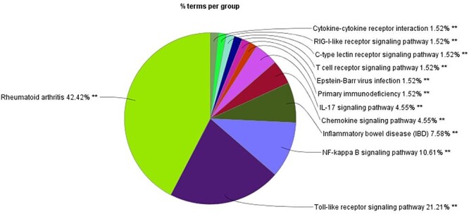
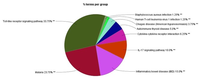
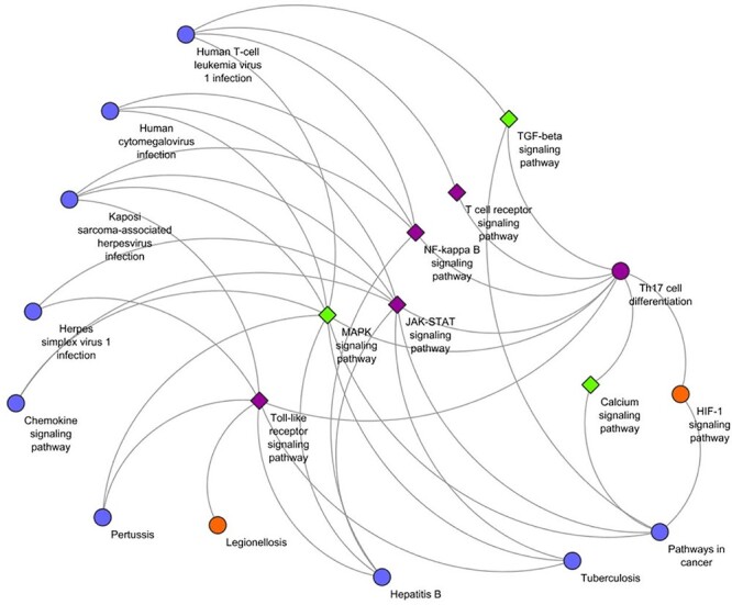
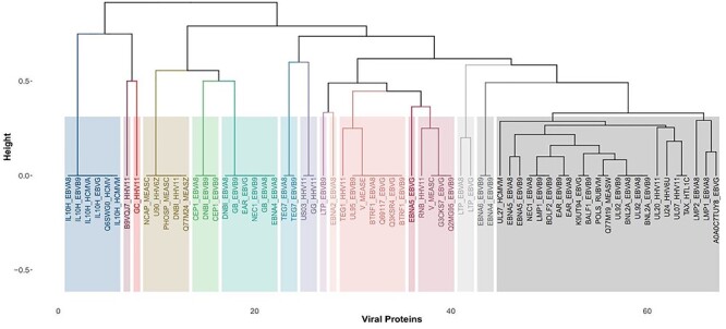
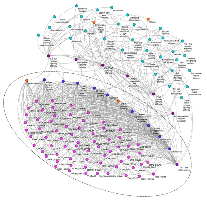
During the course of a viral infection, virus-host protein-protein interactions (PPIs) play a critical role in allowing viruses to replicate and survive within the host. These interspecies molecular interactions can lead to viral-mediated perturbations of the human interactome causing the generation of various complex diseases. Evidences suggest that viral-mediated perturbations are a possible pathogenic etiology in several neurodegenerative diseases (NDs). These diseases are characterized by chronic progressive degeneration of neurons, and current therapeutic approaches provide only mild symptomatic relief; therefore, there is unmet need for the discovery of novel therapeutic interventions. In this paper, we initially review databases and tools that can be utilized to investigate viral-mediated perturbations in complex NDs using network-based analysis by examining the interaction between the ND-related PPI disease networks and the virus-host PPI network. Afterwards, we present our theoretical-driven integrative network-based bioinformatics approach that accounts for pathogen-genes-disease-related PPIs with the aim to identify viral-mediated pathogenic mechanisms focusing in multiple sclerosis (MS) disease. We identified seven high centrality nodes that can act as disease communicator nodes and exert systemic effects in the MS-enriched Kyoto Encyclopedia of Genes and Genomes (KEGG) pathways network. In addition, we identified 12 KEGG pathways, 5 Reactome pathways and 52 Gene Ontology Immune System Processes by which 80 viral proteins from eight viral species might exert viral-mediated pathogenic mechanisms in MS. Finally, our analysis highlighted the Th17 differentiation pathway, a disease communicator node and part of the 12 underlined KEGG pathways, as a key viral-mediated pathogenic mechanism and a possible therapeutic target for MS disease.

Keywords: multiple sclerosis; neurodegenerative diseases; protein–protein interactions; viral perturbations; virus–host–disease interactions.


Figures

Figure 1
Figure 1
A schematic representation of the multifactorial origin of a group of pathologically distinct but related neurological diseases. NDs are complex disorders that are caused by the combination of environmental and genetic factors; however, how the interplay of these factors contributes to their development still remains unclear. One hypothesis is that NDs might be caused by the combinatorial effects of viral infections, other environmental factors and genetic susceptibility. The combination of these factors contributes to the clinical heterogenicity and histopathological diversity of the central nervous lesion that characterizes these diseases, as they differ in the subset of neurons and anatomical structures that are affected and have both common and distinct pathological abnormalities. Figure contains illustrations obtained from Servier medical art (https://smart.servier.com/), provided free and licensed under the Creative Commons Attribution 3.0 Unported License.
Figure 2
Figure 2
Schematic representation of the construction of the integrated virus–host–ND PPI network, where the virus–host PPI network is merged with a ND-related PPI network. Figure contains illustrations obtained from Servier medical art (https://smart.servier.com/), provided free and licensed under the Creative Commons Attribution 3.0 Unported License.
Figure 3
Figure 3
Illustration of the most commonly used approach for the investigation of virus–host–disease PPIs highlighting recourses and tools that can be utilized at each step. The first step involves the collection of data, and virus–host PPIs can be collected from several databases such as VirHostNet 2.0 [73], Viruses.STRING [74], PHISTO [75], VirusMentha [76] and HPIDB 3.0 [77]. Disease data can be collected from databases such as the STRING: disease query app in Cytoscape [81], MalaCards [78], DisGeNET [79], OMIM [237] and ClinVar [238]. The second step involves the visualization of the integrated network, which can be performed with Cytoscape [85], the igraph package in python and R [86] and the NetworkX package in python [87]. The third step involves network topological analysis that can be performed with several plugins offered by Cytoscape such as NetworkAnalyzer [88] and CytoHubba [89], as well as clustering analysis apps, such as CytoCluster [112] and ClusterViz [113]. The final step involves enrichment analysis that can be performed either on the whole network or on subnetworks using the ClueGO [96] plugin in Cytoscape [98] and enrichR [105], an R interface to the Enrichr database [106, 107]. Pathway enrichment analysis can be performed form several databases including the KEGG [99], Reactome [100] and WikiPathways [101]. Figure contains illustrations obtained from Servier medical art (https://smart.servier.com/), provided free and licensed under the Creative Commons Attribution 3.0 Unported License.
Figure 4
Figure 4
Schematic representation of the methodology applied in this paper to investigate the interaction between virus-host-MS PPIs using a network-based approach with the aim to identify viral-mediated pathogenic mechanisms that might be involved in the development of MS. (A) Reconstruction of the virus-host-MS PPIs network. (B) Subnetwork identification,enrichment analysis and filtering process of the enriched results. (C) Construction and topological analysis of the ND-based MS enriched KEGG pathway to pathway network and comparison with the post filtering enriched pathways obtained from each subnetwork. Figure contains illustrations obtained from Servier medical art (https://smart.servier.com/), provided free and licensed under the Creative Commons Attribution 3.0 Unported License.
Figure 5
Figure 5
Comparison results of the top 200 highest disease-associated proteins between the four NDs: ALS, MS, PD and AD.
Figure 6
Figure 6
Complementary network of the four common NDs KEGG pathways (IBD, HIF-1 signaling pathway, malaria and legionellosis) and their interactions with the 11 complementary nodes/pathways, created using PathwayConnector.
Figure 7
Figure 7
Enriched KEGG pathway analysis results of the 166 unique MS disease proteins, obtained using the ClueGO app in Cystoscope, with the pathways classified into groups and the percentage indicating the number of terms in each group.
Figure 8
Figure 8
KEGG enrichment analysis results of the 45 common disease proteins between MS, SLE, RA and type 1 diabetes, with the pathways classified into groups and the percentage indicating the number of terms in each group.
Figure 9
Figure 9
KEGG enrichment analysis results of the 53 disease proteins that are shared between MS and some of the other autoimmune diseases (SLE, RA, type 1 diabetes), with the pathways classified into groups and the percentage indicating the number of terms in each group.
Figure 10
Figure 10
Community clustering of the MS-enriched KEGG pathways network resulted in the formation of four community clusters (a-d). Orange color nodes represent the four common ND pathways between ALS, MS, PD and AD. Hub–bottleneck nodes are represented in color purple, and blue color nodes represent infectious disease pathways of some of the viruses we included for the reconstruction of our virus–host–MS PPIs network.
Figure 11
Figure 11
Filtering process applied on the enriched analysis results of the three subnetworks.
Figure 12
Figure 12
(A) Pathogen–genes–disease triangle. (B) Comparison of the post-filtering KEGG pathways enriched results that are also MS disease-related terms between the three subnetworks, indicating the 12 KEGG pathways that fall within the pathogen–genes–disease triangle.
Figure 13
Figure 13
Schematic visualization of the complementary network for the 12 KEGG pathways, which are indicated in circular nodes, and the 7 complementary nodes shown in diamond shape. The orange nodes represent nodes that are also part of the four common ND pathways and purple nodes are hub–bottleneck nodes that act as a bridge of communication between nodes/pathways in the MS disease KEGG pathways network. The green nodes are complementary nodes, which are not MS disease-related pathway terms (MS unique, MS shared or NDs common).
Figure 14
Figure 14
Clustering dendrogram of the 67 viral proteins from 8 viral species EBV, HCMV, HHV-6A, HHV-6B, HSV-1, HTLV-1, Measles and Rubella based on target similarity of the final 12 KEGG pathways.
Figure 15
Figure 15
Tissue-specific gene enrichment analysis results of the 200 MS disease-associated proteins, using the GTEx dataset.
Figure 16
Figure 16
Tissue-specific gene enrichment analysis results of the human targets of the 80 viral proteins that interact with the 12 KEGG pathways, 52 GO ISP and 5 Reactome pathways, using the GTEx dataset.
Figure 17
Figure 17
Schematic illustration of the possible viral-mediated pathogenic mechanisms obtained through our pipeline approach indicating how the resulting 67 viral proteins (lilac color) targeting the 12 identified KEGG pathways in the MS-enriched KEGG pathways network, which in turn interact with the hub–bottleneck disease communicator nodes (purple color), can exert systemic effects within the network and lead to the development of MS. The ND common pathways are indicated in orange.

Similar articles

Cited by

References

    1. Migliore L, Coppedè F. . Genetics, environmental factors and the emerging role of epigenetics in neurodegenerative diseases. Mutat. Res. Fundam. Mol. Mech, 2009;667:82–97. - PubMed
    1. Patrick KL, Bell SL, Weindel CG, et al. . Exploring the “multiple-hit hypothesis” of neurodegenerative disease: bacterial infection comes up to bat. FCIMB 2019;9:138. - PMC - PubMed
    1. De Chiara G, Marcocci ME, Sgarbanti R, et al. . Infectious agents and neurodegeneration. Mol Neurobiol 2012;46:614–38. - PMC - PubMed
    1. Alam M, Alam Q, Kamal M, et al. . Infectious agents and neurodegenerative diseases: exploring the links. Curr Top Med Chem 2017;17:1390–9. - PubMed
    1. Zhou L, Miranda-Saksena M, Saksena NK. Viruses and neurodegeneration. Virol J 2013;10:172. - PMC - PubMed

Publication types