. 2021 Jul 8;13(1):125.
doi: 10.1186/s13195-021-00860-1.

Aη-α and Aη-β peptides impair LTP ex vivo within the low nanomolar range and impact neuronal activity in vivo

Affiliations

Aη-α and Aη-β peptides impair LTP ex vivo within the low nanomolar range and impact neuronal activity in vivo

Maria Mensch et al. Alzheimers Res Ther. .

Abstract

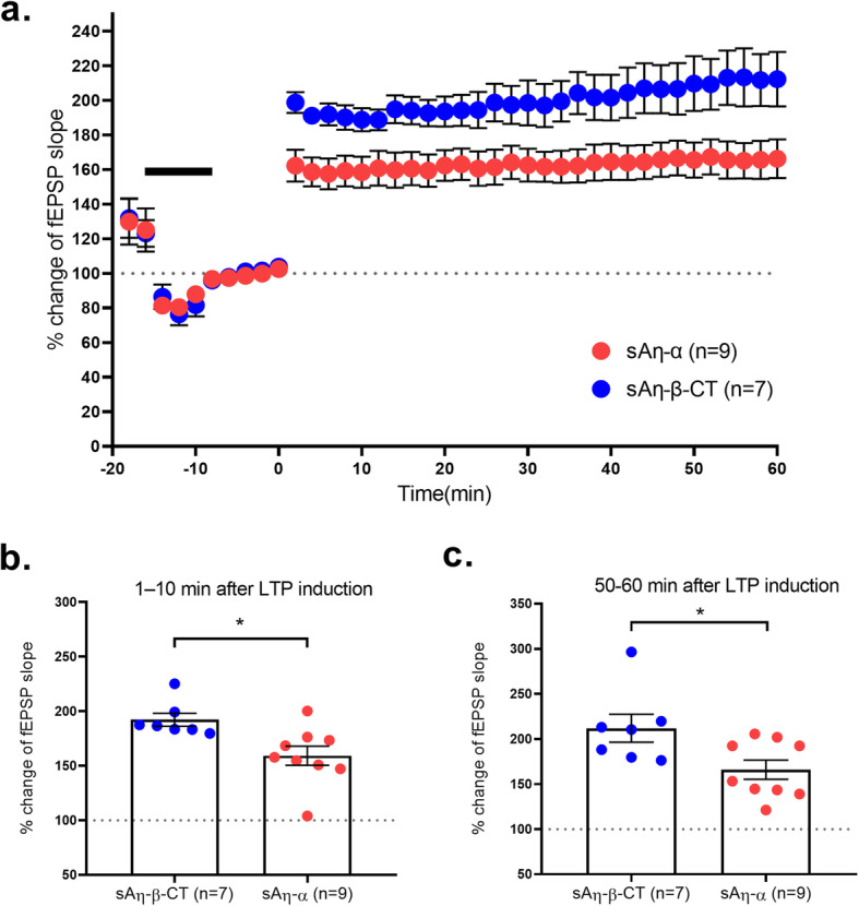
Background: Amyloid precursor protein (APP) processing is central to Alzheimer's disease (AD) etiology. As early cognitive alterations in AD are strongly correlated to abnormal information processing due to increasing synaptic impairment, it is crucial to characterize how peptides generated through APP cleavage modulate synapse function. We previously described a novel APP processing pathway producing η-secretase-derived peptides (Aη) and revealed that Aη-α, the longest form of Aη produced by η-secretase and α-secretase cleavage, impaired hippocampal long-term potentiation (LTP) ex vivo and neuronal activity in vivo.

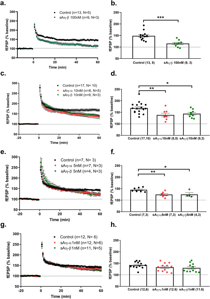
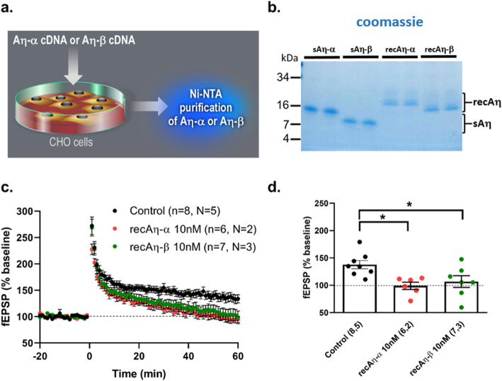
Methods: With the intention of going beyond this initial observation, we performed a comprehensive analysis to further characterize the effects of both Aη-α and the shorter Aη-β peptide on hippocampus function using ex vivo field electrophysiology, in vivo multiphoton calcium imaging, and in vivo electrophysiology.

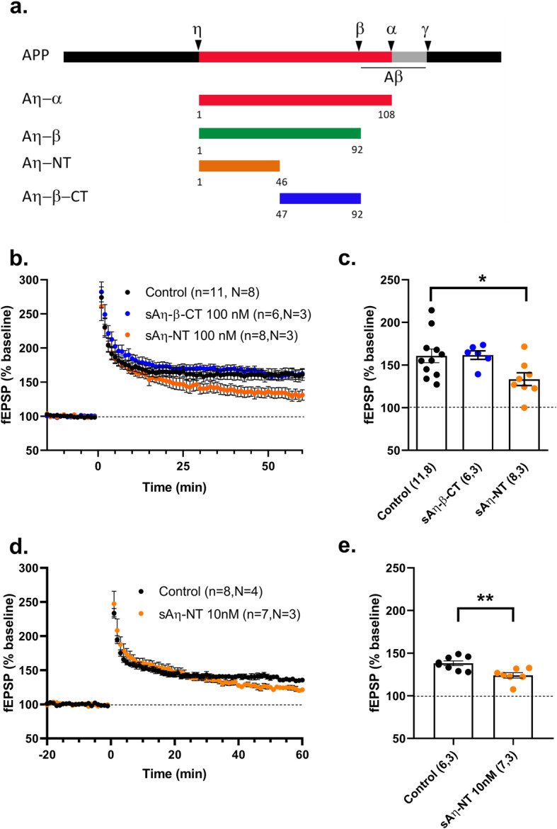
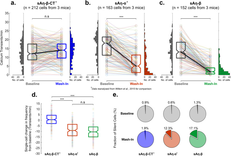
Results: We demonstrate that both synthetic peptides acutely impair LTP at low nanomolar concentrations ex vivo and reveal the N-terminus to be a primary site of activity. We further show that Aη-β, like Aη-α, inhibits neuronal activity in vivo and provide confirmation of LTP impairment by Aη-α in vivo.

Conclusions: These results provide novel insights into the functional role of the recently discovered η-secretase-derived products and suggest that Aη peptides represent important, pathophysiologically relevant, modulators of hippocampal network activity, with profound implications for APP-targeting therapeutic strategies in AD.

Keywords: APP processing; Alzheimer; Electrophysiology; Hippocampus; Synaptic plasticity.


Conflict of interest statement

The authors declare that they have no competing interests.

Figures

Fig. 1
Fig. 1
Synthetic Aη-α and Aη-β acutely inhibit LTP within the low nanomolar range. LTP was analyzed ex vivo at CA3-CA1 synapse in hippocampal slices of RjOrl:SWISS mice. a, c, e, and g Summary graphs of fEPSP slope (% baseline) pre- and post-LTP induction (time 0) in control (aCSF only) or in presence of a 100 nM sAη–β, c 10 nM sAη–α or sAη–β, e 5 nM sAη–α or sAη–β, and g 1 nM sAη–α or sAη–β, throughout the recording. b, d, f, and h Summary of fEPSP magnitude 45–60 min after LTP induction as fEPSP (% baseline) for data shown in a, c, e, and g, respectively (n= slices, N= mice), *p< 0.05, **p< 0.01, ***p< 0.001. Detailed statistics are shown in Supplementary Table S1
Fig. 2
Fig. 2
Ten nanomolar of soluble recombinant Aη-α and Aη-β lower LTP. a Diagram explaining the production of soluble HIS-tagged recAη-α and recAη-β samples used in c and d (see the “Methods” section for details of Ni-NTA purification and sample quantification). b Commassie stain of 1 μg of Aη peptides. Synthetic Aη peptides or recombinant CHO cell-expressed glycosylated purified Aη peptides were separated in SDS-PAGE and stained with GelCode Blue. c, d LTP was analyzed ex vivo at CA3-CA1 synapse in hippocampal slices of RjOrl:SWISS mice. Summary graphs of c fEPSP slope (% baseline) pre- and post-LTP induction (time 0) and d fEPSP magnitude 45–60 min after LTP induction in control (aCSF only) or in presence of 10 nM recAη–α or recAη–β throughout the recording (n= slices, N= mice), *p< 0.05. Detailed statistics are shown in Supplementary Table S2
Fig. 3
Fig. 3
N-terminal of Aη is necessary and sufficient for LTP impairment. a Diagram showing APP processing (secretases cleavage sites are shown) and boundaries of shorter synthetic peptides used to identify the active site. be LTP was analyzed ex vivo at CA3-CA1 synapse in hippocampal slices of RjOrl:SWISS mice. Summary graphs of b, d fEPSP slope (% baseline) pre- and post-LTP induction (time 0) and c, e fEPSP magnitude 45–60 min after LTP induction in control (aCSF only) or in presence of b, c 100 nM sAη–NT or sAη–β-CT or d, e of 10 nM sAη–NT, throughout the recording (n= slices, N= mice), *p< 0.05. Detailed statistics are shown in Supplementary Table S3
Fig. 4
Fig. 4
Synthetic Aη-α and Aη-β acutely modulate neuronal activity in vivo. a The median frequency of calcium transients in the CA1 region of hippocampi of C57Bl/6 mice did not differ significantly (n.s) from baseline following superfusion (wash-in, blue) of sAη–β–CT (control peptide). Note the high degree of similarity in distribution of calcium transient frequencies as denoted by the upright histograms. The frequency of calcium transients for each individual neuron before (baseline) and after superfusion (wash-in) of sAη–β–CT is overlaid. b Superfusion of sAη–α (wash-in, red) induced a significant decrease in median calcium transient frequency and a positive skew in the distribution of calcium transient frequencies towards hypoactivity. The frequency of calcium transients for each individual neuron before (baseline) and after superfusion (wash-in) of sAη–α is overlaid. c Superfusion of sAη–β (wash-in, green) also induced a significant decrease in median calcium transient frequency and a positive skew in the distribution of calcium transient frequencies towards hypoactivity. The frequency of calcium transients for each individual neuron before (baseline) and after superfusion (wash-in) of sAη–β is overlaid. d Across all individual cells studied, the median change in calcium transient frequency following superfusion of sAη–α (red) and sAη–β (green) was not statistically different (n.s), but were significantly lower relative to the null change associated with sAη–β–CT (blue). The change in calcium transient frequency for each individual neuron after superfusion (wash-in) of each peptide is overlaid. e While the proportion of silent neurons (i.e. showing absence of calcium transients) were small during baseline conditions (grey charts), and following sAη–β–CT superfusion (wash-in, blue), superfusion of sAη–α (wash-in, red) and sAη–β (wash-in, green) was associated with a dramatic increase in the number of inactive cells. For each boxplot, the central line denotes the median with bottom and top demarcations indicating the 25th and 75th percentiles, respectively. Data for sAη–α and sAη-β-CT were previously published [3] and presented and reanalyzed in this figure for comparison purposes (denoted as). ***p< 0.001. Detailed statistics are shown in Supplementary Table S4
Fig. 5
Fig. 5
sAη–α acutely lowers LTP in vivo. a LTP was measured in vivo in the dentate gyrus of Sprague-Dawley rats. Summary graphs of fEPSP slope (% baseline) pre- and post-LTP induction (time 0) upon 10 min of intra-hippocampal injection of 1 μM of control peptide (sAη–β–CT) or sAη–α. Injection time is shown by the black bar. Note a transient decline and partial recovery of baseline responses upon the injection and a subsequent LTP induction. b Summary of fEPSP magnitude 1–10 min after LTP induction as fEPSP (% baseline) for data shown in a. c Summary of fEPSP magnitude 50–60 min after LTP induction as fEPSP (% baseline) for data shown in a. All recordings were done in vivo in Sprague Dawley rats with n= number of rats. *p< 0.05 Detailed statistics are shown in Supplementary Table S5

References

    1. van der Kant R, Goldstein LSB. Cellular functions of the amyloid precursor protein from development to dementia. Dev Cell. 2015;32(4):502–515. doi: 10.1016/j.devcel.2015.01.022. - DOI - PubMed
    1. García-González L, Pilat D, Baranger K, Rivera S. Emerging alternative proteinases in APP metabolism and Alzheimer’s disease pathogenesis: a focus on MT1-MMP and MT5-MMP. Front Aging Neurosci. 2019;11:244. doi: 10.3389/fnagi.2019.00244. - DOI - PMC - PubMed
    1. Willem M, Tahirovic S, Busche MA, Ovsepian SV, Chafai M, Kootar S, Hornburg D, Evans LDB, Moore S, Daria A, Hampel H, Müller V, Giudici C, Nuscher B, Wenninger-Weinzierl A, Kremmer E, Heneka MT, Thal DR, Giedraitis V, Lannfelt L, Müller U, Livesey FJ, Meissner F, Herms J, Konnerth A, Marie H, Haass C. eta-Secretase processing of APP inhibits neuronal activity in the hippocampus. Nature. 2015;526(7573):443–447. doi: 10.1038/nature14864. - DOI - PMC - PubMed
    1. Baranger K, Marchalant Y, Bonnet AE, Crouzin N, Carrete A, Paumier JM, Py NA, Bernard A, Bauer C, Charrat E, Moschke K, Seiki M, Vignes M, Lichtenthaler SF, Checler F, Khrestchatisky M, Rivera S. MT5-MMP is a new pro-amyloidogenic proteinase that promotes amyloid pathology and cognitive decline in a transgenic mouse model of Alzheimer’s disease. Cell Mol Life Sci. 2016;73(1):217–236. doi: 10.1007/s00018-015-1992-1. - DOI - PMC - PubMed
    1. Selkoe DJ. Alzheimer’s disease is a synaptic failure. Science. 2002;298(5594):789–791. doi: 10.1126/science.1074069. - DOI - PubMed

Publication types

Substances