. 2021 Sep;31(9):1024-1027.
doi: 10.1038/s41422-021-00526-5. Epub 2021 Jul 8.

Hsp70 chaperones TDP-43 in dynamic, liquid-like phase and prevents it from amyloid aggregation

Affiliations

Hsp70 chaperones TDP-43 in dynamic, liquid-like phase and prevents it from amyloid aggregation

Jinge Gu et al. Cell Res. 2021 Sep.
No abstract available


Conflict of interest statement

The authors declare no competing interests.

Figures
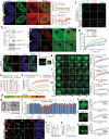
Fig. 1
Fig. 1. Hsp70 chaperones TDP-43 in liquid-like phase and prevents it from pathological aggregation.
a, b Representative images and the intensity profile along the indicated line of HeLa cells expressing RFP-Hsp70 with TDP-43-HA in response to stress (250 μM of arsenite, 30 min). Arrows, co-localization of Hsp70 with TDP-43 NBs. See also Supplementary information, Figs. S1 and S2. c Western blot analysis confirming expression of Hsc70EGFP-KI in the stable KI cell line. The anti-Hsp70s antibody recognizes both Hsp70 and Hsc70. d, e Representative images of Hsc70EGFP-KI and TDP-43-HA in control (no stress) or stressed Hsc70EGFP-KI cells (250 μM of arsenite, 30 min). Arrows, co-localization of Hsc70EGFP-KI with stress-induced TDP-43 NBs. f In vitro co-LLPS of TDP-43-MBP (50 μM) and Hsp70 (10 μM) at indicated condition. See also Supplementary information, Fig. S3. g FRAP analyses of TDP-43-MBP LDs formed in the in vitro LLPS assay. n = 6. See also Supplementary information, Fig. S5. h, i Representative images showing the effect of si-Hsp70s on the assembly of TDP-43 NBs in stressed HeLa cells. G3BP, a marker for SGs. See also Supplementary information, Fig. S7. jm Representative images showing stress-induced EGFP-TDP-43 NBs in HeLa cells (j) and the regions (dashed circle) photo-bleached in the FRAP assays in km at indicated time. The fluorescent intensity (FI) recovery curves are shown on the right, n = 8. n The CCK-8 assay showing enhanced TDP-43 cytotoxicity by si-Hsp70s in prolonged stress. n = 3. o The thioflavine T (ThT) fluorescence assay of TDP-43 LCD (20 μM) with different concentrations of Hsp70 as indicated. n = 3. p Schematic of the main functional domains in TDP-43. q Negative-staining TEM images of the ThT samples at 30 h in o. r Residue-specific intensity changes of signals in the 2D 1H-15N HSQC spectra of 15N-labeled TDP-43 LCD with different concentrations of Hsp70 as indicated. The CR in the TDP-43 LCD is indicated in the dashed box. See also Supplementary information, Fig. S9. su The FRAP assay evaluating the impact of OE of Hsp70s on the dynamics of different types of EGFP-TDP-43-K181E NIs. The regions (dashed circles) of the same size are photo-bleached. n = 20 each group, pooled results of five independent biological repeats. See also Supplementary information, Fig. S14a–e. v, w Representative images of EGFP-TDP-43 K181E NIs co-expressed with RFP (v) or RFP-Hsp70 (w) in 293T cells that are co-immunostained for pTDP-43 (Ser409/410). x, y Quantification of the percentage of cells showing pTDP-43+ NIs (x) and the relative size of pTDP-43+ NIs (normalized to area labeled by DAPI) (y). n = ~25 cells in x and 6 in y. See also Supplementary information, Fig. S14f, g. Data are means ± SEM; Two-way ANOVA in g, km, su, Student’s t-test in n, x, y; *P < 0.05 and ***P < 0.001; ns, not significant. Scale bars, 5 μm in a, b, d, e, hj, su, 10 μm in f, v, w, 1 μm in km and 200 nm in q.

Similar articles

Cited by

References

    1. Wang C, et al. Mol Cell. 2020;79:443–58. doi: 10.1016/j.molcel.2020.06.019. - DOI - PubMed
    1. Chen-Plotkin AS, Lee VM, Trojanowski JQ. Nat Rev Neurol. 2010;6:211–20. doi: 10.1038/nrneurol.2010.18. - DOI - PMC - PubMed
    1. Ramaswami M, Taylor JP, Parker R. Cell. 2013;154:727–36. doi: 10.1016/j.cell.2013.07.038. - DOI - PMC - PubMed
    1. Freibaumeibaum BD, Chitta RK, High AA, Taylor JP. J Proteome Res. 2010;9:1104–20. doi: 10.1021/pr901076y. - DOI - PMC - PubMed
    1. Estestes PS, Boehringer A, Zwick R, Tang JE, Grigsby B, Zarnescu DC. Hum Mol Genet. 2011;20:2308–21. doi: 10.1093/hmg/ddr124. - DOI - PMC - PubMed

Publication types