. 2021 Jun 21:2021:9952060.
doi: 10.1155/2021/9952060. eCollection 2021.

The Research on the Treatment of Metastatic Skin Cutaneous Melanoma by Huanglian Jiedu Decoction Based on the Analysis of Immune Infiltration Analysis

Affiliations

The Research on the Treatment of Metastatic Skin Cutaneous Melanoma by Huanglian Jiedu Decoction Based on the Analysis of Immune Infiltration Analysis

Ding Li et al. Evid Based Complement Alternat Med. .

Abstract

Objective: To explore the potential mechanism of Huanglian Jiedu Decoction (HJD) treatment and prevention of metastatic Cutaneous Melanoma (CM) occurrence and metastasis based on network pharmacological methods and immune infiltration analysis.

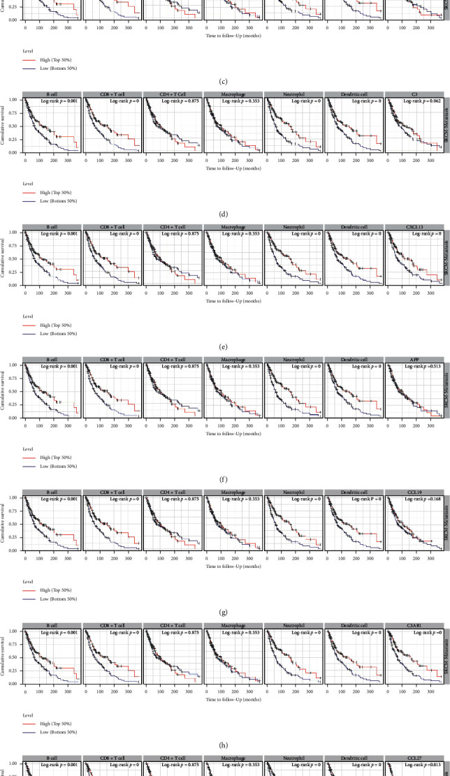
Methods: The GEO database was used to obtain metastatic CM disease targets, the TCMSP database and the HERB database were used to obtain HJD action targets, core genes were screened by protein interaction network, and the potential mechanism of HJD in the treatment of metastatic CM was explored by enrichment analysis, prognostic analysis and immune infiltration analysis.

Results: HJD treatment of metastatic CM involved 60 targets, enrichment analysis showed that HJD treatment of metastatic CM involved Chemokine signaling pathway, NF-kappa B signaling pathway, and Fluid shear stress and atherosclerosis, etc. Prognostic analysis revealed that HJD had a certain ability to improve the prognosis of metastatic CM patients. Immune infiltration analysis showed that HJD could inhibit the immune cell infiltration of metastatic CM patients by acting on related targets.

Conclusions: Our study identified the potential mechanism of HJD in the treatment of metastatic CM through network pharmacology, and revealed the mechanism of HJD in the prevention of Skin Cutaneous Melanoma metastasis through immune infiltration analysis and prognostic analysis.


Conflict of interest statement

The authors declare that they have no conflicts of interests.

Figures

Figure 1
Figure 1
The result of differentially analysis is visualized by the heatmap. (a) Part (GSE7553), (b) part (GSE8401), (c) part (GSE15605), (d) part (GSE46517). The gradual color ranging from blue to red represents the changing process from down-to up-regulation.
Figure 2
Figure 2
Merging and visualization based on the results of the 4 data sets difference analysis. The gradual color ranging from green to red represents the changing process from down-to up-regulation.
Figure 3
Figure 3
HJD-drug-target network including 1 Chinese herbal formula, 4 Chinese medicines and 376 targets.
Figure 4
Figure 4
The intersection between HJD and DEGs. A total of 60 genes have been screened out.
Figure 5
Figure 5
Construction the PPI network through STRING database (a). Rank based on the number of nodes in the PPI network (b). Top 10 core genes was screened out based on “MCC” calculation method (c). Correlation analysis between the expression of 10 core genes. The expression of C3 and C3AR1, the expression of CXCL13 and CCL19, the expression of CCL21 and CXCL12, and the expression of CCL19 and C3 were significantly positively correlated (d).
Figure 6
Figure 6
Enrichment analysis of HJD treat METASTATIC CM. Cellular components, biological processes, and molecular functions analysis (a). KEGG pathway analysis (b).
Figure 7
Figure 7
Correlation of core genes expression with immune infiltration level in METASTATIC CM (TIMER). (a) The expression of CXCL12 is positively correlated with the level of B cell (partial.cor = 0.136, p=1.15e − 02), CD8+ T cell (partial.cor = 0.344, p=9.29e − 11), CD4+ T cell (partial.cor = 0.28, p=1.24e − 07), macrophage (partial.cor = 0.467, p=2.16e − 20), neutrophil (partial.cor = 0.408, p=1.62e − 15) and dendritic cell (partial.cor = 0.444, p=4.92e − 18) immune infiltration. Negatively correlated with the level of immune infiltration of purity (cor = -0.615, p=2.59e − 38). (b) The expression of CXCL9 is positively correlated with the level of B cell (partial.cor = 0.201, p=1.73e − 04), CD8+ T cell (partial.cor = 0.679, p=8.20e − 47), CD4+ T cell (partial.cor = 0.255, p=1.71e − 06), macrophage (partial.cor = 0.255, p=1.27e − 06), neutrophil (partial.cor = 0.68, p=6.05e − 49) and dendritic cell (partial.cor = 0.672, p=1.48e − 46) immune infiltration. Negatively correlated with the level of immune infiltration of purity (cor = -0.585, p=5.42e − 34). (c) The expression of CCL21 is positively correlated with the level of B cell (partial.cor = 0.361, p=4.46e − 12), CD8+ T cell (partial.cor = 0.103, p=6.04e − 02), CD4+ T cell (partial.cor = 0.291, p=4.07e − 08), macrophage (partial.cor = 0.1, p=6.20e − 02), neutrophil (partial.cor = 0.104, p=5.05e − 02) and dendritic cell (partial.cor = 0.158, p=3.27e − 03) immune infiltration. Negatively correlated with the level of immune infiltration of purity (cor = -0.491, p=6.32e − 23). (d) The expression of C3 is positively correlated with the level of B cell (partial.cor = 0.205, p=1.24e − 04), CD8+ T cell (partial.cor = 0.227, p=2.58e − 05), CD4+ T cell (partial.cor = 0.378, p=3.75e − 13), macrophage (partial.cor = 0.318, p=1.04e − 09), neutrophil (partial.cor = 0.33, p=2.23e − 10) and dendritic cell (partial.cor = 0.383, p=1.88e − 13) immune infiltration. Negatively correlated with the level of immune infiltration of purity (cor = -0.586, p=4.21e − 34). (e) The expression of CXCL13 is positively correlated with the level of B cell (partial.cor = 0.327, p=4.30e − 10), CD8+ T cell (partial.cor = 0.597, p=7.04e − 34), CD4+ T cell (partial.cor = 0.273, p=2.61e − 07), macrophage (partial.cor = 0.188, p=3.87e − 04), neutrophil (partial.cor = 0.591, p=2.21e − 34) and dendritic cell (partial.cor = 0.579, p=3.18e − 32) immune infiltration. Negatively correlated with the level of immune infiltration of purity (cor = -0.656, p=4.02e − 45). (f) The expression of APP is positively correlated with the level of B cell (partial.cor = 0.031, p=5.60e − 01), CD8+ T cell (partial.cor = 0.015, p=7.88e − 01), macrophage (partial.cor = 0.166, p=1.85e − 03), neutrophil (partial.cor = 0.11, p=3.87e − 02)and purity (cor = 0.096, p=7.08e − 02) immune infiltration. Negatively correlated with the level of immune infiltration of CD4+ T cell (partial.cor = -0.008, p=8.82e − 01) and dendritic cell (cor = -0.052, p=3.38e − 01). (g) The expression of CCL19 is positively correlated with the level of B cell (partial.cor = 0.328, p=4.10e − 10), CD8+ T cell (partial.cor = 0.286, p=9.27e − 08), CD4+ T cell (partial.cor = 0.402, p=7.93e − 15), macrophage (partial.cor = 0.049, p=3.59e − 01), neutrophil (partial.cor = 0.25, p=2.01e − 06) and dendritic cell (partial.cor = 0.379, p=3.57e − 13) immune infiltration. Negatively correlated with the level of immune infiltration of purity (cor = -0.59, p=1.17e − 34). (h) The expression of C3AR1 is positively correlated with the level of B cell (partial.cor = 0.186, p=5.01e − 04), CD8+ T cell (partial.cor = 0.503, p=6.36e − 23), CD4+ T cell (partial.cor = 0.234, p=1.15e − 05), macrophage (partial.cor = 0.634, p=8.46e − 41), neutrophil (partial.cor = 0.668, p=1.10e − 46) and dendritic cell (partial.cor = 0.692, p=2.69e − 50) immune infiltration. Negatively correlated with the level of immune infiltration of purity (cor = -0.588, p=2.53e − 34). (i) The expression of CCL27 is positively correlated with the level of B cell (partial.cor = 0.075, p=1.65e − 01), CD4+ T cell (partial.cor = 0.09, p=9.72e − 02), macrophage (partial.cor = 0.06, p=2.66e − 01), neutrophil (partial.cor = 0.0.067 p=2.10e − 01), dendritic cell (partial.cor = 0.025, p=6.51e − 01) and purity (cor = 0.007, p=9.01e − 01) immune infiltration. Negatively correlated with the level of immune infiltration of CD8+ T cell (partial.cor = -0.008, p=8.84e − 01). (j) The expression of GAL is positively correlated with the level of purity (cor = 0.07, p=1.88e − 01) immune infiltration. Negatively correlated with the level of immune infiltration of B cell (partial.cor = -0.027, p=6.17e − 01), CD8+ T cell (partial.cor = -0.169, p=1.91e − 03), CD4+ T cell (partial.cor = -0.098, p=6.96e − 02), macrophage (partial.cor = -0.003, p=9.58e − 01), neutrophil (partial.cor = -0.202, p=1.43e − 04) and dendritic cell (partial.cor = -0.132, p=1.43e − 02).
Figure 8
Figure 8
(a) The gene CXCL12 existed deep deletion, arm-level deletion, diplod and arm-level gain in METASTATIC CM. (b) The gene CXCL9 existed arm-level deletion, diploid, arm-level gain and high amplication. (c) The gene CCL21 existed deep deletion, arm-level deletion, diplod, arm-level gain and high amplication in METASTATIC CM. (d) The gene C3 existed arm-level deletion, diplod, arm-level gain and high amplication in METASTATIC CM. (e) The gene CXCL13 existed arm-level deletion, diplod, arm-level gain and high amplication in METASTATIC CM. (f) The gene APP existed deep deletion, arm-level deletion, diplod and arm-level gain in METASTATIC CM. (g) The gene CCL19 existed deep deletion, arm-level deletion, diplod, arm-level gain and high amplication in METASTATIC CM. (h) The gene C3AR1 existed deep deletion, arm-level deletion, diplod, arm-level gain and high amplication in METASTATIC CM. (i) The gene CCL27 existed deep deletion, arm-level deletion, diplod, arm-level gain and high amplication in METASTATIC CM. (j) The gene GAL existed deep deletion, arm-level deletion, diplod, arm-level gain and high amplication in METASTATIC CM.
Figure 9
Figure 9
The prognostic value of core genes and immune cells in METASTATIC CM (TIMER). (a) METASTATIC CM patients with low expression of CXCL12 have a poor prognosis (p value = 0.12). (b) METASTATIC CM patients with low expression of CXCL9 have a poor prognosis (p value = 0). (c) METASTATIC CM patients with low expression of CCL21 have a poor prognosis (p value = 0.807). (d) METASTATIC CM patients with high expression of C3 have a poor prognosis (p value = 0.062). (e) METASTATIC CM patients with low expression of CXCL13 have a poor prognosis (p value = 0). (f) METASTATIC CM patients with high expression of APP have a poor prognosis (p value = 0.513). (g) METASTATIC CM patients with low expression of CCL19 have a poor prognosis (p value = 0.168). (h) METASTATIC CM patients with low expression of C3AR1 have a poor prognosis (p value = 0). (i) METASTATIC CM patients with high expression of CCL27 have a poor prognosis (p value = 0.813). (j) METASTATIC CM patients with low expression of GAL have a poor prognosis (p value = 0.001). Low-abundance B cells (p value = 0.001), CD8 + cells (p value = 0), macrophage (p value = 0.353), neutrophils (p value = 0) and dendritic cells (p value = 0) and high-abundance CD4+T cell (p value = 0.875) have a poor prognosis.

Similar articles

References

    1. Marzagalli M., Ebelt N. D., Manuel E. R. Unraveling the crosstalk between melanoma and immune cells in the tumor microenvironment. Seminars in Cancer Biology. 2019;59:236–250. doi: 10.1016/j.semcancer.2019.08.002. - DOI - PubMed
    1. Siegel R. L., Miller K. D., Fuchs H. E., Jemal A. Cancer statistics, 2021. CA: A Cancer Journal for Clinicians. 2021;71(1):7–33. doi: 10.3322/caac.21654. - DOI - PubMed
    1. Gómez-Abenza E., Ibáñez-Molero S., García-Moreno D., et al. Zebrafish modeling reveals that SPINT1 regulates the aggressiveness of skin cutaneous melanoma and its crosstalk with tumor immune microenvironment. Journal of Experimental & Clinical Cancer Research. 2019;38(1):p. 405. doi: 10.1186/s13046-019-1389-3. - DOI - PMC - PubMed
    1. Chapman P. B., Hauschild A., Robert C., et al. Improved survival with vemurafenib in melanoma with BRAF V600E mutation. New England Journal of Medicine. 2011;364(26):2507–2516. doi: 10.1056/NEJMoa1103782. - DOI - PMC - PubMed
    1. Hodi F. S., O’Day S. J., McDermott D. F., et al. Improved survival with ipilimumab in patients with metastatic melanoma. New England Journal of Medicine. 2010;363(8):711–723. doi: 10.1056/NEJMoa1003466. - DOI - PMC - PubMed

LinkOut - more resources