Wnt7a promotes muscle regeneration in branchiomeric orbicularis oris muscle
- PMID: 34239670
- PMCID: PMC8255207
Wnt7a promotes muscle regeneration in branchiomeric orbicularis oris muscle
Abstract
Background: The orbicularis oris muscle exhibits a deficiency in cleft lip patients. Compared with the somite-derived limb muscles, the regeneration performance of the branchiomeric orofacial muscle has seldom been investigated.
Objective: This study aims to explore the possibility of augmenting the orbicularis oris muscle through the stimulus of Wnt7a.
Methods: Adult rat orbicularis oris muscle and tibialis anterior muscle were injected with recombinant human Wnt7a protein. The muscles were harvested at different time points after Wnt7a delivery. Muscle regeneration-related activity, including cell proliferation, stem cell proportion, myofiber plasticity, and total fiber number, was examined.
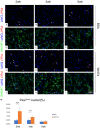
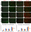
Results: Adult rat orbicularis oris muscle and tibialis anterior muscle exhibit similar regeneration-related activities after Wnt7a administration. Recombinant human Wnt7a administration resulted in enhanced cell proliferation, stem cell expansion, and fiber type remodelling in rat orbicularis oris muscle. In addition, newly formed myofibers were detected, contributing to an increased total fiber number.
Conclusion: Wnt7a induces vigorous regeneration in rat orbicularis oris muscle. This study helps lay a foundation for developing biotherapies to combat orofacial muscle deficiency.
Keywords: Skeletal muscle; cell proliferation; myosin heavy chain; regeneration.
IJCEP Copyright © 2021.
Conflict of interest statement
None.
Figures


Similar articles
-
Tensile force impairs lip muscle regeneration under the regulation of interleukin-10.J Cachexia Sarcopenia Muscle. 2024 Dec;15(6):2497-2508. doi: 10.1002/jcsm.13584. Epub 2024 Oct 1. J Cachexia Sarcopenia Muscle. 2024. PMID: 39351645 Free PMC article.
-
Wnt7a induces satellite cell expansion, myofiber hyperplasia and hypertrophy in rat craniofacial muscle.Sci Rep. 2018 Jul 13;8(1):10613. doi: 10.1038/s41598-018-28917-6. Sci Rep. 2018. PMID: 30006540 Free PMC article.
-
Co-delivery of Wnt7a and muscle stem cells using synthetic bioadhesive hydrogel enhances murine muscle regeneration and cell migration during engraftment.Acta Biomater. 2019 Aug;94:243-252. doi: 10.1016/j.actbio.2019.06.025. Epub 2019 Jun 19. Acta Biomater. 2019. PMID: 31228633 Free PMC article.
-
Development of the orbicularis oris muscle in normal and cleft lip and palate human fetuses using three-dimensional computer reconstruction.Plast Reconstr Surg. 1988 Mar;81(3):336-45. doi: 10.1097/00006534-198803000-00004. Plast Reconstr Surg. 1988. PMID: 3277211 Review.
-
Orofacial Muscles: Embryonic Development and Regeneration after Injury.J Dent Res. 2020 Feb;99(2):125-132. doi: 10.1177/0022034519883673. Epub 2019 Nov 1. J Dent Res. 2020. PMID: 31675262 Free PMC article. Review.
Cited by
-
Wnt/β-catenin signaling pathway as an important mediator in muscle and bone crosstalk: A systematic review.J Orthop Translat. 2024 Jun 20;47:63-73. doi: 10.1016/j.jot.2024.06.003. eCollection 2024 Jul. J Orthop Translat. 2024. PMID: 39007034 Free PMC article. Review.
References
-
- Schreurs M, Suttorp CM, Mutsaers HAM, Kuijpers-Jagtman AM, Von den Hoff JW, Ongkosuwito EM, Carvajal Monroy PL, Wagener FADTG. Tissue engineering strategies combining molecular targets against inflammation and fibrosis, and umbilical cord blood stem cells to improve hampered muscle and skin regeneration following cleft repair. Med Res Rev. 2020;40:9–26. - PMC - PubMed
-
- Dado DV, Kernahan DA. Anatomy of the orbicularis oris muscle in incomplete unilateral cleft lip based on histological examination. Ann Plast Surg. 1985;15:90–98. - PubMed
-
- Lazzeri D, Viacava P, Pollina LE, Sansevero S, Lorenzetti F, Balmelli B, Funel N, Gatti GL, Naccarato AG, Massei A. Dystrophic-like alterations characterize orbicularis oris and palatopharyngeal muscles in patients affected by cleft lip and palate. Cleft Palate Craniofac J. 2008;45:587–591. - PubMed
-
- Ridgway EB, Estroff JA, Mulliken JB. Thickness of orbicularis oris muscle in unilateral cleft lip: before and after labial adhesion. J Craniofac Surg. 2011;22:1822–1826. - PubMed
-
- de Chalain T, Zuker R, Ackerley C. Histologic, histochemical, and ultrastructural analysis of soft tissues from cleft and normal lips. Plast Reconstr Surg. 2001;108:605–611. - PubMed
LinkOut - more resources
Full Text Sources
