

Comparison of unenhanced magnetic resonance imaging and ultrasound in detecting very small hepatocellular carcinoma
- PMID: 34239704
- PMCID: PMC8239491
- DOI: 10.4254/wjh.v13.i6.699
Comparison of unenhanced magnetic resonance imaging and ultrasound in detecting very small hepatocellular carcinoma
Abstract
Background: In hepatocellular carcinoma (HCC), detection and treatment prior to growth beyond 2 cm are important as a larger tumor size is more frequently associated with microvascular invasion and/or satellites. In the surveillance of very small HCC nodules (≤ 2 cm in maximum diameter, Barcelona clinical stage 0), we demonstrated that the tumor markers alpha-fetoprotein and PIVKA-Ⅱ are not so useful. Therefore, we must survey with imaging modalities. The superiority of magnetic resonance imaging (MRI) over ultrasound (US) to detect HCC was confirmed in many studies. Although enhanced MRI is now performed to accurately diagnose HCC, in conventional clinical practice for HCC surveillance in liver diseases, unenhanced MRI is widely performed throughout the world. While, MRI has made marked improvements in recent years.
Aim: To make a comparison of unenhanced MRI and US in detecting very small HCC that was examined in the last ten years in patients in whom MRI and US examinations were performed nearly simultaneously.
Methods: In 394 patients with very small HCC nodules, those who underwent MRI and US at nearly the same time (on the same day whenever possible or at least within 14 days of one another) at the first diagnosis of HCC were selected. The detection rate of HCC with unenhanced MRI was investigated and compared with that of unenhanced US.
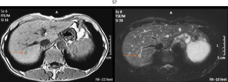
Results: The sensitivity of unenhanced MRI for detecting very small HCC was 95.1% (97/102, 95% confidence interval: 90.9-99.3) and that of unenhanced US was 69.6% (71/102, 95% confidence interval: 60.7-78.5). The sensitivity of unenhanced MRI for detecting very small HCC was significantly higher than that of unenhanced US (P < 0.001). Regarding the location of HCC in the liver in patients in whom detection by US was unsuccessful, S7-8 was identified in 51.7%.
Conclusion: Currently, unenhanced MRI is a very useful tool for the surveillance of very small HCC in conventional clinical follow-up practice.
Keywords: Comparison of magnetic resonance imaging and ultrasound; Magnetic resonance imaging; Surveillance of very small hepatocellular carcinoma; Ultrasound; Unenhanced magnetic resonance imaging.
©The Author(s) 2020. Published by Baishideng Publishing Group Inc. All rights reserved.
Conflict of interest statement
Conflict-of-interest statement: There are no conflicts of interest to disclose.
Figures


References
-
- Forner A, Llovet JM, Bruix J. Hepatocellular carcinoma. Lancet. 2012;379:1245–1255. - PubMed
-
- Stravitz RT, Heuman DM, Chand N, Sterling RK, Shiffman ML, Luketic VA, Sanyal AJ, Habib A, Mihas AA, Giles HC, Maluf DG, Cotterell AH, Posner MP, Fisher RA. Surveillance for hepatocellular carcinoma in patients with cirrhosis improves outcome. Am J Med. 2008;121:119–126. - PubMed
-
- Tarao K, Nozaki A, Komatsu H, Komatsu T, Taguri M, Tanaka K, Chuma M, Numata K, Maeda S. Real impact of tumor marker AFP and PIVKA-II in detecting very small hepatocellular carcinoma (≤ 2 cm, Barcelona stage 0) - assessment with large number of cases. World J Hepatol. 2020;12:1046–1054. - PMC - PubMed
-
- Colli A, Fraquelli M, Casazza G, Massironi S, Colucci A, Conte D, Duca P. Accuracy of ultrasonography, spiral CT, magnetic resonance, and alpha-fetoprotein in diagnosing hepatocellular carcinoma: a systematic review. Am J Gastroenterol. 2006;101:513–523. - PubMed
LinkOut - more resources
Full Text Sources
