. 2021 Jun 28;1(6):865-878.
doi: 10.1021/jacsau.1c00078. Epub 2021 May 25.

Iron Chelator Transmetalative Approach to Inhibit Human Ribonucleotide Reductase

Affiliations

Iron Chelator Transmetalative Approach to Inhibit Human Ribonucleotide Reductase

Kavita Gaur et al. JACS Au. .

Abstract

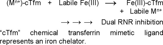
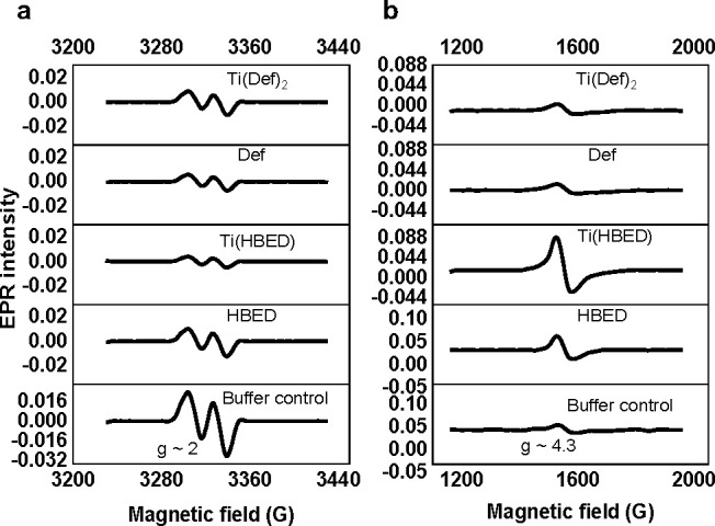
Efforts directed at curtailing the bioavailability of intracellular iron could lead to the development of broad-spectrum anticancer drugs given the metal's role in cancer proliferation and metastasis. Human ribonucleotide reductase (RNR), the key enzyme responsible for synthesizing the building blocks of DNA replication and repair, depends on Fe binding at its R2 subunit to activate the catalytic R1 subunit. This work explores an intracellular iron chelator transmetalative approach to inhibit RNR using the titanium(IV) chemical transferrin mimetic (cTfm) compounds Ti(HBED) and Ti(Deferasirox)2. Whole-cell EPR studies reveal that the compounds can effectively attenuate RNR activity though seemingly causing different changes to the labile iron pool that may account for differences in their potency against cells. Studies of Ti(IV) interactions with the adenosine nucleotide family at pH 7.4 reveal strong metal binding and extensive phosphate hydrolysis, which suggest the capacity of the metal to disturb the nucleotide substrate pool of the RNR enzyme. By decreasing intracellular Fe bioavailability and altering the nucleotide substrate pool, the Ti cTfm compounds could inhibit the activity of the R1 and R2 subunits of RNR. The compounds arrest the cell cycle in the S phase, indicating suppressed DNA replication, and induce apoptotic cell death. Cotreatment cell viability studies with cisplatin and Ti(Deferasirox)2 reveal a promising synergism between the compounds that is likely owed to their distinct but complementary effect on DNA replication.


Conflict of interest statement

The authors declare no competing financial interest.

Figures

Scheme 1
Scheme 1
Figure 1
Figure 1
Metal ion coordination to the chemical transferrin mimetic (cTfm) chelators. (a) Physiologically relevant (pH 7.4) Fe(III) coordination by human serum transferrin (sTf) and the cTfm chelators HBED and Def. (b) ORTEP diagram of the neutral compound Ti(Def)2 (Ti[C42H26N6O8]) with ellipsoids set at 50% probability (hydrogen atoms were omitted for clarity). (c) The pH-dependent speciation of the Ti(IV) HBED at a 1:1 metal/ligand ratio and micromolar concentrations. (d) The pH-dependent speciation of the Ti(IV) deferasirox at a 1:2 metal/ligand ratio and micromolar concentrations. The complete speciation diagrams can be found in Figure S1.
Figure 2
Figure 2
Whole-cell EPR spectra for the 3 h treatment of Jurkat cells with 50 μM [Ti(Def)2]2–, Def, [TiO(H+–HBED)]1–, and HBED2– and also the buffer control. The EPR signal corresponding to (a) the tyrosyl radical of the RNR enzyme (g ≈ 2) and to the (b) intracellular high-spin Fe(III) (S = 5/2; g ≈ 4.3). Experimental conditions: microwave frequency, 9.338 GHz; microwave power, 2 mW; magnetic field modulation amplitude, 0.5 mT for the tyrosyl radical and 1 mT for high-spin Fe(III) (S = 5/2), temperature: 20 K. All EPR spectra were baseline-corrected.
Figure 3
Figure 3
Effect of compounds (written without charge states for simplicity) on Jurkat cells. (a) Cell cycle analyses using flow cytometry at 3, 10, and 24 h of treatment with 29 μM Def, 18.8 μM [Ti(Def)2]2–, and 26.3 μM HBED and [TiO(H+–HBED)]. (b) Time-dependent apoptosis assay performed using flow cytometry after treating the cells with 20 μM cisplatin, 50 μM [Ti(Cit)3]8–, 19 μM [Ti(Def)2]2–, 27 μM [TiO(H+–HBED)], 29 μM Def, 27 μM HBED2–, and buffer control for 24 and 72 h and then staining with annexin V–Alexa Fluor 488/propidium iodide (PI). * p-value ≤ 0.05 vs the corresponding control group.
Figure 4
Figure 4
Isobolographic analysis of the antiproliferative activity of the 1:1 and 1:2 [Ti(Def)2]2–/cisplatin combinations against Jurkat cells for 48 h.
Figure 5
Figure 5
31P NMR spectra for (a) Ti6O7(ATP)5(H2O)34 and (b) ATP at pH 7.4 and 5 mM concentration based on ATP content. Also refer to Table 2 as well as Figures S14 (a zoom-in of this figure) and S15.
Scheme 2
Scheme 2. Proposed Mechanism of Action for Ti(IV) cTfm as Combined R1 and R2 RNR Inhibitors
(a) Decreasing the LIP via transmetalation would prevent Fe binding of the R2 site and enzyme activation. (b) Ti(IV) modification of the nucleotide pool would inhibit substrate binding to the catalytic site of the R1 subunit. Created with BioRender.com.

References

    1. Weinberg R.The biology of cancer, 2nd ed.; Garland Science: New York, 2013; p 960.
    1. Cairo G.; Bernuzzi F.; Recalcati S. A precious metal: Iron, an essential nutrient for all cells. Genes Nutr. 2006, 1 (1), 25–39. 10.1007/BF02829934. - DOI - PMC - PubMed
    1. Lane D. J. R.; Merlot A. M.; Huang M. L. H.; Bae D. H.; Jansson P. J.; Sahni S.; Kalinowski D. S.; Richardson D. R. Cellular iron uptake, trafficking and metabolism: Key molecules and mechanisms and their roles in disease. Biochim. Biophys. Acta, Mol. Cell Res. 2015, 1853 (5), 1130–1144. 10.1016/j.bbamcr.2015.01.021. - DOI - PubMed
    1. Sutherland R.; Delia D.; Schneider C.; Newman R.; Kemshead J.; Greaves M. Ubiquitous cell-surface glycoprotein on tumor cells is proliferation-associated receptor for transferrin. Proc. Natl. Acad. Sci. U. S. A. 1981, 78 (7), 4515.10.1073/pnas.78.7.4515. - DOI - PMC - PubMed
    1. Philpott C. C.; Ryu M.-S. Special delivery: distributing iron in the cytosol of mammalian cells. Front. Pharmacol. 2014, 5, 173.10.3389/fphar.2014.00173. - DOI - PMC - PubMed