Review
. 2021 Sep 15:7:693-726.
doi: 10.1146/annurev-vision-100419-111350. Epub 2021 Jul 9.

Optical Coherence Tomography and Glaucoma

Affiliations
Review

Optical Coherence Tomography and Glaucoma

Alexi Geevarghese et al. Annu Rev Vis Sci. .

Abstract

Early detection and monitoring are critical to the diagnosis and management of glaucoma, a progressive optic neuropathy that causes irreversible blindness. Optical coherence tomography (OCT) has become a commonly utilized imaging modality that aids in the detection and monitoring of structural glaucomatous damage. Since its inception in 1991, OCT has progressed through multiple iterations, from time-domain OCT, to spectral-domain OCT, to swept-source OCT, all of which have progressively improved the resolution and speed of scans. Even newer technological advancements and OCT applications, such as adaptive optics, visible-light OCT, and OCT-angiography, have enriched the use of OCT in the evaluation of glaucoma. This article reviews current commercial and state-of-the-art OCT technologies and analytic techniques in the context of their utility for glaucoma diagnosis and management, as well as promising future directions.

Keywords: OCT; glaucoma; imaging; optical coherence tomography.


Figures

Figure 1
Figure 1
Stratus-OCT (Carl Zeiss Meditec, Dublin, California) B-scan of (a) the peripapillary retina with retinal layers labeled and (b) the macular retina. Abbreviations: INL, inner nuclear layer; IPL, inner plexiform layer; NFL, nerve fiber layer; OCT, optical coherence tomography; ONL, outer nuclear layer; OPL, outer plexiform layer; RPE, retinal pigment epithelium.
Figure 2
Figure 2
Cirrus HD-OCT (Carl Zeiss Meditec, Dublin, California) report for a subject with one glaucomatous eye (OD) and one nonglaucomatous eye (OS). (a) The RNFL thickness map (①) displays RNFL thickness around the optic disc through a color map, with the thicker RNFL measurements in red and yellow and the thinner RNFL measurements in green and blue. RNFL thickness measurements by quadrant (S, I, N, and T) and clock hour are shown. (②) Values within the normal range of age-matched controls are in green (within normal limits). Values that fall outside the normal range for their age are displayed in yellow if p < 5% and ≥ 1% (borderline) or in red if p < 1% (outside normal limits). RNFL thickness in the superior and inferior quadrants is abnormal in OD compared with OS, in which the inferior quadrant is borderline. The RNFL deviation map (③) shows RNFL thickness deviations from the normative database overlaid on an en face image. Borderline RNFL thickness measurements are shown in yellow, and RNFL thicknesses outside normal limits are shown in red. Substantial glaucomatous thinning is seen in red OD. Abbreviations: C/D ratio, cup-to-disc ratio; HD-OCT, high-definition optical coherence tomography; I, inferior; N, nasal; NA, not applicable; ONH, optic nerve head; RNFL, retinal nerve fiber layer; S, superior; T, temporal.
Figure 3
Figure 3
Example of a GPA report (Carl Zeiss Meditec, Dublin, California) of a glaucomatous eye that has shown progression in peripapillary RNFL thickness (format modified for publication). The deviation map (①) shows areas of change that are statistically significant for the first time (orange) and subsequent areas of statistically significant change (red). The location and shape of these areas of change indicate the likelihood that the RNFL thinning actually represents glaucoma and its progression. In other words, if the location and shape are consistent with glaucoma, then it is likely that the detected changes are due to glaucoma. Below the deviation maps, graphs show the average RNFL, the superior RNFL, and the inferior RNFL thicknesses over time (②). Again, the first statistically significant drop in RNFL thickness is indicated by an orange dot, and subsequent significant reductions are indicated by red dots. These are trend analyses, as opposed to the event analyses shown in the deviation maps. The average cup-to-disc ratio graph indicates whether there is a statistically significant increase in cup-to-disc ratio. If the RNFL is decreasing and the cup-to-disc ratio is increasing, then this is good corroboratory evidence that the change measured is real. Below these graphs is the RNFL thickness profile (③), on which the baseline profiles (B_1 and B_2) are overlaid on today’s visit (C). This is a useful graph in that the locations and degrees of thinning can be seen, and if they are in areas that would be expected to change with glaucoma, then this increases the likelihood that those changes are real glaucomatous changes. The table of RNFL and ONH summary parameters (④) presents the same data shown in panel a and highlights statistically significant change in orange and red. Abbreviations: GPA, guided progression analysis; INF, inferior; NAS, nasal; ONH, optic nerve head; RNFL, retinal nerve fiber layer; SS, signal strength; SUP, superior; TEMP, temporal.
Figure 4
Figure 4
SS-OCT images displaying the pores (blue) and the surrounding beams (yellow) in the microstructure of the lamina cribrosa of (a,b) healthy and (c,d) glaucomatous eyes. Although the differences in microstructure between glaucomatous and healthy eyes are not visually apparent, using a segmentation analysis that quantifies the microstructure, Wang et al. (2013) found a greater beam thickness–to–pore diameter ratio and greater variability in pore diameter in glaucomatous eyes than in healthy eyes. Figure adapted from Wang et al. (2013); copyright 2013 IOVS, Association for Research in Vision Science. Abbreviation: SS-OCT, swept-source optical coherence tomography.
Figure 5
Figure 5
(a) AngioPlex OCT-angiography (Carl Zeiss Meditec, Dublin, California) image of a glaucomatous eye. (b) Peripapillary superficial vessel density (mm/mm2) in the central, inner, outer, and full regions is quantified in the table. (c,d) Region of decreased vessel density correlates with region of RNFL thinning as seen on (c) the RNFL thickness map and (d) the RNFL deviation map from the Cirrus HD-OCT (Carl Zeiss Meditec) report. Abbreviations: ETDRS, Early Treatment Diabetic Retinopathy Study; HD-OCT, high-definition OCT; OCT, optical coherence tomography; RNFL, retinal nerve fiber layer.
Figure 6
Figure 6
The artery (Ainf) and vein (Vinf) at the dotted line on the (a) en face visible-light OCT image are seen in the (b) B-scan, from which (c) their respective spectroscopic OCT signals are fitted to the hemoglobin absorption profile to determine retinal arterial and venous blood oxygen saturations. Image courtesy of Z. Ghassabi, M. Wu, I. Rubinoff, Y. Wang, B. Davis, B. Tayebi, G. Wollstein, J. Schuman, H. Zhang & H. Ishikawa (unpublished study). Abbreviation: OCT, optical coherence tomography.
Figure 7
Figure 7
Convolutional neural networks. Filter functions reduce a portion of the depicted input image (box) into feature maps (gray squares), which are representations of the presence of features in the input image (①). Pooling layers downscale feature information by summarizing parts of the feature map (②). Forward feeding of information to subsequent layers produces the final output layer, which ultimately yields the desired classification (③). Abbreviations: N, nasal; T, temporal.

References

    1. Adhi M, Liu JJ, Qavi AH, Grulkowski I, Lu CD, et al. 2014. Choroidal analysis in healthy eyes using swept-source optical coherence tomography compared to spectral domain optical coherence tomography. Am. J. Ophthalmol 157:1272–81.e1 - PubMed
    1. Alnawaiseh M, Lahme L, Müller V, Rosentreter A, Eter N. 2018. Correlation of flow density, as measured using optical coherence tomography angiography, with structural and functional parameters in glaucoma patients. Graefes Arch. Clin. Exp. Ophthalmol 256:589–97 - PubMed
    1. Ang BCH, Lim SY, Dorairaj S. 2020. Intra-operative optical coherence tomography in glaucoma surgery—a systematic review. Eye 34:168–77 - PMC - PubMed
    1. Arend O, Plange N, Sponsel WE, Remky A. 2004. Pathogenetic aspects of the glaucomatous optic neuropathy: fluorescein angiographic findings in patients with primary open angle glaucoma. Brain Res. Bull 62:517–24 - PubMed
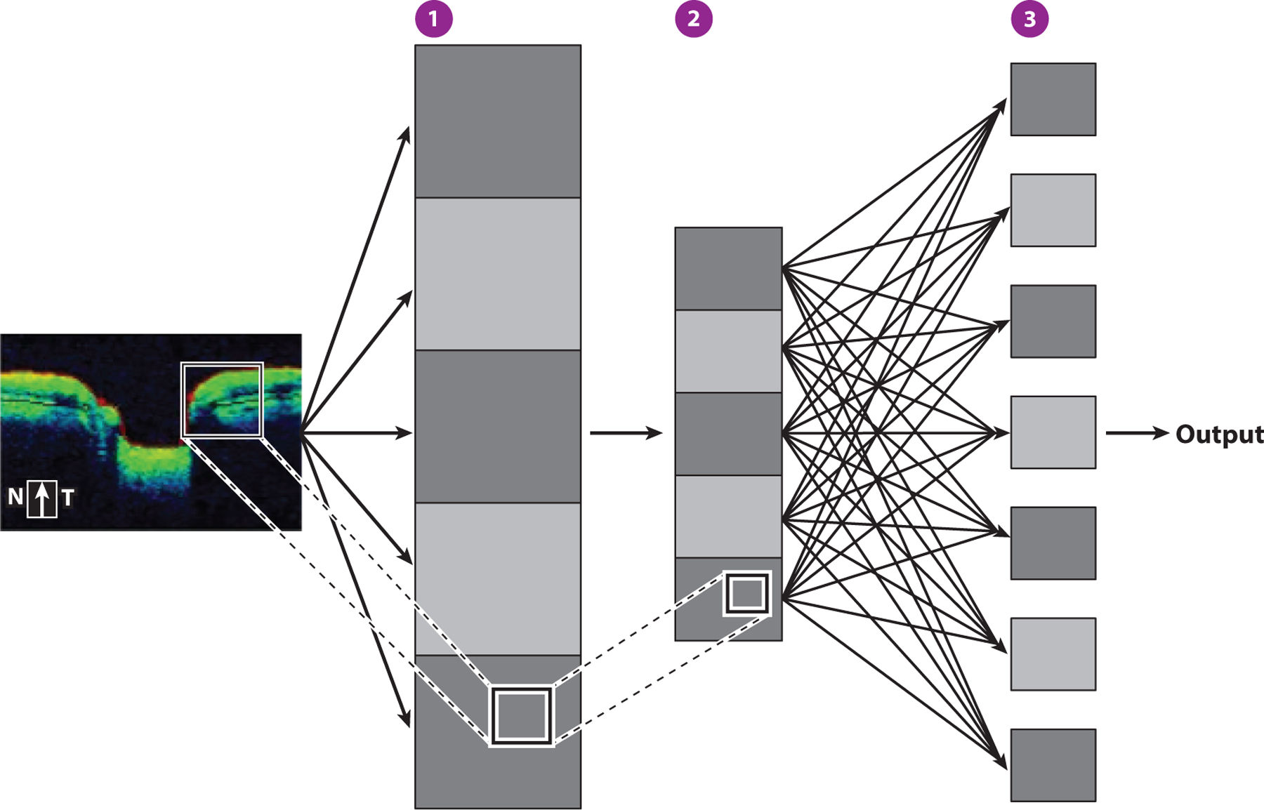
    1. Asaoka R, Murata H, Hirasawa K, Fujino Y, Matsuura M, et al. 2019. Using deep learning and transfer learning to accurately diagnose early-onset glaucoma from macular optical coherence tomography images. Am. J. Ophthalmol 198:136–45 - PubMed

Publication types

MeSH terms