

Pulmonary fibrosis and its related factors in discharged patients with new corona virus pneumonia: a cohort study
- PMID: 34243776
- PMCID: PMC8267229
- DOI: 10.1186/s12931-021-01798-6
Pulmonary fibrosis and its related factors in discharged patients with new corona virus pneumonia: a cohort study
Abstract
Background: Thousands of Coronavirus Disease 2019 (COVID-19) patients have been discharged from hospitals Persistent follow-up studies are required to evaluate the prevalence of post-COVID-19 fibrosis.
Methods: This study involves 462 laboratory-confirmed patients with COVID-19 who were admitted to Shenzhen Third People's Hospital from January 11, 2020 to April 26, 2020. A total of 457 patients underwent thin-section chest CT scans during the hospitalization or after discharge to identify the pulmonary lesion. A total of 287 patients were followed up from 90 to 150 days after the onset of the disease, and lung function tests were conducted about three months after the onset. The risk factors affecting the persistence of pulmonary fibrosis were identified through regression analysis and the prediction model of the persistence of pulmonary fibrosis was established.
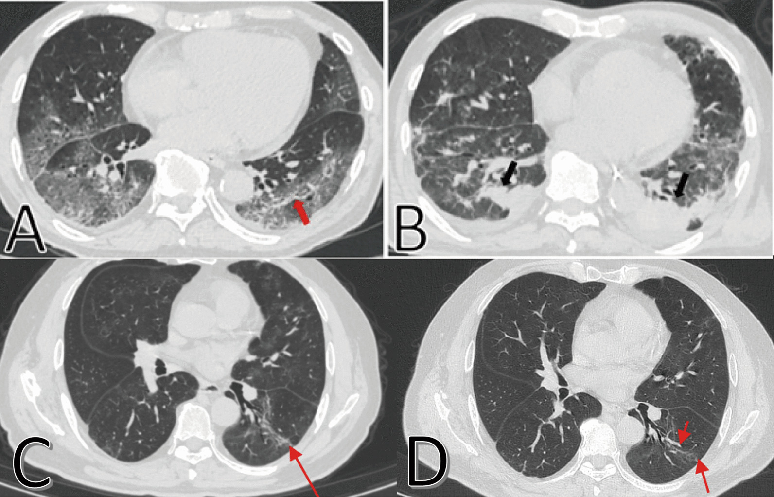
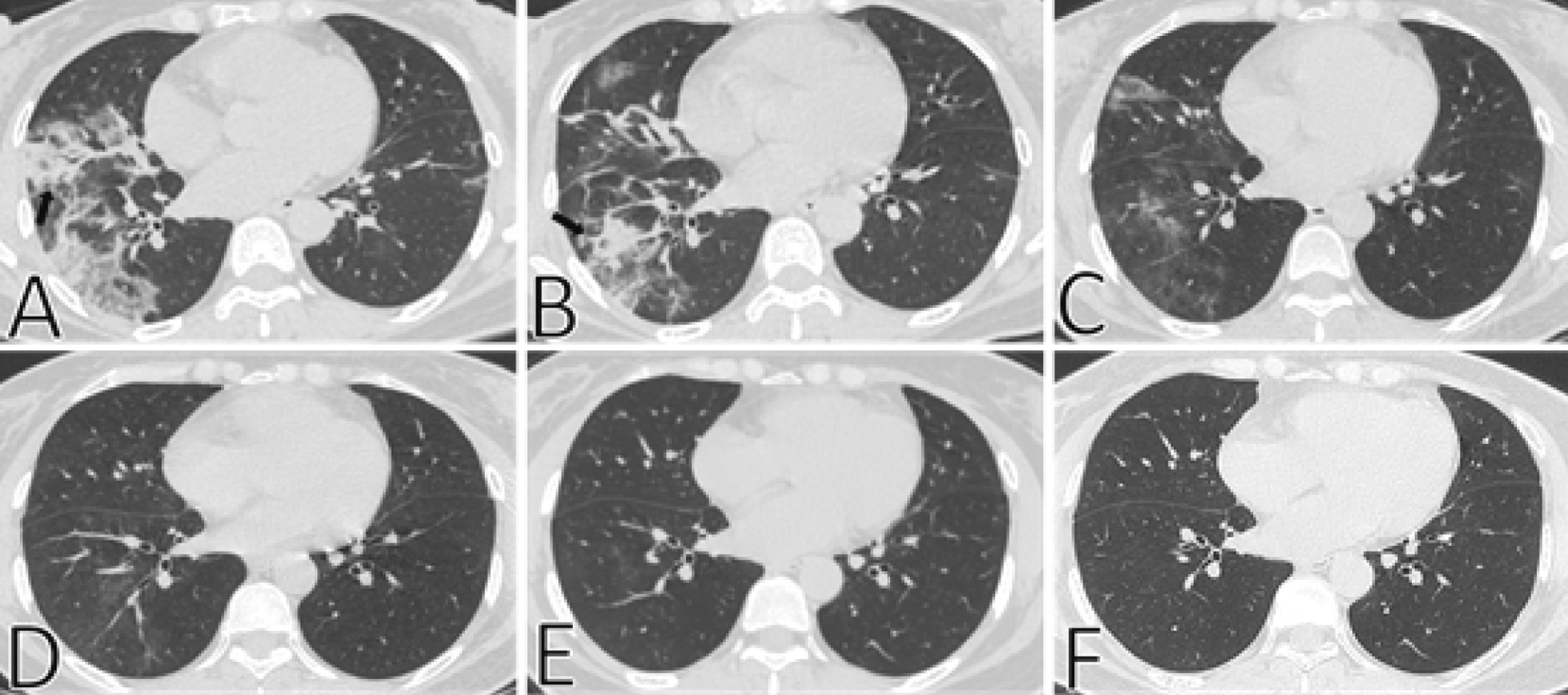
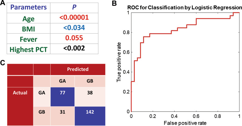
Results: Parenchymal bands, irregular interfaces, reticulation and traction bronchiectasis were the most common CT features in all COVID-19 patients. During the 0-30, 31-60, 61-90, 91-120 and > 120 days after onset, 86.87%, 74.40%, 79.56%, 68.12% and 62.03% patients developed with pulmonary fibrosis and 4.53%, 19.61%, 18.02%, 38.30% and 48.98% patients reversed pulmonary fibrosis, respectively. It was observed that Age, BMI, Fever, and Highest PCT were predictive factors for sustaining fibrosis even after 90 days from onset. A predictive model of the persistence with pulmonary fibrosis was developed based-on the Logistic Regression method with an accuracy, PPV, NPV, Sensitivity and Specificity of the model of 76%, 71%, 79%, 67%, and 82%, respectively. More than half of the COVID-19 patients revealed abnormal conditions in lung function after 90 days from onset, and the ratio of abnormal lung function did not differ on a statistically significant level between the fibrotic and non-fibrotic groups.
Conclusions: Persistent pulmonary fibrosis was more likely to develop in patients with older age, higher BMI, severe/critical condition, fever, a longer viral clearance time, pre-existing disease and delayed hospitalization. Fibrosis developed in COVID-19 patients could be reversed in about a third of the patients after 120 days from onset. The pulmonary function of less than half of COVID-19 patients could turn to normal condition after three months from onset. An effective prediction model with an average area under the curve (AUC) of 0.84 was established to predict the persistence of pulmonary fibrosis in COVID-19 patients for early diagnosis.
Keywords: COVID-19 recovered patient; Lung function; Persistent consequences; Pulmonary fibrosis; Risk factor.
© 2021. The Author(s).
Conflict of interest statement
The authors declare no competing interests.
Figures
References
-
- Huang C, Wang Y, Li X, Ren L, Zhao J, Hu Y, Zhang L, Fan G, Xu J, Gu X, Cheng Z, Yu T, Xia J, Wei Y, Wu W, Xie X, Yin W, Li H, Liu M, Xiao Y, Gao H, Guo L, Xie J, Wang G, Jiang R, Gao Z, Jin Q, Wang J, Cao B. Clinical features of patients infected with 2019 novel coronavirus in Wuhan. China Lancet. 2020;395(10223):497–506. - PMC - PubMed
-
- https://covid19.who.int/. Data last updated: 2020/7/14, 10:42am CEST.
-
- Antonio GE, Wong KT, Hui DS, Wu A, Lee N, Yuen EH, Leung CB, Rainer TH, Cameron P, Chung SS, Sung JJ, Ahuja AT. Thin-section CT in patients with severe acute respiratory syndrome following hospital discharge: preliminary experience. Radiology. 2003;228(3):810–815. doi: 10.1148/radiol.2283030726. - DOI - PubMed
MeSH terms
Grants and funding
LinkOut - more resources
Full Text Sources
Medical
Miscellaneous
