Review
. 2021 Nov;46(11):902-917.
doi: 10.1016/j.tibs.2021.06.003. Epub 2021 Jul 7.

Mitochondrial compartmentalization: emerging themes in structure and function

Affiliations
Review

Mitochondrial compartmentalization: emerging themes in structure and function

Joseph C Iovine et al. Trends Biochem Sci. 2021 Nov.

Abstract

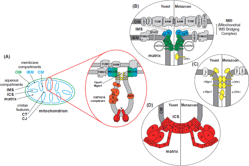
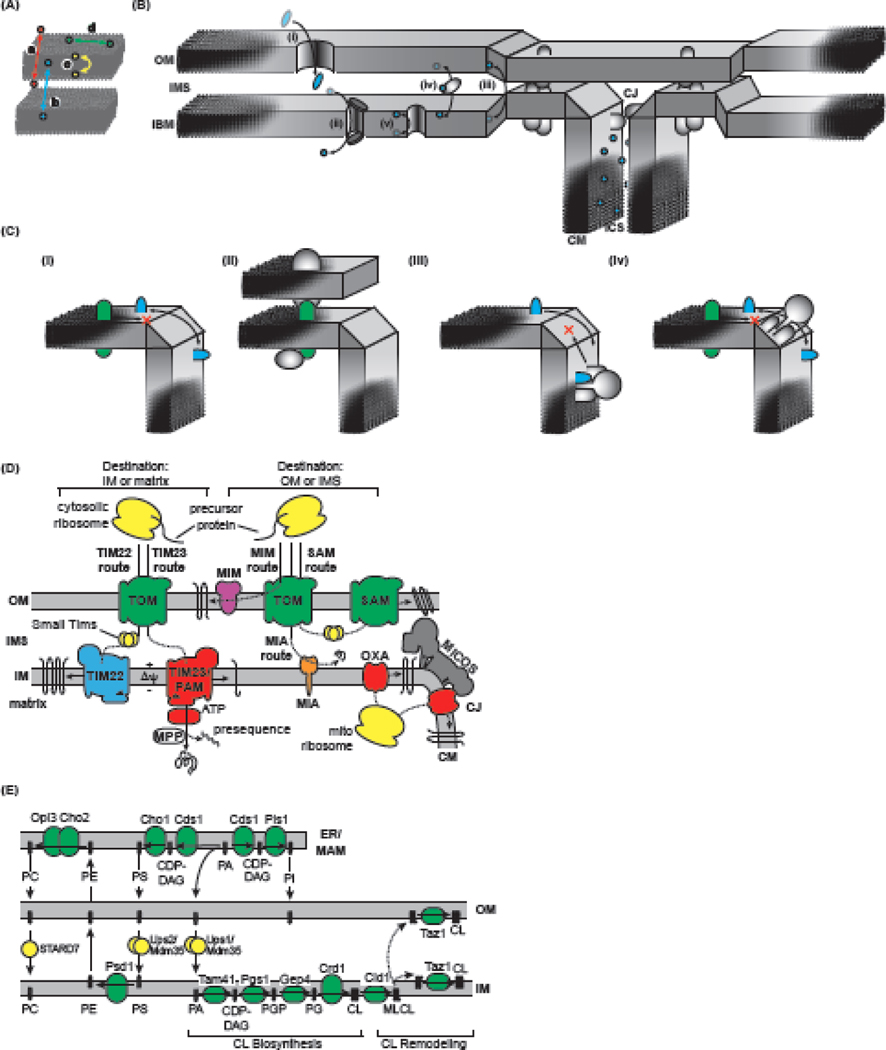
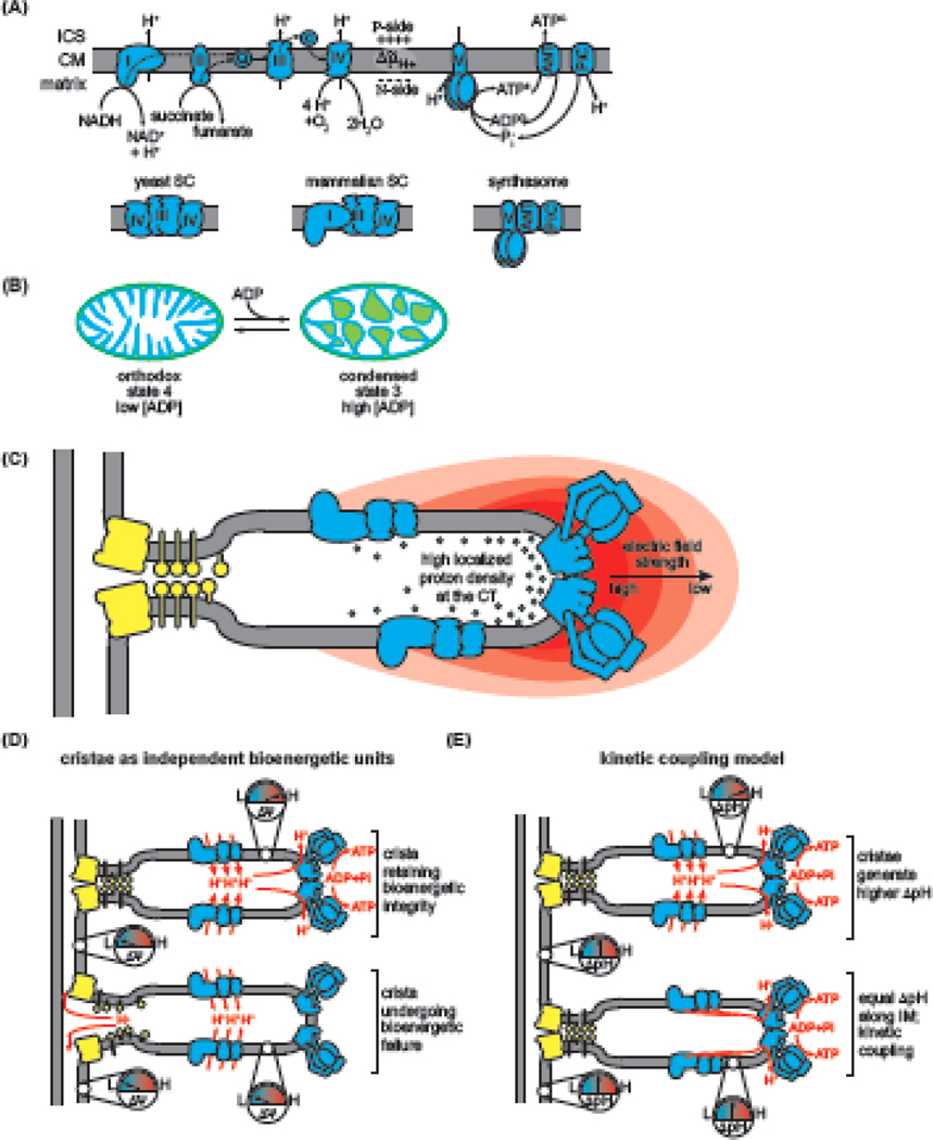
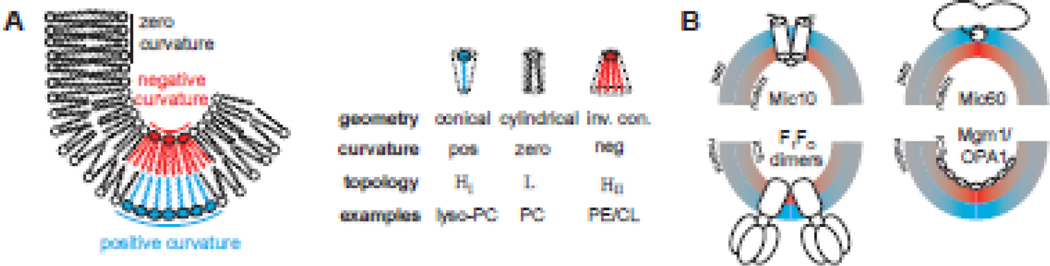
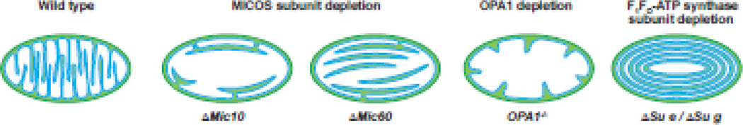
Within cellular structures, compartmentalization is the concept of spatial segregation of macromolecules, metabolites, and biochemical pathways. Therefore, this concept bridges organellar structure and function. Mitochondria are morphologically complex, partitioned into several subcompartments by a topologically elaborate two-membrane system. They are also dynamically polymorphic, undergoing morphogenesis events with an extent and frequency that is only now being appreciated. Thus, mitochondrial compartmentalization is something that must be considered both spatially and temporally. Here, we review new developments in how mitochondrial structure is established and regulated, the factors that underpin the distribution of lipids and proteins, and how they spatially demarcate locations of myriad mitochondrial processes. Consistent with its pre-eminence, disturbed mitochondrial compartmentalization contributes to the dysfunction associated with heritable and aging-related diseases.

Keywords: bioenergetics; cristae; macromolecular trafficking; mitochondria; morphogenesis; ultrastructure.


Conflict of interest statement

Declaration of interests None declared by authors.

Figures

Figure 1.
Figure 1.. Mitochondrial Ultrastructure and Morphogenic Complexes.
A) Major mitochondrial compartments. Subcompartments are established by the outer membrane (OM; green) and inner membrane (IM; cyan), parsed into the inner boundary membrane (IBM) and cristae membrane (CM). Aqueous compartments include the intermembrane space (IMS) (between OM and IBM), the intracristal space (ICS) (enclosed by cristae), and matrix. Major cristae features include the crista junction (CJ) and the crista tip (CT). B-D) Assemblies that establish IM morphology. The subunits of each complex are shown for yeast (left) and for metazoans (right). B) The MICOS Complex establishes and stabilizes CJs. MICOS is composed of six (yeast) or seven (metazoan) known subunits, each named as MicX (X numbering based on molecular weight). The MIC60 subcomplex (cyan) consists of the central Mic60 (mitofilin) subunit and peripheral regulatory proteins Mic19/25, mediating a number of interactions (dashed lines) with IBM and OM proteins. The MIC10 subcomplex (green) contains the central Mic10 subunit, apolipoprotein O-related Mic26/27, which antagonistically regulate Mic10 assembly, and Mic12, which mediates MIC60-MIC10 subcomplex association. C) Mgm1/Opa1 Assemblies are GTPases that exist as long forms (l-Mgm1/l-Opa1) with an N-terminal transmembrane anchor, or as short forms (s-Mgm1/s-Opa1) that are proteolytically processed by IM proteases Pcp1 (yeast) or OMA1/YME1 (metazoan). Oligomers of l-Mgm1/l-Opa1 assemble in a cardiolipin-dependent manner to stabilize connections of cristae walls. D) The F1FO-ATP synthase consists of the F1 sector [catalytic headpiece (α3β3) and central stalk (γ,δ,ε)] and FO sector [rotor (c ring, a, 8), peripheral stalk (b, d, h/F6, OSCP), and supernumerary subunits (f, i/j/6.8, e, g, k)]. Homodimer assembly is mediated by subunits e, g and k (DAPIT in metazoans). Higher-order assembly occurs as extended dimer rows along cristae ridges.
Figure 2.
Figure 2.. Determinants of Organellar Compartmentalization.
A) Defined physical compartments include those that exist (a) between membrane-separated aqueous compartments, (b) between membranes, (c) between leaflets of a given membrane, and (d) between lateral zones of a membrane. B) Inter-compartment transit routes include (i) passive or (ii) active transporters that allow movement of molecules between aqueous compartments; (iii) transient and/or regulated contact sites between membranes, (iv) carrier proteins that ferry lipids between distinct membranes; and (v) transporters that mediate transbilayer lipid diffusion. C) Lateral compartmentalization between the inner boundary membrane (IBM) and cristae membrane (CM) originates from several nonexclusive mechanisms, including (i) restricted diffusion related to membrane curvature, (ii) compartment-specific localization by protein interactions, (iii) active sorting by crista junction (CJ) complexes (e.g., MICOS), and (iv) assembly of otherwise mobile subunits into large quaternary assemblies. IMS, intermembrane space; ICS, intracristal space. D) Protein trafficking routes include the Translocon of the Outer Mitochondrial Membrane (TOM) and Sorting and Assembly Machinery (SAM) complexes of the outer membrane (OM), and the Translocons of the Inner Mitochondrial Membrane 23 and 22 (TIM23 and TIM22), the Presequence Translocase-Associated Motor (PAM) complex, the Mitochondrial Intermembrane Space Assembly (MIA) and Oxidase Assembly (OXA) translocase. Targeted polypeptides (precursors) contain targeting information as cleavable presequences or in the protein itself that is recognized by cognate transport machineries to sort proteins to their correct destinations. E) Mitochondrial lipid homeostasis requires the uptake of lipids that are mostly produced in the mitochondrialassociated membrane (MAM) subcompartment of the ER. CDP-DAG, cytidine diphosphate glycerol; Cho, PS synthase; Cds1, CDP-DAG synthase; Opi3/Cho2, PE methyltransferases; PI, phosphatidylinositol; Pis1, PI synthase; Psd1, PS decarboxylase 1.
Figure 3.
Figure 3.. Mitochondrial Compartmentalization and Energy Transduction.
A) The OXPHOS system comprises the electron transport chain of respiratory complexes CI-CIV, which accept reducing equivalents and pass them through a series of redox centers, vectorially pumping H+ toward the intracristal space (ICS) at CI, CIII, and CIV to establish the Δμ˜H+ across the cristae membrane (CM). ATP synthesis involves the F1FO-ATP synthase, transporters that mediate ADP/ATP exchange (ANT in mammals, Aac in yeast), and H+/Pi symport (PiC), together defining the synthasome supercomplex (SC). The Δμ˜H+ provides a reservoir of H+ that flows toward the matrix across the membrane-bound rotary motor of the FO sector of ATP synthase, driving ATP synthesis on the catalytic F1 sector. Below, structural organization of the yeast and metazoan respiratory SCs and the metazoan synthasome. B) Inner membrane (IM) plasticity coupled to the metabolic state shown by the transition from orthodox topology (matrix expanded, ICS contracted) to condensed topology (matrix contracted, ICS expanded) when respiration rate increases upon ADP addition. C) Cristae geometry enhances the electrochemical proton potential, illustrated by a greater electric field (gradient of red arcs) greater localized proton density (green spheres) at the highly curved regions of the crista tip (CT) (adapted from [27]). D) Hetero-potential model [106]. Upper, the Δψ of individual cristae are electrochemically insulated from the inner boundary membrane (IBM) and from each other, which enhances the energetic reserves of cristae. Lower, bioenergetic crisis in individual cristae are isolated from other crista. E) Models of ΔpH distribution. Upper, cristae are modeled as domains of enhanced ΔpH relative to IBM. Lower, ΔpH does not differ significantly between CM and IBM, but OXPHOS efficiency is enhanced by lateral membrane diffusion of protons between proton gradient sources and sinks (kinetic coupling model of [107]).
Box 2, Figure I.
Box 2, Figure I.
Membrane curvature determinants include (A) lipids and (B) membrane-interactive proteins that promote local positive (cyan) and negative (red) membrane curvature.
Box 3, Figure I.
Box 3, Figure I.
Schematic illustration of wild type mitochondrial ultrastructure with lamellar cristae in comparison with those deficieint in MICOS subunits, OPA1, and dimer-stabilizing subunits of F1FO-ATP synthase as indicated.

References

    1. Cogliati S. et al. (2016) Mitochondrial Cristae: Where Beauty Meets Functionality. Trends Biochem Sci 41 (3), 261–273. - PubMed
    1. Quintana-Cabrera R. et al. (2018) Who and how in the regulation of mitochondrial cristae shape and function. Biochem Biophys Res Commun 500 (1), 94–101. - PubMed
    1. Giacomello M. et al. (2020) The cell biology of mitochondrial membrane dynamics. Nat Rev Mol Cell Biol 21 (4), 204–224. - PubMed
    1. Li W. et al. (2020) Dynamic organization of intracellular organelle networks. Wiley Interdiscip Rev Syst Biol Med, e1505. - PubMed
    1. Rasmussen N. (1995) Mitochondrial structure and the practice of cell biology in the 1950s. J Hist Biol 28 (3), 381–429. - PubMed

Publication types

MeSH terms

Substances

LinkOut - more resources