. 2021 Jul 9;7(28):eabg5060.
doi: 10.1126/sciadv.abg5060. Print 2021 Jul.

Mechanical forces regulate endothelial-to-mesenchymal transition and atherosclerosis via an Alk5-Shc mechanotransduction pathway

Affiliations

Mechanical forces regulate endothelial-to-mesenchymal transition and atherosclerosis via an Alk5-Shc mechanotransduction pathway

Vedanta Mehta et al. Sci Adv. .

Abstract

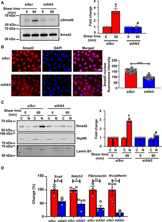
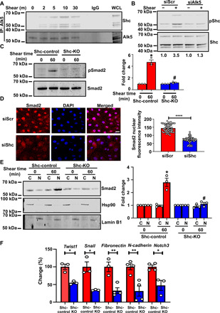
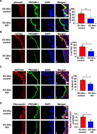
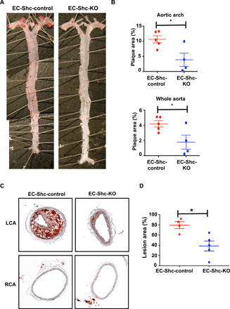
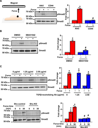
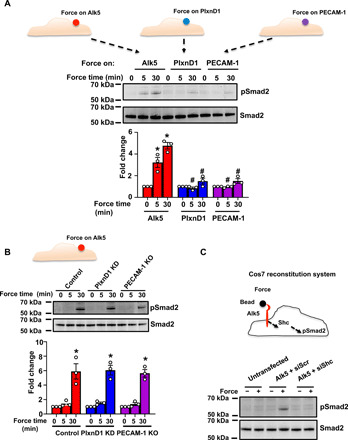
The response of endothelial cells to mechanical forces is a critical determinant of vascular health. Vascular pathologies, such as atherosclerosis, characterized by abnormal mechanical forces are frequently accompanied by endothelial-to-mesenchymal transition (EndMT). However, how forces affect the mechanotransduction pathways controlling cellular plasticity, inflammation, and, ultimately, vessel pathology is poorly understood. Here, we identify a mechanoreceptor that is sui generis for EndMT and unveil a molecular Alk5-Shc pathway that leads to EndMT and atherosclerosis. Depletion of Alk5 abrogates shear stress-induced EndMT responses, and genetic targeting of endothelial Shc reduces EndMT and atherosclerosis in areas of disturbed flow. Tensional force and reconstitution experiments reveal a mechanosensory function for Alk5 in EndMT signaling that is unique and independent of other mechanosensors. Our findings are of fundamental importance for understanding how mechanical forces regulate biochemical signaling, cell plasticity, and vascular disease.


Figures

Fig. 1
Fig. 1. Shear stress induces EndMT via Alk5.
(A) Mouse ECs were transfected with scrambled (Scr) or Alk5 siRNA and exposed to fluid shear stress (12 dynes cm−2) using a parallel plate system for 60 min. Phosphorylation of Smad2 was determined by Western blotting and quantified using Image Studio Lite v.5.2. (B) BAECs were transfected with Scr or Alk5 siRNA and exposed to fluid shear stress (12 dynes cm−2) using a parallel plate system for 60 min. Nuclear translocation of Smad2 was determined by immunofluorescence staining and quantified as mean fluorescence intensity on ImageJ. DAPI, 4′,6-diamidino-2-phenylindole. (C) Mouse ECs were transfected with Scr or Alk5 siRNA and exposed to fluid shear stress (12 dynes cm−2) using a parallel plate system for 60 min. The cells were then separated into cytosolic and nuclear fractions before analysis by Western blotting with the Smad2 antibody. Western blots, performed using antibodies against the cytoplasmic protein Hsp90 and nuclear protein Lamin B1, indicated that the extracts are virtually free from cross-contamination. Hsp90 served as the loading control for cytoplasmic extracts and Lamin B1 for nuclear extracts. (D) Mouse ECs were transfected with either Scr or Alk5 siRNA and exposed to atheroprone flow for 48 hours using a cone-and-plate viscometer. Quantitative polymerase chain reaction (qPCR) was performed to quantify the expression of the EndMT markers Snail, fibronectin, Notch3, and N-cadherin. Scale bar, 20 μm; n = 4. Data are presented as means ± SEM. P values were obtained by two-tailed Student’s t tests using GraphPad Prism. Phosphorylated are indicated by “p-.“ *P < 0.05, **P < 0.01, ****P < 0.001, and #P < 0.05 relative to the respective shear time point of Scr siRNA.
Fig. 2
Fig. 2. Shc associates with Alk5 and promotes shear stress–induced EndMT.
(A) Coimmunoprecipitation of Alk5 with Shc after shear stress. n = 3. IgG, immunoglobulin G. WCL, whole cell lysate. (B) Scr or Alk5 siRNA–transfected ECs were exposed to chronic oscillatory shear stress; Shc was immunoprecipitated and its Y239,240 phosphorylation was assayed. n = 3. (C) ECs from Shc-Control and Shc-KO mice were exposed to shear stress, and phosphorylation of Smad2 was determined. (D) Scr or Shc siRNA–transfected BAECs were exposed to shear stress; nuclear translocation of Smad2 was determined by immunofluorescence staining and quantified as mean fluorescence intensity on ImageJ. (E) ECs from Shc-control and Shc-KO mice were exposed to shear stress and separated into cytosolic and nuclear fractions. Western blots, performed using antibodies against the cytoplasmic protein Hsp90 and nuclear protein Lamin B1, indicated that the extracts are virtually free from cross-contamination. Hsp90 served as the loading control for cytoplasmic extracts and Lamin B1 for nuclear extracts. (F) ECs from Shc-Control and Shc-KO mice were exposed to chronic shear stress; qPCR was performed to quantify the expression of the EndMT markers Twist1, Snail, fibronectin, N-cadherin, and Notch3. Scale bar, 20 μm; n = 4. Data are presented as means ± SEM. P values were obtained by two-tailed Student’s t tests using GraphPad Prism. *P < 0.05; **P < 0.01, ****P < 0.001, and #P < 0.05 relative to the respective shear time point of Scr siRNA.
Fig. 3
Fig. 3. Shc mediates EndMT in disturbed shear stress regions in vivo.
(A to D) Sections of the LCA from mice that underwent partial carotid ligation in the LCA, followed by 3 weeks of high-fat diet feeding, were immunostained with the phosphorylated form of the EndMT intermediary Smad2 and EndMT markers ACTA2, Notch3, and fibronectin. Staining was also performed with PECAM-1 and DAPI to identify the endothelium and nuclei, respectively. Positive cells (defined as coexpressing PECAM-1 and the respective EndMT marker) were quantified and shown by arrowheads. Scale bars, 10 μm; n = 4 EC-Shc-control mice and 4 EC-Shc-KO mice. Data are presented as means ± SEM. P values were obtained using two-tailed Student’s t tests using GraphPad Prism. *P < 0.05; **P < 0.01.
Fig. 4
Fig. 4. Shc regulates atherosclerotic plaque formation in areas of atheroprone shear stress.
(A) Representative en face preparations of whole aortas showing atherosclerosis in EC-Shc-control and EC-Shc-KO mice after 8 weeks of high-fat diet feeding, visualized by Oil Red O staining. Photo credit: Jianhua Huang, University of North Carolina at Chapel Hill. (B) Quantification of the percentage plaque area in whole aortas and aortic arches from EC-Shc-control and EC-Shc-KO mice. n = 5 EC-Shc-control mice and 4 EC-Shc-KO mice. (C) Representative images of plaque deposition in the LCA after partial carotid ligation and 3 weeks of high-fat diet feeding in EC-Shc-control and EC-Shc-KO mice. The RCA served as an unoperated control. (D) Quantification of the percentage lesion area in the LCAs from EC-Shc-control and EC-Shc-KO mice after partial carotid ligation and high-fat diet feeding. n = 4 EC-Shc-control mice and 5 EC-Shc-KO mice. Data are presented as means ± SEM. P values were obtained using two-tailed Student’s t tests using GraphPad Prism. *P < 0.05.
Fig. 5
Fig. 5. Force on Alk5 specifically induces mechano-EndMT via Shc.
(A) Mouse ECs were incubated with anti-Alk5 or CD44 (negative control) antibody–coated beads and subjected to force (10 pN). Phosphorylation of Smad2 was determined by Western blotting and quantified using Image Studio Lite v.5.2. n = 3 (B and C) Mouse ECs were treated with the (B) Alk5 kinase inhibitor SB431542 or (C) varying concentrations of a TGFβ-neutralizing antibody (Ab), incubated with anti-Alk5–coated beads and subjected to force before analysis of the phosphorylation of Smad2. n = 3. Data are presented as means ± SEM. P values were obtained using two-tailed Student’s t test using GraphPad Prism. DMSO, dimethyl sulfoxide. (D) Shc-control and Shc-KO ECs were incubated with anti-Alk5–coated beads and subjected to force application before analysis of the phosphorylation of Smad2. n = 3. Data are presented as means ± SEM. P values were obtained using two-tailed Student’s t test using GraphPad Prism. Phosphorylated proteins are indicated by “p-.” *P < 0.05 relative to the no-force condition, and #P < 0.05 relative to the respective force application time point with Alk5.
Fig. 6
Fig. 6. Alk5 is unique and sufficient to induce EndMT signaling in response to force.
(A) Mouse ECs were incubated with anti-Alk5, anti-PLXND1, or anti–PECAM-1 antibody–coated beads and subjected to force (10 pN) for the indicated time periods. Phosphorylation of Smad2 was determined by Western blotting and quantified using Image Studio Lite v.5.2. n = 3. (B) Mouse ECs transfected with control siRNA or siRNA against PlxnD1, or PECAM-1 KO cells were incubated with anti-Alk5–coated beads and subjected to force for the indicated times before analysis of the phosphorylation of Smad2. n = 3. KD, knockdown. (C) Cos7 cells were left untransfected or transfected with pcDNA-Alk5 and either Scr or Shc siRNA. The cells were incubated with anti-Alk5–coated beads and subjected to force before analysis of the phosphorylation of Smad2 (n = 3). *P < 0.05 relative to the no-force condition, and #P < 0.05 relative to the respective force application time point with Alk5.

References

    1. Hahn C., Schwartz M. A., Mechanotransduction in vascular physiology and atherogenesis. Nat. Rev. Mol. Cell Biol. 10, 53–62 (2009). - PMC - PubMed
    1. Davies P. F., Barbee K. A., Volin M. V., Robotewskyj A., Chen J., Joseph L., Griem M. L., Wernick M. N., Jacobs E., Polacek D. C., DePaola N., Barakat A. I., Spatial relationships in early signaling events of flow-mediated endothelial mechanotransduction. Annu. Rev. Physiol. 59, 527–549 (1997). - PubMed
    1. Davies P. F., Flow-mediated endothelial mechanotransduction. Physiol. Rev. 75, 519–560 (1995). - PMC - PubMed
    1. Givens C., Tzima E., Endothelial mechanosignaling: Does one sensor fit all? Antioxid. Redox Signal. 25, 373–388 (2016). - PMC - PubMed
    1. Medici D., Shore E. M., Lounev V. Y., Kaplan F. S., Kalluri R., Olsen B. R., Conversion of vascular endothelial cells into multipotent stem-like cells. Nat. Med. 16, 1400–1406 (2010). - PMC - PubMed