. 2021 Oct;70(10):2333-2343.
doi: 10.2337/db20-1212. Epub 2021 Jul 8.

Insulin Resistance in Skeletal Muscle Selectively Protects the Heart in Response to Metabolic Stress

Affiliations

Insulin Resistance in Skeletal Muscle Selectively Protects the Heart in Response to Metabolic Stress

Dandan Jia et al. Diabetes. 2021 Oct.

Abstract

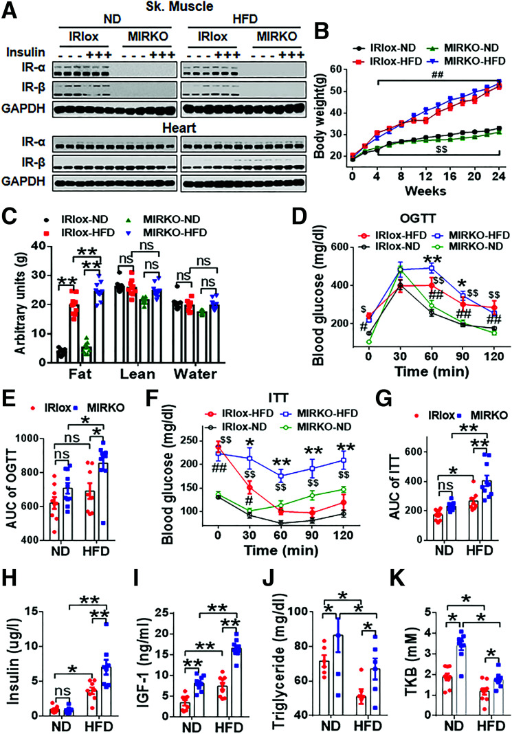
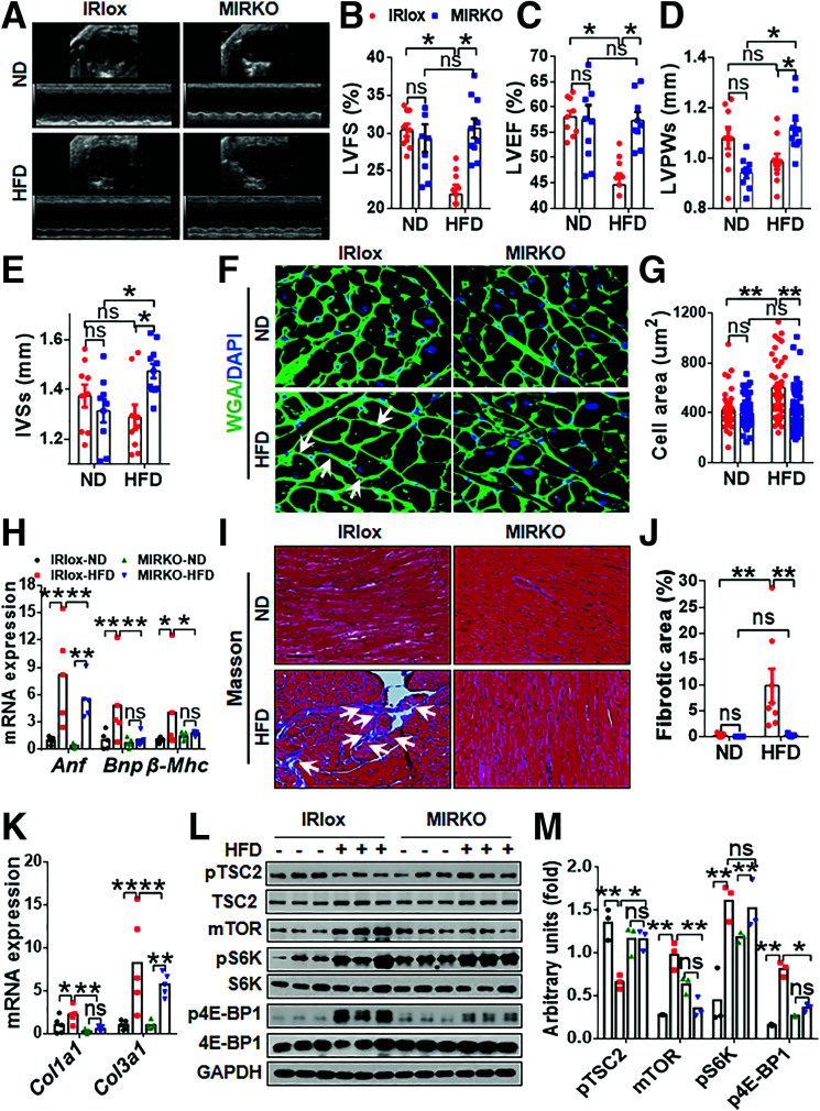
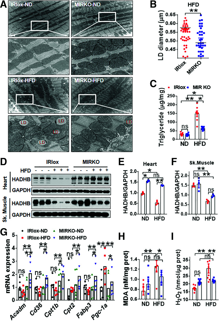
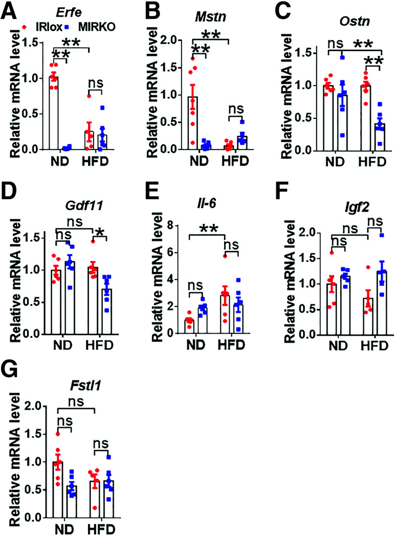
Obesity and type 2 diabetes mellitus (T2DM) are the leading causes of cardiovascular morbidity and mortality. Although insulin resistance is believed to underlie these disorders, anecdotal evidence contradicts this common belief. Accordingly, obese patients with cardiovascular disease have better prognoses relative to leaner patients with the same diagnoses, whereas treatment of T2DM patients with thiazolidinedione, one of the popular insulin-sensitizer drugs, significantly increases the risk of heart failure. Using mice with skeletal musclespecific ablation of the insulin receptor gene (MIRKO), we addressed this paradox by demonstrating that insulin signaling in skeletal muscles specifically mediated cross talk with the heart, but not other metabolic tissues, to prevent cardiac dysfunction in response to metabolic stress. Despite severe hyperinsulinemia and aggregating obesity, MIRKO mice were protected from myocardial insulin resistance, mitochondrial dysfunction, and metabolic reprogramming in response to diet-induced obesity. Consequently, the MIRKO mice were also protected from myocardial inflammation, cardiomyopathy, and left ventricle dysfunction. Together, our findings suggest that insulin resistance in skeletal muscle functions as a double-edged sword in metabolic diseases.


Figures

Figure 1
Figure 1
MIRKO aggravates DIO-induced insulin resistance, leading to hyperinsulinemia and glucose and insulin intolerance. The MIRKO and IRlox control mice were fed with either ND or HFD for 24 weeks, followed by analysis of metabolic parameters including the following. A: Evaluation of the insulin receptor knockout efficiency by Western blot analysis of IR-α and IR-β expression in skeletal muscle (Sk. Muscle) and heart (n = 3 per group). B: Analysis of weight gain in response to feeding with ND and HFD (n = 10 per group). C: Evaluation of whole-body composition using a quantitative MRI system (n = 10 per group). D and E: Oral glucose tolerance tests (OGTT) and quantification of area under the curve (AUC) (n = 8–10). F and G: Insulin tolerance tests (ITT) and quantification of area under the curve (n = 8–10). HK: ELISA of the levels of insulin (H), IGF-1 (I), triglycerides (J), and total ketone bodies (TKB) (K) in the serum of MIRKO and IRlox control mice (n = 8–10). #IRlox HFD vs. IRlox ND, $MIRKO HFD vs. MIRKO ND, *MIRKO HFD vs. IRlox HFD in B, D, and F. Data are expressed as means ± SEM. ns, nonsignificant. *P < 0.05, **P < 0.01; #P < 0.05; ##P < 0.01; $P < 0.05; $$P < 0.01.
Figure 2
Figure 2
MIRKO mitigates DIO-induced LV dysfunction and adverse remodeling. A: Representative images of M-mode echocardiography of MIRKO and IRlox control mice fed with ND and HFD. (n = 8–10). BE: Quantitative analysis of echocardiographic parameters, including LVFS (B), LVEF (C), LVPWs (D), and IVSs (E) (n = 8–10). F and G: Representative images of wheat germ agglutinin staining (WGA) of LV (F) and quantitative analysis of myocardial area (G) of heart sections from MIRKO and the IRlox control mice in response to DIO (n = 3 per group). H: RT-qPCR analysis of the mRNA expression levels of myocardial hypertrophy biomarkers, including Anf, Bnp, and β-Mhc (n = 5 per group). I and J: Analysis of cardiac fibrosis by Masson’s trichrome staining (200× magnification) (I) and quantitative analysis of fibrotic area (%) (J) (n = 3 per group). K: RT-qPCR analysis of the mRNA expression levels of collagen I (Col1a1) and III (Col3a1) (n = 5 per group). L: Western blot analysis of signal transduction pathways mediated by mTORC1 and its effector proteins in the heart, including TSC2, S6K, and 4E-BP1. M: Quantification of panel L. Data are expressed as means ± SEM. ns, nonsignificant. *P < 0.05; **P < 0.01.
Figure 3
Figure 3
MIRKO selectively prevents DIO-induced insulin resistance in heart. A: Western blot analysis of AKT phosphorylation (Thr308 and Ser473) and GSK3β (Ser9) in skeletal muscle (Sk.Muscle) and heart tissues from MIRKO and IRlox control (Ctl) mice after injection of 1.0 units/kg body weight i.p. insulin for 15 min (n = 3 per group). B and C: Quantitative analysis of the ratio of phosphorylation (Phosphor) and total protein expression of AKT and GSK3β in A. Data are expressed as means ± SEM. ns, nonsignificant. *P < 0.05; **P < 0.01.
Figure 4
Figure 4
MIRKO prevents DIO-induced inflammation and apoptosis in the heart. A: Western blot analysis of key biomarkers for inflammation and apoptosis in the heart, including NLRP3, TXNIP, STING (cGAS, STING, pTBK1, TBK1), and apoptotic pathways (Bax, Bcl-2, C-cas3, Cyt c) (n = 3 per group). B and C: Quantitative analysis of the protein expression levels of panel A. D: Quantitative RT-PCR analysis of the mRNA levels of anti-inflammatory (Il-4) and proinflammatory (Tnf, Nfkb, Il-1b, Il-6) cytokines in the heart (n = 5–6). E and F: Representative images (E) and quantitative analysis (F) of apoptosis in heart sections stained with TUNEL in response to DIO. Arrows highlight apoptotic cells. n = 3 per group. Data are expressed as means ± SEM. ns, nonsignificant. *P < 0.05; **P < 0.01.
Figure 5
Figure 5
MIRKO promotes FAO by preventing DIO-induced mitochondrial dysfunction in the heart. A: Representative images of transmission electron microscopy performed in heart tissues with highlight of LD. Scale bars represent 1 μm (n = 2 per group). B: Quantitative analysis of LD diameter (n = 59) from multiple TEM images in the LV of MIRKO and IRlox control mice in response to DIO (n = 2 per group). C: Quantitative analysis of triglyceride content in the heart tissues from MIRKO and IRlox control mice in response to DIO (n = 7 per group). D: Immunoblot analysis of mitochondrial trifunctional protein β-subunit (HADHB) in the heart and skeletal muscle (Sk. Muscle) tissues from MIRKO and IRlox control mice in response to DIO (n = 3 per group). E and F: Quantitative analysis of HADHB level in panel D (n = 3 per group). G: Quantitative RT-PCR analysis of the mRNA expression levels of key biomarkers involved in mitochondrial FAO including Acadm, Cd36, Cpt1b, Cpt2, Fabp3, and Pgc-1α (n = 6 per group). H and I: Analysis of levels of malondialdehyde (MDA), a by-product of lipid peroxidation (H), and H2O2, an indicator of oxidative stress (I) in the heart tissues (n = 6–8). Data are expressed as means ± SEM. ns, nonsignificant; prot, protein. *P < 0.05; **P < 0.01.
Figure 6
Figure 6
MIRKO mimics the effect of DIO on mRNA expression of major myokines in skeletal muscle. AG: Quantitative RT-PCR analysis of the mRNA expression of major myokines implicated in cardiac protection, including myonectin (Erfe) (A), myostatin (Mstn) (B), osteocrin (Ostn) (C), growth differentiation factor 11 (Gdf11) (D), interleukin-6 (Il-6) (E), insulin-like growth factor 2 (Igf2) (F), and follistatin-like 1 (Fstl1) (G) in the skeletal muscle of MIRKO and the IRlox control mice in response to DIO (n = 5–8). Data are expressed as means ± SEM. ns, nonsignificant. *P < 0.05; **P < 0.01.

References

    1. Riehle C, Abel ED. Insulin signaling and heart failure. Circ Res 2016;118:1151–1169 - PMC - PubMed
    1. Guo S. Insulin signaling, resistance, and the metabolic syndrome: insights from mouse models into disease mechanisms. J Endocrinol 2014;220:T1–T23 - PMC - PubMed
    1. Kenny HC, Abel ED. Heart failure in type 2 diabetes mellitus. Circ Res 2019;124:121–141 - PMC - PubMed
    1. Brüning JC, Michael MD, Winnay JN, et al. . A muscle-specific insulin receptor knockout exhibits features of the metabolic syndrome of NIDDM without altering glucose tolerance. Mol Cell 1998;2:559–569 - PubMed
    1. Lavie CJ, Alpert MA, Arena R, Mehra MR, Milani RV, Ventura HO. Impact of obesity and the obesity paradox on prevalence and prognosis in heart failure. JACC Heart Fail 2013;1:93–102 - PubMed

Publication types

MeSH terms