. 2021 Jul 9;373(6551):231-236.
doi: 10.1126/science.abg2264.

An isoform of Dicer protects mammalian stem cells against multiple RNA viruses

Affiliations

An isoform of Dicer protects mammalian stem cells against multiple RNA viruses

Enzo Z Poirier et al. Science. .

Abstract

In mammals, early resistance to viruses relies on interferons, which protect differentiated cells but not stem cells from viral replication. Many other organisms rely instead on RNA interference (RNAi) mediated by a specialized Dicer protein that cleaves viral double-stranded RNA. Whether RNAi also contributes to mammalian antiviral immunity remains controversial. We identified an isoform of Dicer, named antiviral Dicer (aviD), that protects tissue stem cells from RNA viruses-including Zika virus and severe acute respiratory syndrome coronavirus 2 (SARS-CoV-2)-by dicing viral double-stranded RNA to orchestrate antiviral RNAi. Our work sheds light on the molecular regulation of antiviral RNAi in mammalian innate immunity, in which different cell-intrinsic antiviral pathways can be tailored to the differentiation status of cells.


Conflict of interest statement

Competing interests: CRS has an additional appointment as Professor in the Faculty of Medicine at Imperial College London and owns stock options and/or is a paid consultant for Bicara Therapeutics, Montis Biosciences, Oncurious NV, Bicycle Therapeutics and Sosei Heptares, all unrelated to this work. The remaining authors declare no competing interests.

Figures

Figure 1
Figure 1. aviD is an isoform of Dicer that efficiently cleaves dsRNA.
(A) Dicer PCR amplicons using vehicle (Neg), a plasmid coding for Dicer, or mouse small intestine cDNA templates. In addition to a canonical product corresponding to full-length Dicer, an in-frame transcript missing exons 7 and 8 (nucleotides 705 to 1346 of the coding sequence) was detected. This corresponds to an isoform termed antiviral Dicer (aviD) lacking the Hel2i domain of the helicase (white). (B) Immunoblots from wild type Dicer+/+aviD+/+ or Dicer—/—aviD—/— mouse ES cell, HEK293T cell or Dicer+/+aviD+/+ human iPSC lysates before (pre-IP) or after immunoprecipitation with a Dicer/aviD specific antibody. Recombinant Flag-tagged Dicer and aviD were included as controls. (C) Recombinant Flag-tagged Dicer, Dicer catalytically-deficient [Dicer(CD), used as a negative control] or aviD was incubated with synthetic Cy5-labelled dsRNA at 37oC for the indicated time. The reactions were resolved on a denaturing polyacrylamide gel, visualised by Cy5 in-gel fluorescence and Dicer vs aviD cleavage quantitated by densitometry. (D) Increasing concentrations of recombinant LGP2 were added to the in vitro dicing reaction as in (C) and incubated 3 h at 37oC. After densitometric quantitation, siRNA amount was normalized to the amount of siRNA produced in a reaction without LGP2. (E) Immunopurified Flag-tagged Dicer, Dicer catalytically-deficient [Dicer(CD)] or aviD, were incubated with let-7a pre-miRNA at 37oC for 20 min. The reactions were resolved on a denaturing polyacrylamide gel, visualised by Cy5 in-gel fluorescence and Dicer vs aviD cleavage quantitated by densitometry. Data in C-E are pooled from three independent experiments and are plotted as mean ± SEM. ns, not significant; **p < 0.01, ***p < 0.001 (two-way ANOVA [C,D] and Mann-Whitney test [E]).
Figure 2
Figure 2. aviD can mediate antiviral RNAi.
(A-B) HEK293T Dicer—/—aviD—/— cells complemented with Dicer (Dicer+/+aviD—/—) or aviD (Dicer—/—aviD+/+) were infected with SINV (A) or ZIKV (B) at MOI 0.1. Supernatant was collected at the indicated times points and viral content determined by plaque assay. (C-E) HEK293T Dicer—/—aviD—/— cells induced by doxocycline to express Flag-Dicer (C), aviD (D), or catalytically-deficient aviD [aviD(CD)] (E) were infected with SINV-GFP. Flow cytometry was used to monitor the expression of Dicer/aviD via anti-Flag staining and SINV replication via GFP fluorescence. (F) Representative contour plots from 16 h post infection. Boxes represent Flag positive cells defined on the basis of the uninduced controls (top left plot). (G) Dicer+/+aviD—/—or Dicer—/—aviD+/+ HEK293T cells were transfected with siRNA targeting Ago2 (siAgo2) or with control siRNA (siCt) and infected with ZIKV at MOI 0.1. Supernatant was collected at the indicated time points and viral content was determined by plaque assay. (H) Immunofluorescence of Dicer—/—aviD+/+ Dicer+/+aviD—/— HEK293T cells expressing ACE2 infected with SARS-CoV-2 and stained for SARS-CoV-2 N protein (magenta) and dsRNA (white). Scale bar: 20 μm. Graph shows percentage of infected cells. Data in A-H are from one of three independent experiments. Data points represent mean ± SEM, n = 3 biological replicates. ns, not significant; *p < 0.05, **p < 0.01, ***p < 0.001. (two-way ANOVA [A-E,G], unpaired t-test [H]).
Figure 3
Figure 3. aviD is enriched in tissue stem cells.
(A) Small intestine from an Lgr5-GFP reporter mouse was fixed, sectioned and probed for aviD mRNA (magenta) by fluorescence in situ hybridisation. The aviD probe was designed to detect the exon-exon junction specific to aviD and cannot detect Dicer mRNA (Fig. S5A). Lgr5+ stem cells were identified with anti-GFP (white) and nuclei were visualized by DNA staining (Hoechst, blue). Scale bar: 30 μm. Percentage of stem (Lgr5+) or differentiated (Lgr5) cells expressing aviD mRNA was determined on 17 images with at least three villi each. Data points represent mean and error bars are SEM. Mann-Whitney test, ***p < 0.001. (B) aviD or Dicer mRNA was measured by PrimeFlow cytometry in stem (Lgr5+) or differentiated (Lgr5) cells from small intestine or skin isolated from Lgr5-GFP reporter mice or in stem or differentiated cells from hippocampus distinguished by the presence or absence of Sox2 mRNA, respectively.
Figure 4
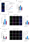
Figure 4. aviD thwarts viral infection in stem cells.
(A) Individual Dicer+/+aviD+/+, Dicer+/+aviD—/— or Dicer—/—aviD+/+ brain organoids were infected with ZIKV and organoid area was monitored for 4 days. Immunofluorescent stanining and confocal microscopy on organoid sections was carried out to identify stem cells by Sox2 expression (green) and infected cells by ZIKV glycoprotein expression (magenta). Scale bar: 100 μm. (B) Production of viral particles from ZIKV-infected organoids was determined by transferring individual organoids into fresh medium at day 3 post infection and collecting the supernatant 24 hours thereafter to determine viral content by plaque assay. (C) Percentage of ZIKV-infected stem cells was measured at 4 days post infection by immunofluorescence on organoids sections. (D) dsRNA in infected stem cells was visualised by immunofluorescence on organoid sections after 4 days of infection. Images show dsRNA (gray) in ZIKV-infected (magenta) stem cells (green). Scale bar: 20 μm. (E) Stem cell division rate was measured by pulsing organoids with EdU at day 3 for 1 h and chasing for 24 h. Organoids section were analysed by immunofluorescence, with Sox2 staining identifying stem cells and EdU staining marking cells in S phase at time of pulsing. (F) Dicer+/+aviD+/+, Dicer+/+aviD—/— or Dicer—/—aviD+/+ brain organoids expressing ACE2 were infected with SARS-CoV-2 for 48 h. Percentage of infected stem cells was determined by immunofluorescence on sections stained for the stem cell marker Sox2 (green) and for the SARS-CoV-2 N protein (magenta). Scale bar: 100 μm. In A-F, mean and SEM are shown. [A,B] n = 16 organoids per condition. [C,D,E] n = 8 organoids per condition, 8 with highest fold change in area at day 4 for Dicer+/+aviD+/+ and Dicer—/—aviD+/+, 8 with lowest fold change in area for Dicer+/+aviD—/—. [F] n = 11 organoids per condition. ns, not significant; *p < 0.05. ***p < 0.001 (two-way ANOVA [A], Mann-Whitney test [B-F]).

Comment in

  • Boosting stem cell immunity to viruses.
    Shahrudin S, Ding SW. Shahrudin S, et al. Science. 2021 Jul 9;373(6551):160-161. doi: 10.1126/science.abj5673. Science. 2021. PMID: 34244399 Free PMC article. No abstract available.
  • Dicing viral RNA in stem cells.
    Zlotorynski E. Zlotorynski E. Nat Rev Mol Cell Biol. 2021 Sep;22(9):586. doi: 10.1038/s41580-021-00406-1. Nat Rev Mol Cell Biol. 2021. PMID: 34326514 Free PMC article.

References

    1. Goubau D, Deddouche S, Reis e Sousa C. Cytosolic Sensing of Viruses. Immunity. 2013;38:855–869. - PMC - PubMed
    1. Witteveldt J, Knol LI, Macias S. MicroRNA-deficient mouse embryonic stem cells acquire a functional interferon response. eLife. 2019;8:e44171. - PMC - PubMed
    1. D’Angelo W, Gurung C, Acharya D, Chen B, Ortolano N, Gama V, Bai F, Guo Y-L. The Molecular Basis for the Lack of Inflammatory Responses in Mouse Embryonic Stem Cells and Their Differentiated Cells. J Immunol. 2017;198:2147–2155. - PMC - PubMed
    1. Hong X-X, Carmichael GG. Innate Immunity in Pluripotent Human Cells: ATTENUATED RESPONSE TO INTERFERON-β. J Biol Chem. 2013;288:16196–16205. - PMC - PubMed
    1. Maillard PV, Veen AG, Poirier EZ, Reis e Sousa C. Slicing and dicing viruses: antiviral RNA interference in mammals. EMBO J. 2019;38 doi: 10.15252/embj.2018100941. - DOI - PMC - PubMed

Publication types

MeSH terms