. 2021 Jul 13;118(28):e2018297118.
doi: 10.1073/pnas.2018297118.

Temporal compartmentalization of viral infection in bacterial cells

Affiliations

Temporal compartmentalization of viral infection in bacterial cells

Audrey Labarde et al. Proc Natl Acad Sci U S A. .

Abstract

Virus infection causes major rearrangements in the subcellular architecture of eukaryotes, but its impact in prokaryotic cells was much less characterized. Here, we show that infection of the bacterium Bacillus subtilis by bacteriophage SPP1 leads to a hijacking of host replication proteins to assemble hybrid viral-bacterial replisomes for SPP1 genome replication. Their biosynthetic activity doubles the cell total DNA content within 15 min. Replisomes operate at several independent locations within a single viral DNA focus positioned asymmetrically in the cell. This large nucleoprotein complex is a self-contained compartment whose boundaries are delimited neither by a membrane nor by a protein cage. Later during infection, SPP1 procapsids localize at the periphery of the viral DNA compartment for genome packaging. The resulting DNA-filled capsids do not remain associated to the DNA transactions compartment. They bind to phage tails to build infectious particles that are stored in warehouse compartments spatially independent from the viral DNA. Free SPP1 structural proteins are recruited to the dynamic phage-induced compartments following an order that recapitulates the viral particle assembly pathway. These findings show that bacteriophages restructure the crowded host cytoplasm to confine at different cellular locations the sequential processes that are essential for their multiplication.

Keywords: bacterial cell compartmentalization; bacteriophage; phage DNA replication; virus assembly; virus infection.


Conflict of interest statement

The authors declare no competing interest.

Figures
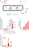
Fig. 1.
Fig. 1.
SPP1 DNA replication and encapsidation. (A) Schematics of the main steps of tailed bacteriophage infection exemplified by SPP1. (B) Quantification of SPP1 DNA synthesis (empty circles) and encapsidation (filled circles) determined by qPCR of total and DNase-protected DNA, respectively, in B. subtilis GSY10004 infected with SPP1lacO64. The amount of SPP1 gene 6 DNA was divided by the amount of B. subtilis gyrA reporter DNA considering that there are 2.7 nucleoids on average per cell (SI Appendix, Fig. S1 C and D) to calculate the number of SPP1 genomes per bacterium. The total number of infectious particles (filled squares; ordinate on the right) was quantified by phage titration of bacteria lysed with 10% chloroform. Note that the DNA molecule encapsidated in SPP1lacO64 particles (∼45.1 kbp) is 1.07-fold longer than the phage genome size (42.3 kbp) (15, 66), a factor that was used to convert phage particle counts into genome equivalents and reciprocally. (Right) A snapshot of phage DNA and particle yields at 30 min p.i., when experiments were stopped because cell lysis initiated. The values in bold were obtained experimentally, and those in gray were calculated based on the figure data. Note that the calculation of ∼46 capsids corresponds to particles that protect viral DNA but are noninfectious (i.e., tailless DNA-filled capsids and eventually defective tailed phage particles). The data are the average from three independent experiments. The biosynthetic effort accounting for phage DNA and virion structural proteins synthesis is depicted underneath the infection time points (Dataset S1). (C) Percentage of encapsidated DNA calculated from the data in B. (D) Phage DNA synthesis at 10 and 25 min p.i. in B. subtilis GSY10004 infected with SPP1lacO64 (15) (red), SPP1lacO64gp40 (defective in initiation of DNA synthesis) (yellow), and SPP1lacO64gp2 (defective in DNA packaging) (green). Total DNA (bars total height) and encapsidated, DNase-protected DNA (overlapping full bars) were quantified by qPCR. The data are the average from three independent experiments.
Fig. 2.
Fig. 2.
Cellular localization of B. subtilis replisome proteins during SPP1 infection. (A) PolC-GFP (Top) and mCFP-DnaX (Bottom) in bacteria noninfected (Left) and infected with SPP1lacO64 (Right) late p.i. (see SI Appendix, Fig. S2B for experimental setup). The B. subtilis strains produce LacI-mCherry constitutively. The signals on the RFP channel are displayed in magenta, while signals on GFP and CFP channels are in green on overlays, respectively. (Scale bar, 2 µm.) The arrowheads show the position of host replication protein foci in noninfected bacteria, and the arrow identifies a focus of mCFP-DnaX that does not colocalize with SPP1 DNA in an infected bacterium. (B) Quantitative analysis of B. subtilis replisome proteins localization relative to the SPP1 DNA focus in bacteria early p.i. according to the gray scale legend on the right. More than 100 cell counts were made for each strain.
Fig. 3.
Fig. 3.
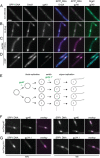
Cellular localization of SPP1 proteins engaged in phage DNA replication (gp40 helicase) and in the switch of genome replication mode (gp34.1 5′-3′ exonuclease). (AD) Localization of SPP1 DNA, mCFP-DnaX (B. subtilis clamp loader subunit), and gp40-mCitrine in noninfected bacteria (A) and in SPP1lacO64-infected bacteria untreated (B), incubated with 200 µM HPUra (C), or with 10 mM DNP (D). Drugs were added at 21 min p.i. to the infected culture that was spotted in an agarose pad containing the same concentration of drug 3 min later. Fluorescence microscopy acquisitions were made between 50 and 60 min p.i. (Scale bar in A, 2 µm.) (E) Scheme of SPP1 replication initiated by a theta mode, leading to an exponential increase of independent circular genome molecules followed by a switch to sigma replication that generates linear concatemers for DNA packaging. SPP1 mutants blocking replication initiation (gp40) or severely affecting the switch from theta-to-sigma mode (gp34.1) are displayed in green. (F and G) Colocalization of gp40-mCitrine (F) and gp34.1-mCitrine (G) with phage DNA in infections with SPP1 mutants defective in gp40 and in gp34.1 production (transcomplementation conditions), respectively. Images at early (Left) and late (Right) stages of infection (SI Appendix, Fig. S2B) are displayed. (Scale bar in F, 2 µm.)
Fig. 4.
Fig. 4.
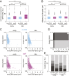
Temporal evolution of the viral DNA compartment. (A) Percentage of cell area occupied by the SPP1 DNA focus in B. subtilis GSY10025 monoinfected (one viral DNA focus per cell) with SPP1lacO64 (blue) or with the DNA packaging-defective mutant SPP1lacO64gp2 (magenta) at early and late times p.i. (SI Appendix, Fig. S2B). More than 800 cells from three independent infections were analyzed. The same datasets were used for all analyses in the figure. (B) Average distance of the SPP1 DNA focus center of mass to the proximal cell pole. Boxplots in A and B show the median of the sample data as a white solid line and draws point as outliers if they are greater than q3+w(q3q1) or less than q1w(q3q1) in which q1 and q3 are the 25th and 75th percentiles of the sample data, respectively, and w=1.5. Statistical significance between conditions in A and in B were calculated using the Mann–Whitney U nonparametric test (****P < 0.0001; ** = 0.001 < P < 0.01; * = 0.01 < P < 0.05; ns = P > 0.05). (C) Surface aspect of the phage DNA focus. The width-to-length ratio (shape factor) of the segmented focus is plotted relative to its area. The vertical and horizontal limits separating all quadrants are defined, respectively, as the median values of the DNA surface and the shape factor from the dataset associated with the infection with SPP1lacO64. Foci shape factors were quantified when DNA encapsidation took place (blue; infection with SPP1lacO64, abbreviated lacO64 in the figure) or was blocked (magenta; infection with SPP1lacO64gp2, abbreviated gp2 in the figure) at early (Left) and late (Right) times p.i. (D) Schematic visual aid of the DNA focus shape (shape cartoon in white) observed in each quadrant of the plot in C. (E) Percentage of viral DNA compartment shape types in the quadrants of C displayed according to the gray scale of D.
Fig. 5.
Fig. 5.
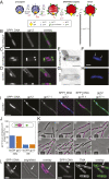
Localization of SPP1 DNA and viral procapsids in infected bacteria. (AF) Ultrastructure of B. subtilis YB886 noninfected and infected with SPP1lacO64gp2. Sections 90-nm thick of bacteria, fixed 35 min p.i., and sectioned at cryogenic temperatures were left untreated (A and B), incubated with 25 U Benzonase (digests DNA) (C and D), or treated with 10 µg/mL trypsin [hydrolyses proteins but phage particles are resistant (43)] (E and F). Sections were labeled with a mouse monoclonal anti-DNA antibody and a rabbit polyclonal anti-procapsid serum visualized with 5 and 15 nm colloidal gold, respectively. (Scale bars in the EM sections, 1 µm.) High-resolution images of the electron micrographs in AF can be found in SI Appendix. (G and H) Epifluorescence of fixed bacteria stained with DAPI from the cultures used for immunoEM experiments. (I and J) Cytological localization of LacI-mCherry that decorates SPP1 DNA (magenta in J) and of mCitrine-gp11 that is incorporated in procapsids (green in J) in B. subtilis GSY10025 noninfected and infected with SPP1lacO64gp2. Gray dotted lines display the cell outline defined from brightfield imaging. (The scale bars represent 2 µm in the fluorescence microscopy images of G and I.) (K) Scheme of procapsid formation and DNA packaging initiation steps during SPP1 viral particle assembly. All components of the SPP1 procapsid and of the DNA packaging apparatus are presented. The major capsid protein gp13 lattice is displayed as a dashed line delimiting the procapsid and by a thin icosahedral outline in the expanded capsid. MCitrine is displayed as a green cylinder in the mCitrine-gp11 fusion protein. Mutants used in this study that block synthesis of different SPP1 proteins engaged in procapsid assembly and DNA packaging are also presented. (L) Time-lapse of filamented B. subtilis GSY10025 (producing mCitrine-gp11) infected with SPP1lacO64gp2. Cell division was inhibited 1 h before infection by the addition of 1 µg/mL PC190723. Signals on the RFP and YFP channels are displayed in magenta and green in overlays, respectively. Time p.i. is displayed on the right top corner of the overlay. The arrow shows dispersion of SPP1 DNA (magenta) in the agarose pad upon cell disruption at 85 min p.i. while clusters of procapsids (green) maintain their integrity. (Scale bar, 5 µm.) (M) Infection of filamented B. subtilis GSY10025 with SPP1lacO64gp6 defective in production of the procapsid portal protein. Portal-less procapsids assembled during infection are not competent to package viral DNA (42). An infected cell late p.i. is presented surrounded by noninfected bacteria. Experimental conditions are as in L.
Fig. 6.
Fig. 6.
Localization of SPP1 DNA and DNA-filled particles in infected bacteria. (A) Scheme of the SPP1 viral particle assembly pathway. MCitrine is displayed as a green cylinder in the gp12-mCitrine fusion protein. (BD) Localization of SPP1 DNA and of capsid auxiliary protein gp12-mCitrine that decorates DNA-filled particles in B. subtilis GSY10024 infected with SPP1lacO64gp2 (B), SPP1lacO64gp12 (C), and SPP1lacO64 (D). Images at late stages of infection (SI Appendix, Fig. S2B) are displayed. Gray dotted lines display cell outlines defined from brightfield imaging. (Scale bar, 2 µm.) (E and G) Thin sections of B. subtilis YB886 infected with SPP1lacO64gp12 (E) and SPP1lacO64 (G) immunolabeled as in Fig. 5 A and B. (Scale bars, 1 µm.) High-resolution images of the electron micrographs in E and G can be found in SI Appendix. (F and H) DAPI staining of fixed bacteria from the cultures used for immunoEM experiments. Arrows in F identify individualized DAPI foci attributed to warehouses. (Scale bar in F, 2 µm.) (I) Infection of B. subtilis GSY10096-filamented cells with SPP1lacO64. The fluorescent reporters of SPP1 DNA, capsids, and tails were LacI-mCherry (magenta in overlays), gp12-mCFP (blue), and gp17.1-mCitrine (green), respectively. Images were taken late p.i. (Scale bar, 5 µm.) (J) Quantification of DNA-filled particles overlapping with the phage DNA compartment of cells monoinfected with SPP1lacO64 and SPP1lacO64gp12 at early and late times p.i. Datasets of >500 cells were analyzed. (K) Snapshots from time-lapse Movie S1 of B. subtilis GSY10024, producing gp12-mCitrine, immobilized in a CellAsic microfluidics system and infected with SPP1lacO64gp12. The first photo shows cells before infection followed by snapshots at time points indicated on the top left corner. They illustrate the temporal and spatial program of SPP1 DNA compartment and viral particles warehouses formation (displayed in magenta and green, respectively). Cells are multi-infected. The white arrow identifies a bacterium undergoing division. The arrowheads show the position of DNA compartments at 67 min p.i. that are disassembled in cell ghosts at 82 min p.i. but prior to full bacterial lysis (110 min p.i.). Images were enhanced as described in the Movie S1 legend. (L) Cell septation through the SPP1 DNA compartment (displayed in magenta) in bacteria producing LacI-mCherry (strain GSY10004) infected with SPP1lacO64. The cell membrane contour is stained with TMA-DPH (rendered in green) on the right side. The white arrow shows septum formation. To enhance the signal from the membrane staining (TMA-DPH), the raw image was denoised (two-dimensional Gaussian filter, radius = 1, 25 pixel) and background was removed using a rolling ball (sigma = 10 pixels) using Fiji (67).

References

    1. Surovtsev I. V., Jacobs-Wagner C., Subcellular organization: A critical feature of bacterial cell replication. Cell 172, 1271–1293 (2018). - PMC - PubMed
    1. Cornejo E., Abreu N., Komeili A., Compartmentalization and organelle formation in bacteria. Curr. Opin. Cell Biol. 26, 132–138 (2014). - PMC - PubMed
    1. Iancu C. V., et al. ., Organization, structure, and assembly of α-carboxysomes determined by electron cryotomography of intact cells. J. Mol. Biol. 396, 105–117 (2010). - PMC - PubMed
    1. McHugh C. A., et al. ., A virus capsid-like nanocompartment that stores iron and protects bacteria from oxidative stress. EMBO J. 33, 1896–1911 (2014). - PMC - PubMed
    1. Schmid M., Speiseder T., Dobner T., Gonzalez R. A., DNA virus replication compartments. J. Virol. 88, 1404–1420 (2014). - PMC - PubMed

Publication types