

Direct RT-PCR amplification of SARS-CoV-2 from clinical samples using a concentrated viral lysis-amplification buffer prepared with IGEPAL-630
- PMID: 34244543
- PMCID: PMC8270935
- DOI: 10.1038/s41598-021-93333-2
Direct RT-PCR amplification of SARS-CoV-2 from clinical samples using a concentrated viral lysis-amplification buffer prepared with IGEPAL-630
Abstract
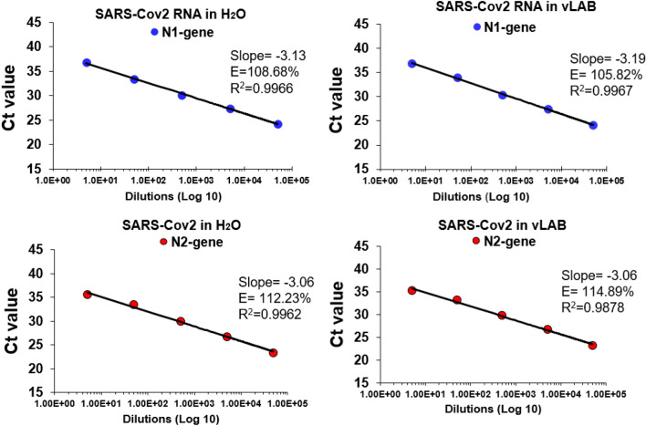
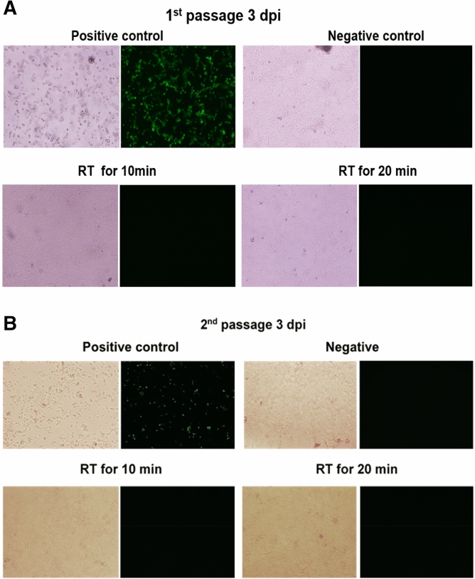
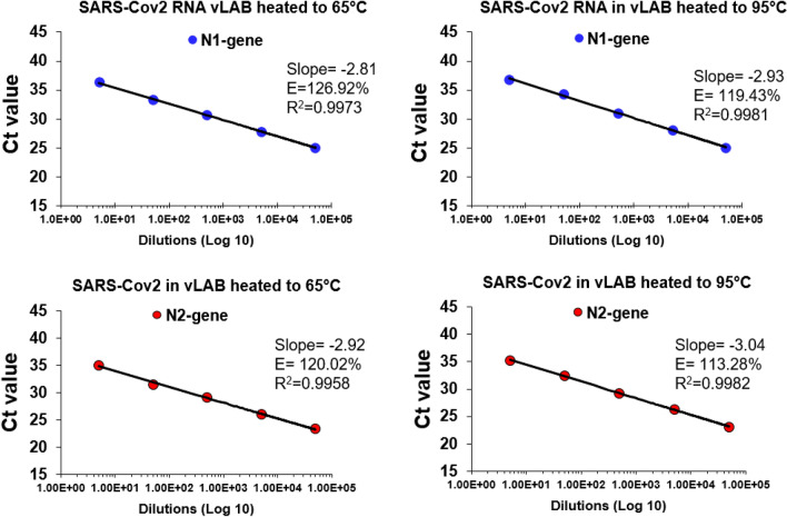
The pandemic of 2019 caused by the novel coronavirus (SARS-CoV-2) is still rapidly spreading worldwide. Nucleic acid amplification serves as the gold standard method for confirmation of COVID-19 infection. However, challenges faced for diagnostic laboratories from undeveloped countries includes shortage of kits and supplies to purify viral RNA. Therefore, it is urgent to validate alternative nucleic acid isolation methods for SARS-CoV-2. Our results demonstrate that a concentrated viral lysis amplification buffer (vLAB) prepared with the nonionic detergent IGEPAL enables qualitative detection of SARS-CoV-2 by direct Reverse Transcriptase-Polymerase Chain Reaction (dRT-PCR). Furthermore, vLAB was effective in inactivating SARS-CoV-2. Since this method is inexpensive and no RNA purification equipment or additional cDNA synthesis is required, this dRT-PCR with vLAB should be considered as an alternative method for qualitative detection of SARS-CoV-2.
© 2021. The Author(s).
Conflict of interest statement
The authors declare no competing interests.
Figures
Similar articles
-
An in-well direct lysis method for rapid detection of SARS-CoV-2 by real time RT-PCR in eSwab specimens.J Virol Methods. 2021 Mar;289:114062. doi: 10.1016/j.jviromet.2021.114062. Epub 2021 Jan 8. J Virol Methods. 2021. PMID: 33428990 Free PMC article.
-
Comparative effects of viral-transport-medium heat inactivation upon downstream SARS-CoV-2 detection in patient samples.J Med Microbiol. 2021 Mar;70(3):001301. doi: 10.1099/jmm.0.001301. Epub 2021 Mar 18. J Med Microbiol. 2021. PMID: 33734960 Free PMC article.
-
Development, deployment and in-field demonstration of mobile coronavirus SARS-CoV-2 Nucleic acid amplification test.J Med Microbiol. 2021 Apr;70(4):001346. doi: 10.1099/jmm.0.001346. J Med Microbiol. 2021. PMID: 33856292 Free PMC article.
-
Sensitivity of ID NOW and RT-PCR for detection of SARS-CoV-2 in an ambulatory population.Elife. 2021 Apr 20;10:e65726. doi: 10.7554/eLife.65726. Elife. 2021. PMID: 33876726 Free PMC article.
-
Pooled Testing Strategies for SARS-CoV-2 diagnosis: A comprehensive review.Diagn Microbiol Infect Dis. 2021 Oct;101(2):115432. doi: 10.1016/j.diagmicrobio.2021.115432. Epub 2021 May 17. Diagn Microbiol Infect Dis. 2021. PMID: 34175613 Free PMC article. Review.
Cited by
-
Monitoring SARS-CoV-2 Surrogate TGEV Individual Virions Structure Survival under Harsh Physicochemical Environments.Cells. 2022 May 27;11(11):1759. doi: 10.3390/cells11111759. Cells. 2022. PMID: 35681454 Free PMC article.
-
Direct Lysis RT-qPCR of SARS-CoV-2 in Cell Culture Supernatant Allows for Fast and Accurate Quantification.Viruses. 2022 Feb 28;14(3):508. doi: 10.3390/v14030508. Viruses. 2022. PMID: 35336915 Free PMC article.
-
Evolution at Spike protein position 519 in SARS-CoV-2 facilitated adaptation to humans.Npj Viruses. 2024 Jul 9;2(1):29. doi: 10.1038/s44298-024-00036-2. Npj Viruses. 2024. PMID: 40295673 Free PMC article.
-
Integrated and streamlined microfluidic device for molecular diagnosis of pathogen through direct PCR amplification.Heliyon. 2025 Jan 22;11(2):e42183. doi: 10.1016/j.heliyon.2025.e42183. eCollection 2025 Jan 30. Heliyon. 2025. PMID: 39925347 Free PMC article.
-
A dual paper-based nucleic acid extraction method from blood in under ten minutes for point-of-care diagnostics.Analyst. 2023 Jun 26;148(13):3036-3044. doi: 10.1039/d3an00296a. Analyst. 2023. PMID: 37265396 Free PMC article.
References
Publication types
MeSH terms
Substances
Grants and funding
LinkOut - more resources
Full Text Sources
Medical
Miscellaneous
